SHORT COMMUNICATION
Oncoplastic Technique for the Elimination
of the Lateral ‘‘Dog Ear’’ During Mastectomy
Krishna B. Clough, MD, Eleanore J. D. Massey, MRCS,
G. K. Mahadev, MD, MRCS, AFRCS, FRCS, Gabriel J. Kaufman, MD,
Claude Nos, MD, and Isabelle Sarfati, MD
The Paris Breast Centre- L’Institut du Sein
n
Abstract: Following a mastectomy, both the cosmetic and functional results can be impaired by the presence of a lat-eral ‘‘dog ear.’’ This is a particular problem in women with a large body habitus giving an increased amount of adipose tis-sue lateral to the breast. The standard approaches to this operation of horizontal or oblique incisions often results in an uncomfortable, unsightly lateral ‘‘dog ear’’. We describe a modification to the standard mastectomy incision that allows extensive excision of the lateral adipose tissue, re-draping the skin over the chest wall, thus eliminating the ‘‘dog ear.’’ The mastectomy is performed through two oblique incisions originating in the axillary skin crease encompassing the nipple areo-lar complex, followed by extensive lateral fat excision. A distance of 2–3 cm is kept between the superior limit of the two incisions. At closure the lateral skin flap is advanced superiomedially on the chest wall without tension. This simple and reproducible technique improves cosmesis and patient satisfaction following modified radical mastectomy by eliminating the lateral ‘‘dog ear.’’ nKey Words: advancement flap, dog ear, mastectomy, oncoplastic surgery
A
modified radical mastectomy can be performed by a transverse incision or an oblique incision. In a patient with large body habitus and an excess axillary fat pad achieving a good functional and cosmetic result can be challenging. Failing to address the axil-lary fat pad at the primary surgery may lead to lateral skin folds, or ‘‘dog ears,’’ which are neither aestheti-cally pleasing nor comfortable (Fig. 1). Patients often complain that this excessive fat pad overhangs the bra causing discomfort. It is the breast surgeons’ responsi-bility to remove the excess fat pad during mastectomy, thereby avoiding a lateral ‘‘dog ear’’ and the need for further surgery, and ensuring optimal patient comfort.Several techniques for addressing this problem have been described and published (1–5). However, these do not always satisfactorily address the issue and may lead to further complications. The fish tail technique (2,3) results in a T-junction which increases the risk of wound tension, delayed wound healing, and necrosis.
We describe a new oncoplastic technique that allows the elimination of the excess tissue with mini-mal skin loss. The two essential principles of this tech-nique are the placing of an incision in the axillary crease and the creation of a lateral skin flap for re-draping the excess skin and fat medially. This medial shift of excess skin eliminates the ‘‘dog ear,’’ enables closure without tension, and leaves enough skin should the patient choose to undergo reconstruction.
DESCRIPTION OF THE TECHNIQUE
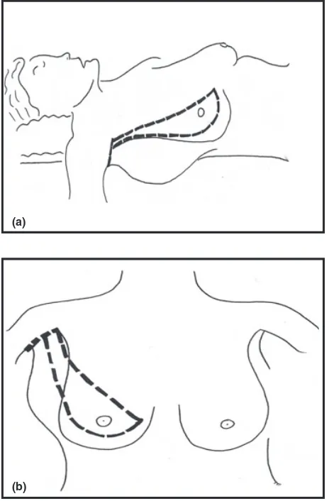
Three mastectomy incisions are marked: two start at the axillary fold. These two oblique lines are ini-tially parallel then diverge as an ellipse to include the breast (Fig. 2a,b). The medial incision initially runs parallel to the lateral edge of pectoralis major then curves over the breast superior to the nipple areolar complex; the lateral incision starts 2–3 cm posteriorly and angles toward the lower outer quadrant, then curves back medially to join the first incision in the lower inner quadrant (Fig. 3). The third incision is placed in the axillary fold, extending from the medial incision posteriorly for as much as is required to medi-alize the excess tissue. The mastectomy is completed Address correspondence and reprint requests to: Krishna B. Clough,
MD, Paris Breast Center-L’Institut du Sein, 7 Avenue Bugeaud, 75116 Paris, France, or e-mail: [email protected].
DOI: 10.1111/tbj.12011
2012 Wiley Periodicals, Inc., 1075-122X/12
in a standard fashion. The tail of the breast is easily visualized. The wide access allows extensive excision of the excess axillary fat, including the fat lying between the superficial fascia and the superficial, or posterior, surface of the latissimus dorsi muscle. The undermining should thus extend well below the ante-rior boarder of the latissimus. The lateral skin flap should be thick enough to avoid any vascular risk (1–3 cm). The lateral flap is then advanced superior-medially so as to re-drape the chest wall and close the defect. Quilting of this flap to the anterior chest wall will reduce the potential cavity and hence reduce the post mastectomy seroma rate (6). The resulting wound resembles an inverted ‘L’ (Fig. 4), with the axillary incision completely hidden under the arm.
Figure 1. Post mastectomy ‘dog ear’ – the mastectomy was per-formed through a standard oblique incision.
(b) (a)
Figure 2. Incisions. (a) Lateral View: The horizontal incision is in the axillary fold. (b) Anterior View: The medial incision runs parallel to the border of pectoralis major before curving medially.
Figure 3. Detail of proximal incisions in relation to the muscles.
Figure 4. Position of the final wound.
The skin is closed in two layers with 3⁄0 Monocryl, with inverted dermal sutures and a subcuticular run-ning suture. Two suction drains are usually placed: one in the axilla and one in the mastectomy cavity.
DISCUSSION
There are two main benefits: functional and cos-metic. The patients no longer return postoperatively complaining of having a ‘new breast’ beneath their arm. Their arm movements are not impeded by the
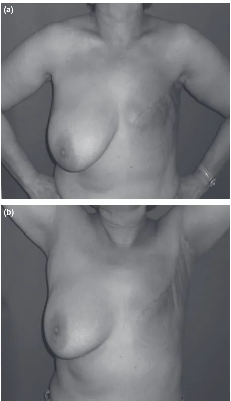
excess volume and they can wear a bra with comfort. The cosmetic outcome is considerably improved by the absence of the ‘‘dog ear’’ and a streamlined silhou-ette. There is no functional disadvantage conferred by these ‘L’ shaped incisions. There is no impingement of the shoulder joint because they do not cross the axil-lary fold (7). They lie in an ideal location for future reconstruction should the patient so wish. As long as the skin flaps are not shaved too thinly, and the obli-que scars are no more than 3 cm apart at their origin allowing closure without tension, these scars heal well giving a good aesthetic result (Fig. 5a,b). One should not perform extensive resection of the axillary skin: the dog ear is avoided by fat excision with medializa-tion of the excess skin. Extensive skin resecmedializa-tion in the axillary fold leads to complications, such as delayed wound healing and wound contracture.
CONCLUSION
Following mastectomy, patients with lateral ‘‘dog ears’’ complain of long-term discomfort, unsightliness, and difficulty wearing a bra. Although many different techniques have been proposed, none of them have emerged as standard, demonstrating the need for a universal solution. This ‘L’ scar technique consistently eliminates the excess lateral tissue. This surgical tech-nique is simple, safe, and reproducible for obese patients requiring mastectomy.
REFERENCES
1. Farrar WB, Fanning WJ. Eliminating the dog-ear in modified radical mastectomy.Am J Surg1988;156:401–402.
2. Nowachi MP, Towpik E, Tcho´rzewska H. Early experience with ‘fish-shaped’ incision for mastectomy. Eur J Surg Oncol
1991;17:615–617.
3. Hussein M, Daltrey IR, Dutta S,et al.Fish-tail plasty: a safe technique to avoid dog-ear deformity.Breast2004;13:206–209.
4. Mirza M, Sinha KS, Forets-Mayer K. Tear-drop incision for mastectomy to avoid dog ear deformity. Ann R Coll Surg Engl
2003;85:131.
5. Devalia H, Chaudhry A, Rainsbury RM,et al.An oncoplastic technique to reduce the formation of lateral ‘dog-ears’ after mastec-tomy.Int Semin Surg Oncol2007;4:29.
6. Kuroi K, Shimozuma K, Taguchi T,et al.Effect of mechani-cal closure of dead space on seroma formation after breast surgery.
Breast Cancer2006;13:260–265.
7. Ed Edlich RF, Calr BA. Predicting scar formation: from ritual practice (Langer’s lines) to scientific discipline (static and dynamic skin tensions).J Emerg Med1998;16:759–60.
(a)
(b)
Figure 5. Mastectomy through an ‘‘L’’ incision. (a) All the excess lateral tissue has been excised. (b) The horizontal incision is in the axillary crease and does not impair arm movement.