ISSN 0973-4562 Volume 9, Number 23 (2014) pp. 13917-13924 © Research India Publications
http://www.ripublication.com
Paper Code: 27334 - IJAER
Identification of
Plasmodium Falciparum
Phase in Red
Blood Cells using Artificial Neural Networks
Kusworo Adi 1), Sri Pujiyanto 2), Rahmat Gernowo 1) Adi Pamungkas 1), Ari Bawono Putranto 1)
1
Department of Physics, the Faculty of Science and Mathematics, Diponegoro University, Indonesia
2
Department of Biology, the Faculty of Science and Mathematics, Diponegoro University, Indonesia
Abstract
Malariae is a medical emergency that must be treated at this time because it has been infecting millions of people. Parasite that causes malariae in human body consists of four types of plasmodium species: P. falciparum, P. vivax, P. ovale, and P.malariae. P. falciparum and P. vivax are the most common type, but the most malignant is P. falciparum. P. falciparum can lead to organ failure and abnormality of the patient's blood. P. falciparum also cause cerebral malariae, if not addressed promptly can lead to death. The analysis carried out by the doctors and the laboratory worker at the moment is still in the conventional manner, namely direct observation using optical microscopy. On the other hand, the need of convenience, practicality and accuracy at this time has become something that is regarded as a necessity in the treatment of malariae. This research was conducted to design the system such as hardware and software that can detect malariae automatically using microscopy imaging. The designed hardware is a modified form of a digital microscopy to acquire images of red blood cell samples, while the software is used to identify the phase of P. falciparum using back propagation artificial neural network. Neural network training process in this research using of 10 images data, consist of 14 Plasmodium falciparum (3 gametocyte phase, 3 schizont phase, 8 trophozoite phase). The neural network testing process using 9 images; consist of 12 plasmodium falciparum (4 gametocyte, 3 schizont, 5 trophozoite). The acuracy of system identification of P. falciparum phase was 87.5%.
1. INTRODUCTION
Malariae is a global problem that currently infecting millions of people in 90 countries annualy. Malariae is caused by a parasite that infects red blood cells that is transmitted through the bite of the anopheles mosquito. In addition, malariae can also be transmitted through blood transfusions [1]. Parasite that causes malariae in human consists of four types of plasmodium species: P. falciparum, P. vivax, P. ovale, and promptly can lead to death [2]. Therefore, malariae is a medical emergency that must be treated at this time. The analysis carried out by the doctors and the laboratory worker at the moment still in the conventional manner, namely direct observation using optical microscopy. On the other hand, the need for convenience, practicality and accuracy has become a necessity in the treatment of malariae.
This research is develop automatic malariae disease identification system to diagnosis of malariae quickly and accurately. Besides, the issue of using local materials and affordable prices became major consideration in this research. In this research we developed detection system of malariae disease with microscopy imaging technique. The designed system consist of two main parts: hardware and software. The hardware development is done by modifying a conventional microscopy into a digital microscopy which its motion and image capture process are controlled by a computer in search for the red blood cells. The software development is done by implementing a method that has been developed previously. In the previuos work, we has been done implemented this methods in tuberculosis bacteria [3,4,5,6]. The morphological method is implemented as the parameter for image pattern recognition. Then the classification process was used Back Propagation Artificial Neural Network (BP-ANN) with three output classes which represent trophozoite phase, schizont phase, or gametocyte phase. Output of this research can provide faster, more practical and accurate analysis result than the conventional microscopy. It is also offer more affordable price than digital microscopy on the market, where digital microscopy on the market only for digital capture process without analysis software and microscopy motion controlled by computer.
2. THEORY
99.7% and a sensitivity of 94%. While identification result of infection stage showed an average specificity of 91.2% and an average sensitivity of 78.8% [11].
Tek et al developed research to detect and identify malariae parasite automatically. The process of image proccessing was begun with improvement of image intensity so that the intensity difference between foreground and background
becoming more clearly. Then the normalization proccess is done based on the average RGB values. The feature extraction is done based on the feature of the color histogram, local area granulometry, and shape measurement vector. The process of classification is done using k-nearest neighbor classifier algorithm. Classification results obtained in the study showed a very high accuracy [12].
Savkare and Narote developed a digital image proccessing system capable of counting and classifying red blood cells infected with malariae. The used segmentation method is thresholding toward the green components using otsu method. Feature extraction was done by using two methods: Feature-Based Shape (Radius, Perimeter, compactness) and Statistical Parameters (Skewness, Kurtosis, Energy), while the classification of plasmodium type used Support Vector Machine
algorithm. The system designed by Savkare and Narote for 20 blood images produced high accuracy [13].
Das et al conducted a study with the aim of characterizing and classifying malariae parasites (Plasmodium vivax and Plasmodium falciparum) microscopically. Image segmentation was done using watershed method and classification using several algorithms: Bayesian learning, and Support Vector Machine (SVM). The results obtained by Das et al showed that the Bayesian learning algorithm has higher accuracy in the classification of the malariae parasite that is 84% compared with the 83.5% accuarcy of SVM algorithm [14].
3. METHOD
Identification of plasmodium falciparum development phase in this research included image acquisition, image enhancement, adaptive color segmentation with thresholding method, feature extraction based on binary pattern, and the classification of back propagation artificial neural network. The block diagram in this study is shown in Figure 3.1.
Figure 3.1. Block Diagram of Research
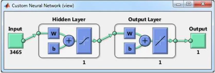
components (Red, Green, Blue) to grayscale. Then the image quality was improved to remove noise using median filter. Once the image quality was enhanced, the next process was image segmentation through thresholding proccess using Otsu method. Segmentation process performed to separate plasmodium and background. The result obtained from image segmentation process was plasmodium falciparum form of binary image. This binary image pattern was then used as input in the process of training and testing in the classification using back propagation artificial neural network algorithm. Artificial neural network used consist of three layers: input layer, hidden layer and output layer. The form of input layer was the binary pattern of morphological plasmodium, while the hidden layer connecting the input layer and the output layer using bipolar sigmoid activation function, and the output layer was the classification result of three plasmodium classes: gametocyte, schizont and
trophozoite. Architecture of the artificial neural network in this study is shown in Figure 3.2.
Figure 3.2. Architecture of back propagation artificial neural network
The training process of artificial neural networks in this study used 10 data of images consist of 14 plasmodium falciparum (3 gametocyte phase, 3 schizont phase, 8 trophozoite phase), while the testing process used 9 images consist of 12 plasmodium falciparum (4 gametocyte, 3 schizont, 5 trophozoite).
4. RESULTS AND DISCUSSION
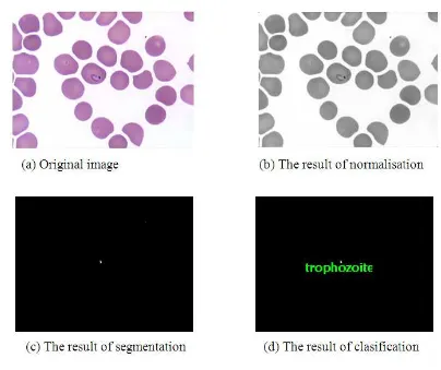
obtained from image segmentation process was plasmodium falciparum form of binary image. Image processing in this study is shown in Figure 4.1.
Figure 4.1. The process of image processing
Figure 4.2. The image of artificial neural network test result
While the whole results of testing the artificial neural network in this study are shown in Table 4.1.
Table 4.1. The test results of classification using artificial neural network algorithm
No. Automatic detection Manual detection
The number of Parasite
Development phase
The number of Parasite
Development phase
1 1 Gametocyte 1 Gametocyte
2 1 Gametocyte 1 Gametocyte
3 1 Gametocyte 1 Gametocyte
4 3 Trophozoite
Schizont Schizont
3 Trophozoite
Schizont Gametocyte
5 1 Trophozoite 1 Trophozoite
6 1 Trophozoite 1 Trophozoite
7 2 Trophozoite
Trophozoite
2 Trophozoite
Trophozoite
8 1 Trophozoite 1 Schizont
Based on 12 development phases of plasmodium falciparum tested on system, 10 phases identified correctly, thus the accuracy of system is 87.5%.
4. CONCLUSION
From the study that has been done, it can be concluded that the developed system is able to perform image acquisition and identify development phase of plasmodium falciparum automatically. The process of image acquisition used modified optical microscopy that is integrated with digital computer. The designed system is able to provide faster, more practical, and accurate analysis results than the conventional microscopy. The identification process of plasmodium falciparum development phase was done using image processing technique and back propagation artificial neural network classification algorithms with an accuracy of 87.5%.
5. ACKNOWLEDGEMENT
This research was funding from Indonesian Directorate General of Higher Education Program in 2014.
6. REFERENCES
[1] National Institute of Allergy and Infectious Diseases (NIAID), 2007, “Understanding Malaria Fighting an Ancient Scourge”, U.S. DEPARTMENT OF HEALTH AND HUMAN SERVICES National Institutes of Health, NIH Publication No. 07-7139.
[2] Ruberto, D. C., Dempster, A., Khan, S., dan Jarra, B, 2002, “Analysis of Infected Blood Cell Images using Morphological Operators”, Image and Vision Computing 20 (2002) 133-146, Elsevier.
[3] Adi, K., Firdausi, K. S., Gernowo, R., Siena, I., dan Putranto, A.B., 2012, "Digital Microscopy Imaging System to Identify Tuberculosis Bactery", National Seminar Proceeding of Insentif Riset Sinas 2012, Bandung 29 - 30 November 2012.
[4] Adi, K., Gernowo, R., Sugiharto, A., Firdausi, K. S., Pamungkas, A., dan Putranto, A. B., 2013, “Tuberculosis (TB) Identification in The Ziehl-Neelsen Sputum Sample in NTSC Channel and Support Vector Machine (SVM) Classification”, International Journal of Innovative Research in Science, Engineering and Technology, Vol. 2/Issue 9
[6] Siena, I., Adi, K., Gernowo, R., Mirnasari, N., 2012, “Development of Algorithm Tuberculosis. Bacteria Identification Using Color. Segmentation and Neural Networks”, IJVIPNS-IJENS, Volume 12, Issue 4, August 2012
[7] Hamouda, A., Khedr, A, Y., dan Ramadan, R.A., 2012, “Automated Red Blood Cell Counting”, International Journal of Computing Science, Vol. 1, No. 2. [8] Maitra, M., Gupta. R. K., dan Mukherjee, M., 2012, “Detection and Counting
of Red Blood Cells in Blood Cell Images using Hough Transform”, International Journal of Computer Applications (0975 – 8887) Volume 53– No.16, September 2012.
[9] Sharif, J. M., Miswan, M. F., Ngadi, M. A., Salam, M. S., dan Mahadi, M., 2012, “Red Blood Cell Segmentation Using Masking and Watershed Algorithm: A Preliminary Study”, International Conference on Biomedical Engineering (ICoBE), Penang, Malaysia, 27-28 February 2012.
[10] Tulsani, H., Saxena, S., dan Yadav, N. 2013., “Segmentation using Morphological Watershed Transformation for Counting Blood Cells”, International Journal of Computer Applications & Information Technology. Vol. 2, Issue III Apr-May 2013 (ISSN: 2278-7720).
[11] Diaz, G., Gonzalez, F. A., dan Romero, E., 2009, “A Semi-Automatic Method for Quantification and Classification of Erythrocytes Infected with Malaria Parasites in Microscopic Images”, Journal of Biomedical Informatics 42 (2009) 296–307.
[12] Tek, F. B., Dempster, A. G., dan Kale, I., 2010, “Parasite Detection and Identification for Automated Thin Blood Film Malaria Diagnosis”, Computer Vision and Image Understanding 114 pp. 21–32.
[13] Savkare, S. S. dan Narote, S. P., 2011, “Automatic Classification of Normal and Infected Blood Cells for Parasitemia Detection”, IJCSNS International Journal of Computer Science and Network Security, Vol.11 No.2, February 2011.