Obesity
An overview of the complex biological effects of expanding adipose tissue
T.w. van Haeften*
Abstract
Obesity has a more complex biology than previously thought. Feeding behaviour is regulated via a integrative circuit of afferent input from the stomach, the pancreas and the intestines via
hormonal inluences and the vagal nerve, which affect hypothalamic nuclei. This will lead to
satiety and inhibition of hunger combined with other effects such as energy expenditure and
growth promoting effects. Moreover, upon suficient caloric intake, triglycerides are taken up
in adipocytes, which in their turn will produce more leptin which inhibits hypothalamic nuclei and which will add to these hypothalamic effects. It has been suggested that more macrophages
enter the adipose tissue which would alter adipocyte secretion. Adipocytes are now known to produce an array of proteins with hormonal effects calledadipo(cyto)kines. In obesity, many of these adipokines are produced in substantially higher amounts with potentially deleterious effcets notably on the risk of atherosclerosis, type 2 diabetes and cancer.
___________________________________________________________________________ * Dept Internal Medicine F 02.126. UMC Utrecht
PO Box 8085 NL 3508 GA Utrecht The Netherlands
INTRODuCTION
Many countries now see a rapidly expanding “pandemic” of obesity.
While this started mainly in the Western world, since some 20 years virtually all countries now see their populations literally growing fatter.
The causes for this are more complex than was initially thought.
A major concern has become the development of “visceral obesity” that is the phenomenon that adipose tissue has enlarged around the “viscera” i.e. within the abdomen.
Visceral obesity may lead to larger risks for a number of diseases, notably cardiovascular (atherosclerotic) disease, diabetes mellitus,
and various forms of cancer (1).
Although the biology behind this is still a matter of intense research, the complexity of this becomes gradually apparent. The unique position of visceral fat as to the portal vein circulation has led to intense research into the relationship of visceral fat and liver function and more generally to the “endocrinological” role of visceral fat tissue.
BIOlOGy Of ADIPOCyTES
Adipose tissue exists mainly of adipocytes, which are formed from precursor cells, so called pre-adipocytes . Adipocytes can rapidly take up fatty acids (FA) and can store fat is triglycerides.
FA circulate in the plasma mainly in the form of triglycerides,bound to Very Low Density Lipoproteins (VLDL) and chylomicrons. Upon activation by insulin, Lipoprotein Lipase (LPL) located in the vascular tissue will exert a lipolytic effect on VLDL-bound triglyceride, forming glycerol and three molecules of FA. FA’sare then taken up into the adipocyte by Fatty Acid Binding Proteins (FABP) which are located at the cell surface.
Within the adipocyte, FA’s and glycerol can form triglyceride,again under stimulation of insulin.Upon uptake of fat, adipocytes become larger , but obesity will not induce further development of preadipocytes to adipocytes.
Inversely, adipocyte triglyceride can rapidly undergo lipolysis in which the FA’s and glycerol are formed; FA’s can then leave the adipocyte. Lipolysis occurs by enzymatic effects of adipose triglyceride lipase (ATGL), hormone sensitive lipase (HSL) and monoglyceride lipase (2). This process is under stimulatory inluence of (nor-)epinephrine by virtue of the activation of hormone sensitive lipase (HSL) (3); insulin is notable in that it has a strong inhibitory effect on HSL and thus on lipolysis. The latter inhibitory mechanism underlines actually the weight losing effect of a calorie-restricted diet:caloric restriction will lead to less insulin production ; thereby less inhibition of lipolysis ensues.
Interestingly, a slight increase in size will induce adipocytes to produce larger quantities of most of its proteins. In apparent contrast
to the situation of obesity, leptin’s major physiological effects are notable when adipocytes are small.
Adipocytes are now known to produce an array of adipocytokines with effects not only on appetite and energy expenditure (leptin) but also on the vasculature (adiponectin, visfatin, resistin, omentin, chemerin, apelin), the immune system (interleukin-6) or have effects on inlammation (Tumor Necrosis Factor-alpha (TNF-alpha), adipsin) (4). Many adipokines may have a role in cell division (leptin, interleukins).
The roles of adiponectin and the effects of leptin for food intake will irst be described, followed by the inluences of other organs on food intake.
Adiponectin
Adiponectin is produced by adipose tissue and skeletal muscle. While larger adipocytes will produce larger quantities of most adipocytokines, the production of adiponectin is lower in larger adipocytes. Adiponectin has anti-inlammatory actions in macrophages via inhibition of nuclear factor kappa-light chain enhancer of B cells (NF-kB) (5). A similar inhibition may also have an anti-atherogenic effect in endothelial cells. By virtue of its effect on endothelial nitric oxide synthase (eNOS), which regulates the production of nitric oxide (NO), adiponectin may diminish proliferation of vascular smooth muscle cells. Some studies point also to a role for adiponectin in myocardial remodeling protecting the myocardium from injury (6).
adipokine. It is of particular note that since adipocytes become larger in obesity, they produce less adiponectin possibly adding to the augmented risk of obese subjects for atherosclerosis and diabetes.
leptin and food intake
Leptin is exclusively produced from adipocytes.Its production is regulated over the course of weeks. Plentitude of food intake will lead to increased production of leptin by the (enlarged) adipocytes.
Leptin acts on leptin receptors present in various tissues (7). Upon binding with leptin, leptin receptors present in the hypothalamus will lead to activation of the melanocortin 4 receptor (MC4) which will decrease hunger (with reduction of food intake) and at the same time will lead to expenditure of energy. Leptin will further diminish neuropeptide Y (NPY) in arcuate nucleus cells, which will then suppress hunger.
In addition, leptin receptors in the hypothalamus are nicely situated to also inluence pituitary function (7,8). Leptin can stimulate gonadotrophin production which activates gonadal function. Upon continued food intake , increasing levels of leptin will then enable the animals gonadal function and ultimately its breeding. Similarly, leptin appears to inluence growth hormone and possibly also Thyrotropin Releasing Hormone (TRH) and thereby Thyroid Stimulating Hormone (TSH) levels and thyroidal function. This constellation would lead to the proposition that feeding itself will activate in the animal a complex hormonal interplay ultimately leading to growth and procreation.
HORMONAl RElATIONS BETwEEN OTHER ORGANS AND fOOD INTAKE
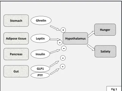
Stomach
GhrelinInterestingly, the effects of leptin are largely opposed by gastric production of ghrelin, a protein which is produced under circumstances of hunger. Ghrelin can stimulate the release of Growth Hormone (GH), hence its name. Upon binding to speciic receptors in the hypothalamus, Ghrelin stimulates Neuropeptide Y (NPY) release and induces food intake (9). It was originally thought that the stomach would secrete ghrelin simply because the stomach was empty and therefore the person was hungry. The thus secreted ghrelin would then stimulate the person via the hypothalamus to go and eat. However, it now appears that in humans this relationship is more complex and that the simple knowledge of a person that “it is time to eat” may be suficient for cerebral stimulation of the stomach to secrete more ghrelin, which indeed in turn will activate the hypothalamus to make the person feel hungry. Therefore, the clinical importance of ghrelin is still under debate.
Pancreas
Insulin secretion Insulin is secreted in low amounts during the fasting state by B cells in the pancreas islets. Upon eating insulin is secreted in larger amounts, both due to the effects of swallowing and the stimulation of food on the intestines and by the mere effect of the rise in plasma glucose. Insulin will then lead to decreases in hepatic glucose production and uptake of glucose and formation of glycogen in muscle and adipose tissue.
has multiple effects on the hypothalamus leading to enhancing satiety after a meal , and inhibition of hunger (10). Indeed , intranasal insulin administration reduces body weight in men (but not in women) (11). Thus, insulin appears to have leptin-like actions on hypothalamic centers although its time course is possibly more rapid. Indirectly, it may enhance energy expenditure possibly via enhanced sympathetic tone.
The gut
The presence of food in the intestines will lead to production of gastric inhibitory polypeptide also known as glucose-independent insulinotropic polypeptide (GIP) and glucagon-like peptide 1 (GLP1) which will activate the B-cells via speciic cell receptors to produce and secrete insulin especially at times that plasma glucose levels are (slightly) elevated (12).
GLP1 and GIPGLP1 has multiple effects presumably on cardiac tissue and especially on food intake. In rodents, GLP1 diminishes food intake by activation of speciic receptors on hypothalamic centers by effects reminiscent of leptin. However, in humans GLP1 is metabolized to inactive metabolites within minutes by dipeptidyl peptidase 4 (DPP4). In humans the effects of GLP1 on food intake have been suggested to be indirect via stimulation of the autonomic nervous system especially the afferent branches of the vagal nerve (13). However, subcutaneous administration of analogues of GLP-1, that are not readily metabolized by DPP4, are effective in reducing hunger and consequently in reducing food intake presumably by direct hypothalamic effects.
In rodents GIP has also an effects on the hypothalamus leading to satiety and less hunger.
In the lower intestines Peptide YY (PYY) is also released in response to food and , just as GLP1, notably in response to protein (14).
Taken together the leptin pathways imply a useful feedback of adipocytes on appetite and food intake. Various hormonal and neuronal effects from the gut further inluence food intake. They also enable the animal to expand energy, and appear to fulill roles in enabling the animal to grow and to breed under circumstances with suficient food.
ROlE Of INflAMMATION IN
ExPANDING ADIPOSE TISSuE
Normal fat tissue contains a low amount of inlammatory cells, notably macrophages of the M2 type and eosinophils. In obesity, these cells are relatively scarce, while other inlammatory cells predominate, notably M1-macrophages, neutrophils , lymphocytes, mast cells and others (15). This apparently leads to augmented secretion of cytokines, among which Tumor Necrosis Factor –alpha (TNF-α), Interleukin-6 (IL-6) and others, which have been reported to impair insulin signaling.
Visceral fat and subcutaneous tissue express a large number of genes in an identical manner. However, subtle differences exist (1), with preponderance of leptin production in subcutaneous fat tissue, while no differences exist in TNF-alpha; complement factors, angiotensinogen and others are produced predominantly in visceral tissue. Many of the deleterious effects of obesity seem to relate mainly to the development of visceral adiposity.
BIOlOGICAl IMPACT Of OBESITy
the risk of atherosclerosis, type 2 diabetes mellitus (Type 2 DM) and of certain types of cancer.These three disorders will be described only briely, since an elaborate review of these complex disorders is outside the scope of this overview.
Atherosclerosis
Epidemiological studies have concluded to a augmented risk for atherosclerotic disease for obese people (1). However, the largest including macrophage activation in the arterial wall, enhanced inlammatory changes, pro-thrombotic changes etc.
Adipokines have multiple effects which can augment the atherosclerotic process. For example, leptin has an NO-dependent vasodilatory effect but it can also upregulate endothelin-1 , a vasoconstrictor (16). It has also been proposed to lead to cellular changes in vascular cells. Resistin has been proposed to activate endothelin (15), and to have multiple effects leading to endothelial dysfunction and possibly proliferative effects on the vasculature (6). Adiponectin has been proposed to exert anti-atherosclerotic effects via its inhibitory effects on TNF-alpha in the vascular wall. Unfortunately, in obesity, less adiponectin is produced in adipocytes. Visfatin is also produced less in obesity.
In obesity, adipocytes grow larger and produce larger amounts of most of the adipokines. It has been proposed that this is related to hypoxia within the adipose cell (16).
Dyslipidemia Obesity is associated with disturbances in plasma lipids, with a notable increase in Low Density Lipoprotein cholesterol (LDL-cholesterol) and plasma triglycerides, and a decrease in High Density Lipoprotein cholesterol (HDL-cholesterol) especially in insulin resistant subjects. Presumably, the underlying insulin resistance leads to slightly elevated lipolysis with production of fatty acids in adipose tissue. Upon uptake in the liver, fatty acids will be bound to triglyceride. In the liver this is process is related to the production of cholesterol and its binding lipoprotein (Apo-B 100); this leads to Very-Low-Density-Lipoprotein (VLDL) formation. VLDL is transported into the blood, and it can release its cholesterol and its triglycerides in the vessel wall, potentially leading to atherosclerosis.
Hypertension .Angiotensinogen and leptin
have been proposed to lead to hypertension. Indeed, although obesity itself clearly relates to hypertension, leptin-deicient humans (who are very obese) are not hypertensive. Leptin can augment sympathetic tone and can upregulate Na/K ATP-ase in the kidney. Large adipocytes produce more angiotensinogen, which would activate aldosterone and thereby lead to hypertension.
Insulin resistance
and to stimulation of glucose uptake. Insulin will also activate IRS-2. Via separate pathway of activating Mitogen Activating Protein Kinase (MAPK), activation of the IRS-1 will also lead to processes involved in expression of other genes notably involved in cellular growth and mitosis.
The activity of IRS1 is of major importance for overall insulin signaling. IRS-1 activity can be decreased by serine phosphorylation by Protein Kinase C (PKC), C-JunKinase (JNK), mammalian target of rapamycin (m-Tor), Suppressor of cytokine signaling-3 (Socs3)and others (17). This will decrease phosphorylation of PI-3K and activateproteasome dependent degradation.
Although the precise mechanisms of the occurrence of insulin resistance is still a matter of intense research, Free Fatty Acids (FFA), interleukin -6 and TNF-alpha have been proposed as causing insulin resistance by inducing transcription of factors leading to serine phosphorylation of IRS-1(and possibly IRS-2) at sites which will decrease the activation of down-stream molecules. The (separate) pathway leading to activation of mitogen activated protein kinase (MAPK) and cellular growth is presumably less or not involved in the process of insulin resistance. This has led to the hypothesis that under states of insulin resistance the MAPK dependent mechanisms have deleterious effects, for example in the myocardium (18). Similarly, it has been proposed that vasoconstrictor effects of insulin is regulated by MAPK dependent signaling while nitric oxide (NO) dependent vasodilator effects are regulated via PI-3K. In states of insulin resistance the MAPK effects would predominate possibly leading to endothelial dysfunction (19).
Type 2 Diabetes
Obesity is strongly associated with Type 2 diabetes.Type 2 diabetes ensues as a consequence of the combination of insulin resistance and pancreas beta cell failure (20). The above mentioned mechanisms for the development of insulin resistance are certainly of importance for the development of diabetes. During the development of type 2 diabetes beta cell mass is probably diminished in part by apoptosis (21).
It is now suspected that a number of adipokines have deleterious effects in beta-cells.
Leptin itself can augment insulin secretion. There is conlicting evidence regarding its potential to affect B cell apoptosis. On one side leptin has been shown to decrease uncoupling protein 2 (UCP2) in Bcells which would decrease apoptosis, but othershave not found an effect on apoptosis (22). However, leptin has multiple effects on gene transcription in various cell types both via Janus Kinase / Signal Transducer and Activator Transcription (JAK/STAT) and MAPK/extracellular signal-regulated kinase (ERK) pathways which may have more subtle effects in apoptosis and/or in B cell function.
Fatty acids, among which notably palmitate, have been shown to be deleterious for B cells and to be able to cause apoptosis. Adiponectin
has been proposed to diminish the deleterious effects of palmitate . In its turn, palmitate appears to lower adiponectin receptor 2 mRNA. Adiponectin may also have effects on gene transcription via ERK pathways (22). Others have proposed that adiponectin (and leptin) increase proliferation of B cells.
been proposed to lead to accumulation of amyloid in islets.
Although a possible role of visfatin is far from clear, it now appears that visfatin may stimulate B cell proliferation; it may have additional effects possibly via gene regulation of PDX1 and the insulin gene (22). Visfatin is notable since its production is decreased in obesity and therefore its potential beneicial effects on B cells might be of less importance in obesity. marked hyperinsulinemia associated with insulin resistance, insulin is suspected to play a major role. Indeed, insulin has proliferative and anti-apoptotic effects in various tissues. Whether this effect is via the actual insulin receptor or via the Insulin-like Growth Factor –I (IGF-1) receptor is so far less clear; insulin can actually be bound to both receptor types (23). The growth promoting properties of IGF-1 are complex, since IGF-1 is bound to a number of binding proteins, the levels of which are partly dependent on plasma insulin.
A number of studies have looked into the potential role of leptin for breast cancer. Leptinappears to augment estrogen production and expression , and it has a proliferative effect on breast tumour cells (23,24). By its MAPK and STAT signaling effects leptin is relevant for cell proliferation and differentiation (23).
Adiponectin has been proposed to protect against carcinogenesis by its insulin sensitizing effects (counteracting hyperinsulinemia), and by inactivating MAPK.
Plasminogen activator inhibitor-1 (PAI-1)
which is also augmented in obesity has also been hypothesized to relate to cancer by promoting tumour growth and/or by effects on the vasculature such as adhesion and angiogenesis (22). Although more adipokines have been named as potentially involved in tumorigenesis, more research will be needed before this would become clearer (22).
CONCluSION
Recent years have seen the exciting discovery of the fundamental role of fat tissue hormonal regulation of food intake, satiety, and energy expenditure, as exempliied by the role of leptin. This has subsequently led to the discovery of several of hormones from other organs , especially the gut , with important effects in the regulation of food intake. Expanding adipose tissue, as is seen in obesity, has now been proposed to be under the inluence of inlammatory cells such as macrophages. This leads presumably to production of deleterious substances such as TNF-alpha, interleukins and others. Several adipokines may be involved in the development of insulin resistance. Moreover, in obesity, adipocytes produce high amounts of deleterious adipokines that may have deleterious effects on the risk of atherosclerosis, type 2 diabetes and cancer. Obese subjects are generally hyperinsulinemic in response to the prevailing insulin resistance. It has been suggested that insulin may have growth proliferative effects involved in tumour growth. Unfortunately, in obesity lower amounts of adiponectin are produced; adiponectin may have protective effects both against the development of atherosclerosis, and cancer and possibly type 2 diabetes.
Legends
feeding behaviour.
Figure 2. Hypothalamic effects of leptin on feeding behaviour are associated with other effects notably on energy expenditure, and complex interactions with growth, and procreation.
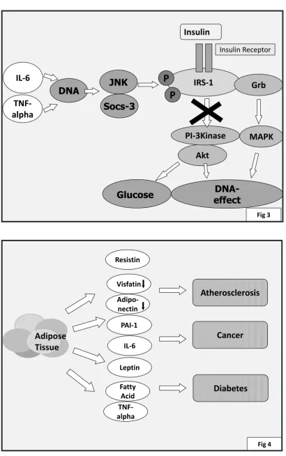
Figure 3 .Insulin resistance: Effects of adipokines on insulin signaling
Figure 4. Schematic representation of potential effects of adipokines on atherosclerosis, cancer and type 2 diabetes mellitus
References
Hajer GR, Van Haeften TW, Visseren FLJ. Adipose tissue dysfunction in obesity, diabetes and cardiovascular diseases. Eur Heart J 2008;29: 2959-2971
Van de Voorde J, Pauwels B, Boydens C, Decaluwe K. Adipocytokines in relation to cardiovascular disease. Metabolism 2013; 62:1513-1521
Arner P. Human fat cell lipolysis: biochemistry, regulation and clinical role. Best Pract Res Clin Endocrinol Metab 2005:19:471-482
Mattu HS, Randeva HS: Role of adipokines in cardiovascular disease. J Endocrinol 2013;216: T17-T36
Mattu HS, Randeva HS. Role of adipokines in cardiovascular disease. J Endocrinol 2013;216:T17-T36
Ouchi N, Shibata R, Walsh K. Cardioprotection by adiponectin. Trends Cardiovasc Medicine 2006; 16: 141-146
Yadav A, Kataria MA, Saini V, Yadav A. Role of leptin and adiponectin in insulin resistance. ClinChim Acta 2013;417:80-84
Ntaios G, Gatselis NK, Makaritsis K, Dalekos GN. Adipokines as mediators of endothelial function and atherosclerosis. Atherosclerosis 2013; 227: 216-221
Tschop M, Smiley DL, Heiman ML. Ghrelin induces adiposity in rodents. Nature 2000: 407;908-913
Yu JH, Kim MS: Molecular mechanisms of appetite regulation. Diabetes Metab J 2012;36: 391-398
Hallschmid M, Benedict C, Schultes B, Fehm HL, Born J, Kern W. Intranasal insulin reduces body fat in men but not in women. Diabetes 2004; 3024-3029
Drucker DJ: Incretin action in the pancreas: potential promise, possible perils, and pathological pitfalls. Diabetes 2013:6:3316-3323
Nishizawa M, Nakabayashi H, Uehara K, Nakagawa A, Uchida K, Koya D: Intraportal GLP1 stimulates insulin secretion predominantly through the hepatoportal-pancreatic vagal relex pathways. Am J Physiol Endocrinol Metab 2013;305:E 376-387
Van der Klaauw AA, Keogh JM, Henning E, Trowse VM, Dhillo WS, Ghatei MA, Farooqi IS: High protein intake stimulates postprandial GLP1 and PYY release. Obesity 2013;21: 1602-1607
Feng B, Zhang T, Xu H. Human adipose dynamics and metabolic health. Ann N Y Acad Sci 2013;1281: 160-177
Aroor AR, McKarns S, Demarco VG, Jia G, Sowers JR: Maladaptive immune and inlammatory pathways lead to cardiovascular insulin resistance. Metabolism 2013: 62:1543-1552
Qi Y, Xu Z, Zhu Q, Thomas C, Kumar R, Feng H, Dostal DE, White MF, Baker KM, Guo S:Myocardial loss of IRS-1 and IRS-2 causes heart failure and is controlled by p38a MAPK during insulin resistance. Diabetes 2013; 3887-3900
Muniyappa R, Sowers JR: Role of insulin resistance in endothelial dysfunction. Rev Endocr Metab Disord 2013; 14:5-12
Stumvoll M, Goldstein BJ, Van Haeften TW: Type 2 diabetes – principles of pathogenesis and therapy. Lancet 2005; 365: 1333-1346
Quan W, Jo EK, Lee MS: Role of pancreatic β-cell death and inlammation in diabetes. Diabetes Obesity Metabol 2013;15 S3: S141-S151
Dunmore SJ, Brown JEP. The role of adipokines in B-cell failure of type 2 diabetes. J Endocrinol 2013;216:T37-T45
De Pergola G, Silvestris F.Obesity as a major risk factor for cancer. J Obesity PMCID: PMC3773450 ; Published online 2013 August 29. doi: 10.1155/2013/291546
Vona-Davis L, Rose DP. Adipokines as endocrine, paracrine, and autocrine factors in breast cancer risk and progression. Endocrine-Related Cancer. 2007;14:189–206
Dieudonne M-N, Bussiere M, Dos Santos E, Leneveu M-C, Giudicelli Y, Pecquery R. Adiponectin mediates antiproliferative and apoptotic responses in human MCF7 breast cancer cells. Biochem Biophys Res Comm 2006;345:271–279
Figures-Stomach Ghrelin
Leptin Hypothalamus Adipose tissue
Pancreas
Gut GLP1 Insulin
PYY
+ Hunger
Satiety
-Fig 1
Adipose tissue Leptin
GH LH
TRH
Hunger
Satiety
Growth Procreation
Thyroid Energy
Expenditure
Hypothalamus
Insulin
IRS-1
Glucose
DNA-effect
TNF-alpha
IL-6
P
Akt
PI-3Kinase
P
Grb
DNA
JNK
Socs-3
MAPK
Insulin ReceptorFig 3
Adipose
Tissue
Fatty Acid
IL-6
TNF-alpha
Adipo-nectin
Atherosclerosis
Visfatin Resistin
Diabetes
Cancer
Leptin PAI-1