Characterization of cell wall oxalate oxidase from maize roots
Mirjana Vuletic´ *, Vesna Hadzˇi-Tasˇkovic´ S
&
ukalovic´
Laboratory of Plant Physiology,Maize Research Institute,Zemun Polje,PO Box89,11081 Beograd-Zemun, Yugosla6ia
Received 11 January 2000; received in revised form 12 May 2000; accepted 12 May 2000
Abstract
Oxalate oxidase activity was detected in the cell wall fraction isolated from maize roots (Zea maysL.). The enzyme was active at acidic pH with optimal activity at pH 3.2. It was thermally extremely stable and resistant to high salt concentration, SDS and pepsin. The enzyme activity was inhibited by sulphydryl reagents 2-mercaptoethanol (2-ME), N-ethyl maleimide (NEM) and dithiotreitol (DTT), but was insensitive to EDTA, KCN and metal ions. Measurements of enzyme activity were performed using colorimetric assay of H2O2, as well as polarographic detection of O2 consumption. Maximal activity was obtained with 5 mM
oxalic acid for the colorimetric method, and 10 mM oxalic acid for the polarographic method. Both methods were applicable in oxalate oxidase characterization, the polarographic method being more suitable under conditions of H2O2interaction with some
of the analyzed substances. © 2000 Elsevier Science Ireland Ltd. All rights reserved.
Keywords:Cell wall; Oxalate oxidase; Root;Zea mays
www.elsevier.com/locate/plantsci
1. Introduction
Oxalate oxidase (oxalate:oxygen oxidoreduc-tase, EC 1.2.3.4) catalyzes the oxidation of
ox-alate by molecular oxygen, yielding CO2 and
H2O2. Such an enzyme activity was demonstrated
in a number of higher plant species and in various plant tissues: in grain sorghum seedlings, roots, leaves and stems [1], barley seedlings [2] and
leaves [3], wheat embryo [4] and leaves [5], Ama
-ranthus leaves [6], beet stem [7], etc. The
charac-teristics of the enzyme varied significantly
depending on the plant species used for the inves-tigation.
The discovery that germin, a developmentally regulated protein in wheat, is in fact an oxalate oxidase [4], focused the scientific attention to this enzyme. Although sorghum oxalate oxidase
activ-ity was found in the 15 000 g supernatant of leaves and roots [1,8], wheat and barley germins are at least partly associated with the cell wall [3 – 5,9]. This enzyme would have a role in
devel-opmental processes by producing H2O2 which is
involved in the oxidative cross-linking of a cell wall polymers [10]. Additionally, increased tran-scription of a gene for germin-like oxalate oxidase in wheat and barley leaves following pathogen attack [3,5], and modulation of germin gene ex-pression in barley roots by salt stress [11] or in wheat seedlings by heavy metal ions [12], sug-gested the involvement of this enzyme in plant response to stress.
In this report, we describe some characteristics of the cell wall-bound oxalate oxidase in maize roots, and compare its activity in different parts of the plant and early stages of development of maize. Enzyme activity was determined by the
standard enzyme assay, measuring H2O2
genera-tion, as well as by measuring the oxygen con-sumption rate. The results obtained using both methods were compared.
* Corresponding author. Tel.: +381-11-3756704; fax: + 381-11-3754994.
E-mail address:[email protected] (M. Vuletic´).
2. Materials and methods
2.1. Plant material
Seeds of maize (Zea mays L., inbred line Oh43)
were germinated on water and transferred after 3 days to 50% Knop [13] nutrient solution. Plants were grown up to 9 days in controlled environ-ment under a 12-h light/dark regime at 22/18°C,
with a light intensity of 40 W m−2 and relative
humidity at 70%. Before experiments, roots were
washed with H2O, dried between filter papers and
immediately used for cell wall isolation. Addition-ally, the cell wall was isolated from leaves and embryonic axis (embryo without scutellum) of im-bibed seedlings.
2.2. Isolation of cell wall
Tissue was homogenized with medium (0.25 M sucrose in 0.05 M Tris – HCl, pH 7.2) in 1:11 ratio (w/v) in a chilled mortar and pestle. The ho-mogenate was squeezed through 0.5 mm nylon
mesh and the filtrate centrifuged at 1000×gfor 10
min in the cold. The pellet was washed twice by resuspension in 10 vol. of the homogenization medium containing 1% (w/v) Triton X-100 and four times with 30 vol. of the same medium with-out Triton X-100. After each wash the pellet was
collected by centrifugation at 1000×g for 10 min.
The final pellet was considered to be the purified cell wall fraction, and was used for most of the experiments. In order to obtain the ionically bound fraction, cell wall was incubated in 1 M
NaCl or 1 M CaCl2 for 30 min with continuous
stirring at 4°C. After centrifugation at 1000×g
for 10 min, the resulting supernatant contained the ionically bound proteins and the pellet the tightly bound cell wall proteins. The enzyme activity was assayed in the dialyzed supernatant, as well as in the pellet which was washed three times and resus-pended in homogenization medium. SDS treat-ment was performed by incubation of the purified cell wall fraction in 0.4% SDS at 37°C for 30 min. After washing three times in the homogenization medium, the resulting pellet was resuspended in the same medium, the enzyme activity assayed, and compared to enzyme activity in the control experiment performed without SDS. Pepsin sus-ceptibility was analyzed by treatment of the cell
wall fraction with 50 mg/ ml−1 of pepsin at pH 2
adjusted with HCl at 37°C for 30 min, and then neutralized with NaOH before measurement of enzyme activity. In the control experiment pepsin was omitted.
2.3. Enzyme assay
Colorimetric assay of oxalate oxidase was car-ried out as described by Pundir [8]. The assay mixture (1 ml), containing 50 mM Na succinate,
pH 3.2 and 50 – 100 mg protein was incubated at
37°C in test tubes wrapped with black paper. In case of experiments where the effect of pH was studied, Na succinate buffers with pHs in the range of 2 – 5 were used. The reaction was started by adding oxalic acid to a final concentration of 10 mM, unless otherwise specified. After 5 min 0.5
ml of color reagent for H2O2 measurement was
added to stop the reaction, and the color was allowed to develop for 30 min at room tempera-ture. The color reagent consisted of 50 mg 4-aminophenazine, 100 mg solid phenol and 500 U of horseradish peroxidase per 100 ml of 0.4 M Na – Pi buffer, pH 7.0. The absorbance of the solution was measured at 520 nm and corrected for absorbance obtained when oxalic acid was omitted from assay mixture. Hydrogen peroxide generated during the reaction was determined by interpolation from a standard curve in the range
from 0.01 to 0.25 mmol H2O2 prepared in 50 mM
succinate buffer. Enzyme activity is expressed as
the amount of H2O2 produced per min and mg of
protein. The effect of various compounds on the enzyme activity was studied by their addition to the reaction mixture before adding oxalic acid. Additional control experiments were performed by substituting the cell wall isolate and oxalic acid in
a standard reaction mixture with H2O2 to a final
concentration of 0.1 mM, in order to test the
possible effect of analyzed compounds with H2O2
measurements. The reaction was performed under standard assay conditions, with or without com-pound added, and its effect calculated as the dif-ference in the absorbances.
2.4. Oxygen consumption
Measurement of oxygen consumption was per-formed using a Clark-type polarographic electrode (Hansatech Ltd., England), at 25°C. Cell wall
assay mixture containing 50 mM Na succinate, pH 3.2, unless otherwise specified, and oxygen con-sumption initiated by the addition of oxalic acid.
The rate of O2 consumption was calculated and
expressed as the amount of O2consumed per min
and mg of protein.
2.5. Protein determination
Protein content was determined according to Lowry et al. [14] using bovine serum albumin as the standard.
3. Results
Oxalate oxidase activity was detected in the cell wall fraction but not in the soluble fraction (20 000 g supernatant) of maize seedlings. The enzyme activity could be detected in the embry-onic axis after 10 h of imbibition, and it increased significantly after 48 h. Such high activity persisted with a maximum at 72 h (Fig. 1). At this time, seedlings were transferred to the nutrient solution, and the activity of the oxalate oxidase in the root and leaf was analyzed. The activity in the leaf cell wall was an order of magnitude lower, compared to the root cell wall activity in the case of 6-day-old plants. Further analysis was performed only on the root cell wall isolates, and the data pre-sented show that there was a gradual decrease of activity with development, 9-day-old plants having only 20% of the maximal activity for the root. In further work, the characterization of the enzyme was done on root cell wall isolates obtained from plants grown for 3 – 5 days on the nutrient solution.
Colorimetric assay showed an optimum of en-zyme activity within 5 min of incubation. A 40% decrease in activity was observed after 10 min of incubation (data not presented). Oxygen consump-tion by cell wall was not observed in the absence of added substrate. The addition of oxalic acid induced oxygen consumption at a constant rate (Fig. 2, insert), no changes being observed over 15 min.
The effect of pH on the activity of the enzyme was studied in the pH range of 2.1 – 5.1 using the succinate buffer. Both assays used yielded similar results showing that maize oxalate oxidase has a pH optimum around 3.2 (Fig. 3). Optimal concen-tration of substrate was determined using both assays as shown in Fig. 4. A total of 5 mM oxalic acid gave maximal reaction rates, in the case of colorimetric assays, and 10 mM in the case of polarographic assays. The relationship between oxalate concentration and reaction rates revealed
Km of 2.54 and 1.31 mM for oxalate obtained by
Fig. 1. Activity of the cell wall oxalate oxidase in early stages of development of maize plant determined in the embryonic axis (embryo without scutellum), roots and leaves. The activ-ity was assayed polarographically and expressed as nmol O2
consumed mg protein−1min−1. The time was measured from
the beginning of the imbibition. The vertical arrow indicates the moment of transfer of seedlings from water to the nutrient solution.
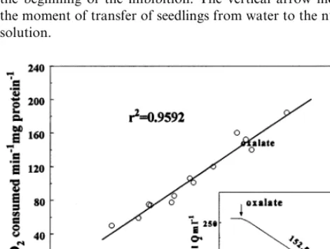
Fig. 2. Oxalate oxidase activities expressed as nmol H2O2
produced mg protein−1 min−1, determined by colorimetric
assay, plotted against the oxalate oxidase activities expressed as nmol O2 consumed mg protein−1 min−1, determined
polarographically, of the same samples. The correlation co-efficient (r2) is presented. Insert: an example of time course of
oxygen consumption induced by the addition of oxalate (figure next to trace is expressed in nmol O2 min−1 mg
Fig. 3. Effect of pH on oxalate oxidase activity assayed by colorimetric method, measuring H2O2 production (), and
by polarographic oxygen consumption measurements ( ). Enzyme activities are expressed as percent of maximal activ-ity9S.E. (indicated by vertical bars).
was used as an alternative method to express the activity of oxalate oxidase.
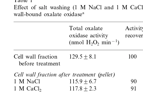
In order to examine the degree of association of oxalate oxidase with cell wall, we subjected purified cell wall fraction to salt washing with 1 M
NaCl or 1 M CaCl2. After both of the treatments,
90% of the enzyme activity was detected in
tightly bound-cell wall fraction (Table 1), demon-strating that binding of oxalate oxidase to the cell wall was more than an ionic association. The heat stability of oxalate oxidase was investigated by heating the cell wall fraction at various tempera-tures in the range of 37 – 80°C for 30 min. Full activity remained at 80°C (data not presented), demonstrating the very good heat-stability of the enzyme. The enzyme was resistant to SDS and pepsin treatment, 90 and 100% of the activity remaining after SDS and pepsin treatment, respec-tively (data not presented).
In order to examine the possible inhibitory ef-fect of some chemical compounds (KCN, 2-mer-captoethanol, dithiotreitol, EDTA, glutathione,
N-ethyl maleimide and L-cysteine) on the activity
of oxalate oxidase we added them to the assay mixture in final concentrations of 0.5 and 5.0 mM and the reaction was performed under standard assay conditions, both detection methods being employed. The effect of these chemical compounds
on the control (H2O2 measurements performed on
a system where external H2O2 was added instead
of oxalic acid and the cell wall preparation
pro-Fig. 4. Effect of oxalate concentration on oxalate oxidase activity assayed by colorimetric method, measuring H2O2
production (), and by polarographic oxygen consumption
measurements ( ). Enzyme activities are expressed as percent of maximal activity9S.E. (indicated by vertical bars).
Table 1
Effect of salt washing (1 M NaCl and 1 M CaCl2) on cell
wall-bound oxalate oxidasea
Total oxalate Activity recovered (%) oxidase activity
(nmol H2O2min−1)
100 129.598.1
Cell wall fraction before treatment
Cell wall fraction after treatment(pellet) 115.996.7
1 M NaCl 90
117.892.3
1 M CaCl2 91
Ionically-bound fraction after treatment(supernatant) 7.990.7
1 M NaCl 6
1 M CaCl2 7.292.1 6
aCell wall was isolated from 5-day-old maize seedlings.
Colorimetric assay was used in enzyme activity determination. One milliliter of the cell wall isolate was used for each of the treatments and the means with 9S.E. of assays in triplicate are shown.
O2 consumption and colorimetric measurements,
respectively. Substrate inhibition was evident with
20 mM oxalic acid, in the case of H2O2
measure-ment, while O2 consumption was not inhibited at
concentrations higher than 10 mM. Because the values obtained by the colorimetric assay were not significantly different with 5 and 10 mM oxalic acid, 10 mM oxalic acid was used in all further experiments. By comparing the specific activities of oxalate oxidase determined in the same cell wall isolates by using two methods in a number of experiments (Fig. 2) a good correlation was
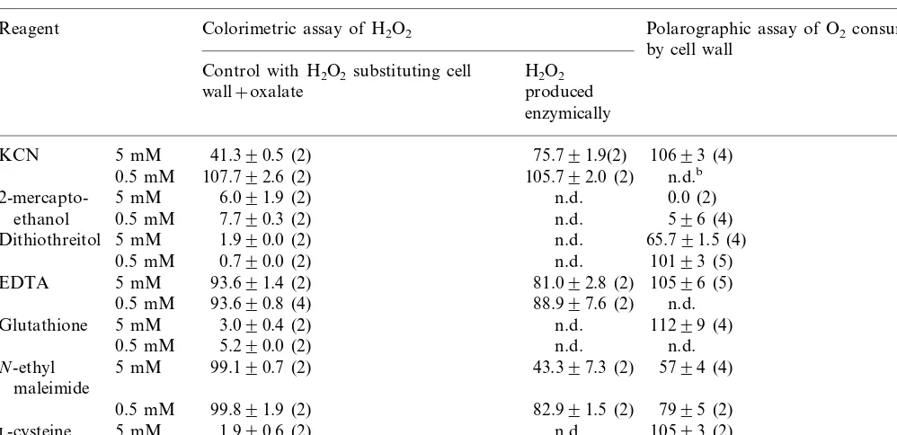
Table 2
Effects of some chemical compounds on the oxalate oxidase activity determined colorimetrically by assaying H2O2 and
polarographically by assaying O2consumptiona
Reagent Colorimetric assay of H2O2 Polarographic assay of O2consumption
by cell wall Control with H2O2substituting cell H2O2
produced wall+oxalate
enzymically
41.390.5 (2) 75.791.9(2)
5 mM 10693 (4)
KCN
107.792.6 (2) 105.792.0 (2)
0.5 mM n.d.b
5 mM 6.091.9 (2) n.d.
2-mercapto- 0.0 (2)
0.5 mM 7.790.3 (2)
ethanol n.d. 596 (4)
1.990.0 (2) n.d.
5 mM 65.791.5 (4)
Dithiothreitol
0.5 mM 0.790.0 (2) n.d. 10193 (5)
93.691.4 (2) 81.092.8 (2)
5 mM 10596 (5)
EDTA
0.5 mM 93.690.8 (4) 88.997.6 (2) n.d.
3.090.4 (2) n.d. 11299 (4)
Glutathione 5 mM
5.290.0 (2) n.d.
0.5 mM n.d.
5 mM 99.190.7 (2)
N-ethyl 43.397.3 (2) 5794 (4)
maleimide
99.891.9 (2) 82.991.5 (2)
0.5 mM 7995 (2)
1.990.6 (2) n.d.
L-cysteine 5 mM 10593 (2)
3.890.4 (2) n.d.
0.5 mM n.d.
aThe results are presented as remaining activity in % 9S.E. with number of experiments in parentheses. bn.d., not determined.
ducing H2O2) along with their effect on oxalate
oxidase activity, measured as H2O2 production
and O2 consumption, are presented in Table 2.
The presence of reagents containing SH groups in
both concentrations strongly affected H2O2
deter-mination in colorimetric assay. Also, higher con-centration of KCN inhibited strongly this reaction (60%), while inhibition by both concentrations of EDTA was not so pronounced (6%). In such
cases, where reagents interfered with H2O2
mea-surements, it was not possible to examine their effect on oxalate oxidase activity measured colori-metrically. By using oxygen consumption rate to express oxalate oxidase activity, inhibitory effect
of KCN, EDTA, glutathione (GSH), L-cysteine
and 0.5 mM DTT was not detected, while higher concentration of DTT inhibited approximately 35% and 2-ME inhibited almost completely en-zyme activity. Only the results of inhibitory effects of NEM were comparable for both methods,
inhi-bition being 20 and 50% with 0.5 and 5.0 mM
NEM, respectively. Thus, in some cases the colori-metric method was not applicable because of the
interaction of various compounds with the H2O2
produced by oxalate oxidase or with reagents used
in the colorimetric assay. This problem was over-come by oxygen consumption measurements.
We also tested the effect of various metal ions and chloride salts on the enzyme activity. Addition of 1 mM NaCl to the assay mixture did not show any effect on the enzyme activity. Also, none of
metal ions tested (Cu2+, Fe3+, Zn2+, Mn2+,
Mg2+, Al3+) stimulated the enzyme activity, nor
reversed the rate of reaction after inhibition by sulphydryl reagent, 2-ME (data not shown).
4. Discussion
In this report we demonstrated the occurrence of oxalate oxidase in cell walls isolated from maize seedlings. A significant increase in activity in the embryonic axis was obtained 2 – 3 days post-imbi-bition, subsequently decreasing in the plant organs that develop. Oxalate oxidase activity associated with cell wall is not unexpected since it was estab-lished that germin, a protein marker of early plant development, is an oxalate oxidase [4], and
wall-bound form accounts for 40% of the total
Also, oxalate oxidase from barley leaves was found to be located in the cell wall [3]. However, contrary to the results obtained for sorghum [1], barley [2] and wheat [15], we did not detect any enzyme activity in the soluble fraction.
Maize oxalate oxidase was shown to be tightly bound to the cell wall since it was not extracted by
either NaCl or CaCl2. It was also shown that the
enzyme is heat stable since it remained fully active at 80°C. Our data show that it is even more stable than the oxalate oxidase purified from barley seedlings, which retained 80% of its activity at 75°C [2]. The resistance of maize enzyme to pepsin proteolysis is similar in characteristics to germin of wheat embryos [16]. Also, the resistance to SDS treatment, observed in our experiments, was shown previously for wheat embryo germin [16] and oxalate oxidase from barley seedlings [17].
Oxalate oxidase was active only at acidic pHs, with a pH optimum around 3.2. Previously, differ-ent values for pH optimum in the acidic range have been reported for different plant species. Our result for maize root oxalate oxidase is close to the results obtained for barley seedlings (pH 3.5) [2] and wheat embryo (pH 3.5) [3], and different from sorghum leaves (pH 5.0) [18] and roots (pH 5.0)
[1]. The Km values obtained in our experiments
were similar toKm obtained forAmaranthusleaves
[6], while they were an order of magnitude higher
than Km obtained for barley root [19] and two
orders higher than for sorghum leaves [18] and beet stem [7].
The insensitivity of the enzyme to the tested metal and chloride ions that we observed is similar to the result obtained with membrane-bound
ox-alate oxidase from Amaranthus leaves [6].
Con-trary to these results, the oxalate oxidase from
sorghum leaves [18] and roots [1] required Fe2+
and Cu2+ for maximal activity, respectively, and
chloride exhibited an inhibitory effect on oxalate oxidase from barley seedlings [2]. EDTA did not inhibit maize root oxalate oxidase. A similar result was obtained for barley seedlings [2] and roots [20], contrary to the strong inhibition of the en-zyme from sorghum roots [1] and leaves [18]. Among the sulphydryl reagents investigated, only 2-ME completely inhibited oxalate oxidase, while DTT and NEM exhibited a less pronounced inhi-bition. Inhibition by 2-ME was also reported for oxalate oxidase in barley seedlings [2] and roots [20] (more than 80%, with 1 mM 2-ME), and in
sorghum roots (28% with 0.5 mM 2-ME) [1], but not in sorghum leaves [18].
Oxalate oxidase activity has been demonstrated in a number of plant species. Of these, only the wheat and barley enzymes have been classified as germin-like oxalate oxidases [4,21] until now. Our experiments indicate that maize root cell wall ox-alate oxidase exhibits most of the characteristics of germins: heat-stability, protease-resistance, SDS-tolerance and similar pH optimum to that of barley and wheat (as opposed to that of sorghum whose oxalate oxidase is not classified as germin). An increase of enzyme activity 48-h post-imbibi-tion, similar to the observed increase in activity in the early stages of wheat embryos [9], suggests its significant role in the early stages of maize devel-opment and also argues in favor of such a contention.
Because oxalate oxidase catalyzes the reaction:
HOOC – COOH+O22CO2+H2O2, consuming
oxygen, we have employed the polarographic method to measure its activity, besides the conven-tional colorimetric method for determination of
the generated H2O2. A comparison of the two
methods has been made using the same cell wall isolates, the results obtained demonstrating a good correlation for optimal assay conditions. However, differences have been observed in the values of the
optimum concentrations, Km, substrate inhibition
and rate dependence on the incubation time. The optimal incubation time in colorimetric reaction of 5 min and decline of activity after prolonging the incubation period to 10 min was also observed in sorghum root oxalate oxidase [1]. Substrate inhibi-tion of oxalate oxidase activity was observed in our experiments above 10 mM oxalic acid in the case of colorimetric assay. A similar inhibition was observed with partially purified enzyme from sor-ghum leaves [18], although at lower concentrations of oxalate (above 0.25 mM). The fact that oxalate oxidase activity assayed polarographically did not show substrate inhibition or decline of activity for periods up to 15 min indicates that the observed phenomena are indeed caused by the colorimetric assay, rather than by inhibition of the enzyme activity. On the other hand, our results show that the colorimetric method is not useful in studies of the effect of several chemical compounds on the activity of oxalate oxidase. In this method, oxalate oxidase activity was assayed by detection of the
peroxidase-de-pendent staining method. KCN is a known in-hibitor of peroxidase [22], and it interfered also in our colorimetric assays. Also, our results demon-strated that this method is not applicable in the studies of the effect of reagents containing SH
groups (L-cysteine, GSH, 2-ME and DTT) on the
oxalate oxidase activity. Their effect on H2O2
mea-surements can be explained by nonenzymic
reac-tion of SH groups with H2O2 [23]. In such
instances oxygen polarography proved to be a more convenient method for measuring the ox-alate oxidase activity.
References
[1] C.S. Pundir, N.K. Kuchhal, Detection of an oxalate oxidase in grain sorghum roots, Phytochemistry 28 (1989) 2909 – 2912.
[2] M. Sugiura, H. Yamamura, K. Hirano, M. Sasaki, M. Morikawa, M. Tsuboi, Purification and properties of oxalate oxidase from barley seedlings, Chem. Pharm. Bull. 27 (1979) 2003 – 2007.
[3] Z. Zhou, Z. Zhang, P.L. Gregersen, J.D. Mikkelsen, E. de Neergaard, D.B. Collinge, H. Thordal-Chrinstensen, Molecular characterization of the oxalate oxidase in-volved in the response of barley to the powdery mildew fungus, Plant Physiol. 117 (1998) 33 – 41.
[4] B.G. Lane, J.M. Dunwell, J.H. Ray, M.R. Schmitt, A.C. Cuming, Germin, a protein marker of early plant devel-opment, is an oxalate oxidase, J. Biol. Chem. 268 (1993) 12239 – 12242.
[5] W.J. Hurkman, C.K. Tanaka, Germin gene expression is induced in wheat leaves by powdery mildew infection, Plant Physiol. 111 (1996) 735 – 739.
[6] L. Goyal, M. Thakur, C.S. Pundir, Purification and properties of a membrane bound oxalate oxidase from
Amaranthus leaves, Plant Sci. 142 (1999) 21 – 28. [7] P. Varalakshmi, K.M. Lathika, K.G. Raghavan, B.B.
Singh, Altered physicochemical characteristics of polyethylene glycol linked beet stem oxalate oxidase, Biotechnol. Bioeng. 46 (1995) 254 – 257.
[8] C.S. Pundir, Purification and properties of oxalate oxi-dase from Sorghum leaves, Phytochemistry 30 (1991) 1065 – 1067.
[9] B.G. Lane, A.C. Cuming, J. Fr(geau, N.C. Carpita, W.J. Hurkman, F. Bernier, E. Dratewka-Kos, T.D. Kennedy, Germin isoforms are discrete temporal markers of wheat
development. Pseudogermin is a uniquely thermostable water-soluble oligomeric protein in ungerminated em-bryos and like germin in germinated emem-bryos, it is incorporated in cell walls, Eur. J. Biochem. 209 (1992) 961 – 969.
[10] P.D. Olson, J.E. Varner, Hydrogen peroxide and lignifi-cation, Plant J. 4 (1993) 887 – 892.
[11] W.J. Hurkman, C.K. Tanaka, Effect of salt stress on germin gene expression in barley roots, Plant Physiol. 110 (1996) 971 – 977.
[12] A. Berna, F. Bernier, Regulation by biotic and abiotic stress of a wheat germin gene encoding oxalate oxidase, a H2O2-producing enzyme, Plant Mol. Biol. 39 (1999)
539 – 549.
[13] D.R. Hoagland, D.I. Arnon, The water-culture method for growing plants without soil, Calif. Agric. Exp. Stn. Circ. 347 (1950) 1 – 39.
[14] O.H. Lowry, N.J. Rosebrough, A.L. Farr, R.J. Randall, Protein measurement with the Folin phenol reagent, J. Biol. Chem. 193 (1951) 265 – 275.
[15] A. Berna, F. Bernier, Regulated expression of a wheat germin gene in tobacco: oxalate oxidase activity and apoplastic localization of the heterologous protein, Plant Mol. Biol. 33 (1997) 417 – 429.
[16] Z.F. Grzelczak, B.G. Lane, Signal resistance of a soluble protein to enzymic proteolysis. An unortodox approach to the isolation and purification of germin, a rare growth -related protein, Can. J. Biochem. Cell Biol. 62 (1984) 1351 – 1353.
[17] Z. Zhang, J. Yang, D.B. Collinge, H. Thordal-Chris-tensen, Ethanol increases sensitivity of oxalate oxidase assays and facilitates direct activity staining in SDS gels, Plant Mol. Biol. Rep. 14 (1996) 266 – 272.
[18] C.S. Pundir, R. Nath, Occurrence of an oxalate oxidase inSorghumleaves, Phytochemistry 23 (1984) 1871 – 1874. [19] J. Chiriboga, Purification and properties of oxalic acid oxidase, Arch. Biochem. Biophys. 116 (1966) 516 – 523. [20] V.P. Kotsira, Y.D. Clonis, Oxalate oxidase from barley
roots: purification to homogeneity and study of some molecular, catalytic and binding properties, Arch. Biochem. Biophys. 340 (1997) 239 – 249.
[21] B. Dumas, A. Sailland, J. -P. Cheviet, G. Freyssinet, K. Pallett, Identification of barley oxalate oxidase as a germin-like protein, C. R. Acad. Sci. Paris 316 (1993) 793 – 798.
[22] W.D. Ellis, H.B. Duford, The kinetics of cyanide and fluoride binding by ferric horseradish peroxidase, Bio-chemistry 7 (1968) 2054 – 2062.
[23] J. Hughes, S. Joshi, D. Ascoli, Elimination of thiol reagent interference during Lowry protein determina-tion, Anal. Biochem. 117 (1981) 1 – 5.