Open access:
www.balimedicaljournal.com
or
www.ojs.unud.ac.id
129
INTRALESIONAL TRIAMCINOLONE ACETONIDE
INJECTION ON EYELID CAPILLARY HEMANGIOMA
(Case Report)
Yuliawati, P., and Rian-Ananta, M.
Department of Ophthalmology, Faculty of Medicine, Udayana University, Bali-Indonesia
Background: Infantile capillary hemangioma or benign hemangio-endotelioma or strawberry nevus
is a most common benign vascular tumor in children. This study aims to report management two cases
of eyelid capillary hemangioma with intralesional injection of triamcinolone acetonide (TA).
Methods: Case report. First case is 11
thmonths old baby, girl, with mass on right superior eyelid
since 5
thmonth old that getting bigger. Mass are soft, blue to purple colored, with size of 30 x 30 x 20
mm, obvious border, refined surface, fixated to beneath structure, and there were dilated blood vessels
visible from anterior surface. Second case is 1
stmonth old baby with mass on left superior eyelid since
a week after birth. On examination found soft mass with blue violet colour, size 30x30x5mm, smooth
surface, fixated, dilated blood vessels at skin surface. There were multiple bright red marks with
variated size such as 2x3x1mm, 2x2 x1mm, 3x2x1mm at eyelid skin surface. Both cases were given
timol 0.5% for two months, but mass were getting bigger until closing visual axis. On both cases there
were mechanical ptosis that close visual axis. Closing of visual axis causes deprivational amblyopia,
so need intervention to reduces deprivation with intralesion TA injection at dose 3 mg/kg body
weight. Results: After TA intralesional injection, tumor size reduced on both cases so visual axis is
opened, without side effect. Conclusion: triamcinolone acetonide intralesional injection on capillary
hemangioma cases reduces tumor size on relatively short time, in the hope to maximally reduce
deprivation amblyopia at visual development age.
Key Words : capillary hemangioma, intralesional injection, triamcinolone acetonide.
INTRODUCTION
Infantile capillary hemangioma or benign hemangio-endotelioma or strawberry nevus is a most common benign vascular tumor in children. Prevalence of capillary hemangioma approximately 1-2% of all children.1,2 Capillary hemangioma composed of vascular endothelial cell proliferation in the fibrous tissue. Tumor manifests when the baby a few weeks old, grew rapidly in the proliferative phase in the first year of life and then experienced regression or involutional phase when children aged 5-7 years.1
Capillary hemangiomas occur during only one-third of babies born, and most cases manifest during 6 months.1,3 Gender predominant more girl suffer from capillary hemangioma of the boy with a ratio 3:1.1,3 These tumors can arise in the mucous membranes, skin, and other soft tissue in the body. The most frequent location of periorbital capillary hemangioma is in quadrant superonasal.1
Correspondence:Yuliawati, P.
Department of Ophthalmology, Faculty of Medicine, Udayana University, Bali-Indonesia Email: [email protected]
Capillary hemangioma is a tumor derived from primitive cells with the ability of differentiation into endothelial cells and pericyte cells.4 Capillary hemangiomas do not require treatment if not cause complications such as ptosis, strabismus, optic nerve compression, proptosis, and threats ambliopia.4,5 Capillary hemangioma management consists of multiple modalities such as corticosteroids (topical, systemic, intralesional injection), beta blockers (topical and systemic), interferon, radiation, and surgery.1,6-8
Intralesional corticosteroid injection was first known to help reduce size of the capillary hemangioma since three decades ago by Azzolini and Nouvenne in 1970.7 Reports of intralesional steroid injection case was first reported by Kushner who reported 4 cases of capillary hemangioma who get injections of triamcinolone acetonide (TA) 40 mg and 6 mg betamethasone sodium phosphate. After one month of observations they obtained reduce tumor mass, except in one patient who had previously been getting corticosteroid injections.9
Open access:
www.balimedicaljournal.com
or
www.ojs.unud.ac.id
130
intralesional injection reduces the size of the eyelidcapillary hemangioma.
CASE REPORT
First case is a 11 month old baby, girl, brought by her parents to the Eye Clinic Sanglah on 5 June 2012, cmplain there is a large mass on her right eyelid arising since five-month-old. Patient was taken to the ophthalmologist and and recommended for intralesional steroid injection, but the patient's parents refused, patient is given only in therapy Timol 0.5% applied twice daily in location of hemangioma. After six months, felt a lump swell up causing the patient can not open his right eyelid. History of spontaneous vaginal birth, full-term gestation, birth weight 2800 grams.
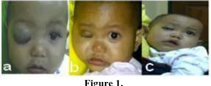
Examination of vital signs are within normal limits. Visual acuity on RE could not be done because the eyelids can not be opened, while the following light (FL +) on LE. Local examination RE shows petal soft mass, purplish blue, size 30x30x20mm, well defined, smooth surface, fixed to the underlying tissues, and there is a dilatation of blood vessels in skin surface. Anterior and posterior segment of RE difficult to evaluated because the eyelid could not be opened. Horizontal palpebral fissure (HPF) is 30 mm, vertical palpebral fissure (VPF) 0 mm, margin reflex distance (MRD) -4 mm, and the margin limbal distance (MLD) 0 mm. Anterior and posterior segment examination LE is within normal limits, measurements of FPH is 30 mm, 12 mm FPV, MRD 5 mm, and 8 mm MLD (Figure 1a).
Parents are advised to check CT angiography to see the tumor flow rate, but the patient's parents refused because he felt his son was too young to do invasive action.
Patients diagnosed with RE eyelid capillary hemangioma and is advised to obtain TA intralesional injection in the Central Surgical Installation Sanglah.
Patients undergoing intra-lesion injection of TA with general anesthesia on 19 June 2012. Dose of TA given is 3mg/kgBB, patient weight of 10 kg, so the dose of the TA injected is 30 mg (3 mL). After injection patients received prescriptions of Paracetamol syrup 3x100mg, Cefixim syrup 2x100mg, Timol 0.5% applied twice daily on the mass.
One month after injection, examination of vital signs within normal limits. Examination of visual acuity gained the impression RLE FL +. On anterior segment examination of RE obtained blefaroptosis, hemangioma mass slightly darker color than surrounding skin, size 25x10x5mm, well defined, smooth surface, soft consistency, fixed to the underlying tissues, and the dilation of blood vessels in the surface of the skin is reduced. Other anterior and posterior segment of RE within normal limits. FPH measurements obtained 30 mm, 2 mm
FPV, MRD -2 mm, and MLD 0 mm (Figure. 1b). Anterior and posterior segment examination LE within normal limits. Patients are advised control three months.
Four months post-injection, examination of vital signs within normal limits. Visual acuity gained the impression RLE FL +. Right eyelid ptosis and eyelid but the size of the right and left about the same. On examination of the anterior segment of the palpebral RE obtained mass decreases, with size 20x5x5mm, eyelid color similar to skin around and did not seem dilation of blood vessels in the skin surface. Other anterior and posterior segment RE within normal limits. FPH measurements obtained 30 mm, 6 mm FPV, MRD 1 mm, and 2 mm MLD. Anterior and posterior segment examination OS within normal limits. FPH measurements obtained 30 mm, 12 mm FPV, MRD 5 mm, and 8 mm MLD (Figure 1c).
[image:2.595.317.525.388.473.2]Patients were scheduled to receive second TA intralesional injection four-six months after the first injection, but the patient's family will be moving to Bandung, so it will control in Cicendo Hospital Bandung. Patients are still advised to use Timol 0.5% twice daily were applied in mass hemangioma.
Figure 1.
a.first case when first came to ophthalmologist; b.a month after TA injection; c.four months after TA
injection
Second case is one month old baby, girl, consulted from dermato venereal department with eyelid hemangioma. Patient complained of arises reddish spots on the upper left eyelid since the baby was two weeks old. Initially start as small red marks, flat surface, and become larger with elevation of the lesion. The patient was taken to a pediatrician and given creams apolar (desonide). After two weeks of usage, red patches did not improve and the more left eyelid swollen and the patient was taken to dermato venereal department Sanglah Hospital on 18 September 2012 and then consulted to oncology division, eye clinic Sanglah Hospital. History of spontaneous vaginal birth, full-term gestation, birth weight 3800 grams.
Open access:
www.balimedicaljournal.com
or
www.ojs.unud.ac.id
131
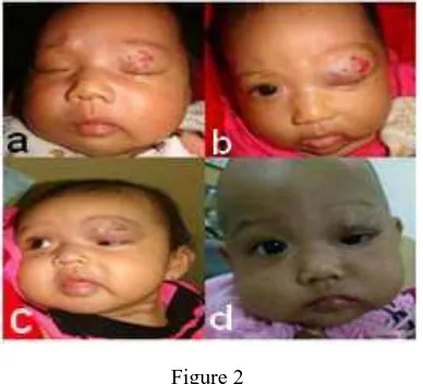
Anterior segment examination of LE obtainedpurplish blue soft mass, size 30x 30x5mm, smooth, soft consistency, fixed to beneath structure, and there is a widening of the blood vessels in the skin surface. Above the surface of the skin there are patches of reddish marks with varying size 2x3x1mm, 2x2x1mm and 3x2x1mm. Other anterior and posterior segments of the LE within normal limits. FPH measurements is 28 mm, FPV8 mm, MRD 3 mm, MLD 5 mm (Figure 2a).
Patients diagnosed with capillary hemangioma OS, and given Timol 0.5% applied twice a day on the surface of the lesion.Two months after the administration of Timol 0.5%, swelling of eyelids felt even heavier. Examination obtained vital signs within normal limits. Impression of visual acuity RLE FL +. Anterior and posterior segment examination RE within normal limits. FPH measurements 28 mm, FPV 11 mm, MRD 5 mm, and MLD 8 mm.
Examination of the anterior segment of the superior eyelid LE obtained are purplish blue soft mass, size 30x30x10mm, smooth, soft consistency, fixed to beneath structure, and there is a widening of the blood vessels in the skin surface. Above the surface of the skin there are patches of reddish marks with varying size 2x3x1mm, 2x2x1mm and 3x2x1mm, difusse with elevated edges. Other anterior and posterior segments of the LE within normal limits. FPH measurements obtained 28 mm, FPV 0 mm, MRD -4 mm, and MLD 0 mm (Figure 2b).
Patients are advised to obtain TA intralesional injection under general anesthesia. Laboratory examination and chest x-ray as a preoperative preparation was done and consulted the pediatric and anesthesia for surgery eligibility. Patients are advised to CT angiography examination under general anesthesia, but the patient's parents refused for fear of the invasive actions performed on the infant child.
TA intralesional injection took place on 9 November 2012. A given dose is 3 mg / kg body weight, so that the patient weight 6 kg, the dose of injection is 18 mg. After injection patients received 3x80mg of Paracetamol syrup, Cefixim syrup 2x100mg, and Timol 0.5% topical twice daily. One month after injection, the patient's vital signs within normal limits. Impression of visual acuity ODS FL +. Anterior and posterior segment examination RE within normal limits. FPH measurements obtained 28 mm, FPV 11 mm, MRD 5 mm, and MLD 8 mm.
Examination of the anterior segment of the superior palpebral LE obtained are purplish blue soft mass, size 20x20x2mm, smooth, soft consistency, fixed, and there is a widening of the blood vessels in the skin surface. Above the surface of the skin there are patches of reddish spots with varying size 2x3x1mm, 2x2x1mm and 3x2x1mm,
difusse with a smoother surface than before injection. Other anterior segment and posterior segment of the LE are in normal range. FPH measurements obtained 28 mm, FPV 5 mm, MRD 1 mm, and MLD 3 mm (Figure 2c). Timol 0.5% with the same dose was continued and patients were advised to control three months later.
Four months after TA intralesional injection, vital signs within normal limits, the impression vision RLE FL (+), anterior and posterior segment examination RE within normal limits. FPH measurements obtained 28 mm, FPV 11 mm, MRD 5 mm, and MLD 8 mm.
Examination of the anterior segment of the superior palpebral LE obtained are purplish blue soft mass, size 20x20x2mm, smooth, soft consistency, fixed, and there is a widening of the blood vessels in the skin surface. Patches on the skin surface has been disappeared. Other anterior segment and posterior segment of the LE in the normal range. FPH measurements obtained 28 mm, FPV 5 mm, MRD 2 mm, and MLD 4 mm (Figure 2d).
Patients are advised to obtain TA intralesional injection replicates, but the family still thinking about it.
DISCUSSION
[image:3.595.324.518.549.725.2]Capillary hemangioma often referred as hemangioma of infacy (HOI) is a benign tumor on the surface or inside the dermis layer that usually undergoing spontaneous involution over increasing age.6 Higher prevalence was found in girls with a ratio 3:1 to boys.1,3,5,7 Capillary hemangioma mostly appear when baby is 6 months old, only a third of cases obtained at birth.1 Prevalence of capillary hemangiomas in white infants are 1-2% at birth and 10 % at one year old baby. Prevalence of capillary hemangiomas in preterm infants with birth weight <1000 grams is 22-30% .3
Figure 2
a. Second case when first came; b.two months after timol 0,5% twice dailly; c.a month after
Open access:
www.balimedicaljournal.com
or
www.ojs.unud.ac.id
132
The first case, eleven month baby, girl, lesionwas first seen when the patient was five months in the form of lumps that grow rapidly in the right superior eyelid with dilation of blood vessels at the skin surface. Second case, one month old baby, girl, lesions first appear at the two-week-old in the form of red spots on the left superior eyelid coupled with superior palpebral edema with dilation of blood vessels at the skin surface that cause the baby to be difficult to open his left eye.
Capillary hemangioma lesions initially appear as flat, firm, with telangiectasias blood vessels in the skin surface. The blood vessels will appear as red macules, within a few weeks the skin will be elevated to form a smooth red nodules that give you an idea as strawberry.1 Tumors will undergo a phase of rapid proliferation untill five years age and then will continue involutional phase. Seventy-five percent of cases will regress spontaneously at children aged seven years.8
The first case was taken to ophthalmologist at the age of 5 months and are advised to obtain intralesional TA injection, but the patient's parents refused. Parents bring their patients back 6 months later, with bigger tumor size, causing the patient can not open her right eyelid. Second case, bright red marks began to appear by two weeks age. Rash accompanied by palpebral edema that grew bigger.
Hemangioma suspicion can be established based on history and clinical examination. Ancillary testing help to diagnose and look for a hemangioma in other locations, especially in the thoracic and abdominal cavity. X-rays, CT scans, MRI, and angiography were instrumental in helping the diagnosis of hemangiomas, which are located in the body cavity. If the location of the hemangioma is located on the surface of the body, clinical examination adequate to establish the diagnosis of capillary hemangioma. Invasive procedure such as a biopsy is becoming obsolete after the development of other inspection methods are not invasive and is done only in confusing cases.1
Diagnosis of capillary hemangioma is made from the history, clinical examination and ancillay testing. The first case lesion are right superior palpebral mass, defined border, blue violet color, size 30x30x20mm, smooth surface, soft, fixed to the underlying tissues, and there is a dilation of blood vessels on the skin surface. Second case lesion is mass on left superior blue violet color, soft, size 30x30x5 mm, smooth, fixed to beneath structure, and there is a blood vessels dilatation in skin surface. Above the surface of the skin there are multiple red marks with varying size 2x3x1mm, 2x2x1mm and 3x2x1mm.
Investigations such as CT angiography was advised on both patients to look at the flow rate of the tumor mass and the blood vessels that act as feeding vessel, but it was rejected by both the
patient's parents because invasive feared would cause unwanted effects and trauma for the patient.
CT angiography is an semi invasive radiological examinations using CT scan with contrast help to produce a picture of the body's blood vessels. Iodine as a contrast material is inserted through the venous catheter. CT scan performed while contrast material flow in the blood vessels. CT angiography can be used to see vascular abnormalities in organs and regions such as the brain, neck, heart, chest, abdomen, pelvis, legs, feet, arms, and hands. Capillary hemangioma picture will appear as significant and homogeneous enhancement with the main feeding artery. CT scan can help determine the extent of the lesion and its relationship with the around structure.9
Most cases of hemangioma will undergo involution or complete regression without treatment with good cosmetic results. Spontaneous regression was reported by 75% by the time children from 7 years.2 Medical intervention given if the tumor size is quite large, causing the closing of the visual axis, anisometropia, strabismus, optic nerve compression, which can cause proptosis and finally exposure keratopathy.1
Both cases the tumor size was large enough to cause closing of the visual axis as is feared will cause anisometropia, amblyopia, and strabismus.10 The first case, patient came when the tumor size was quite large and closed the visual axis. FPH obtained from measurements is 30 mm, FPV 0 mm, MRD -4 mm, and MLD 0 mm. From these measurements have shown that the visual axis are closed and intralesional TA injection was needed to shrink the tumor mass and immediately open the visual axis. Four months after intralesional TA injection, the visual axis is already open (FPH 30 mm, FPV 6 mm, MRD 1 mm, MLD 2 mm), but still needed re-injection to shrink the tumor mass and opening wider visual axis.
The second, measurements showed FPH 28 mm, FPV 8 mm, MRD 3 mm, and MLD 5 mm. Visual axis is still open, patients are advised to get topical Timol 0,5% twice daily. Two months after starting therapy Timol, tumor size continues to expand and cause the closure of the total visual axis closure (FPH 28 mm, FPV 0 mm, MRD -4 mm, and MLD 0 mm). TA intralesional injection required to open the visual axis. Four months after TA intralesional injection, visual axis began to open (FPH 28 mm, FPV 5 mm, MRD 1 mm, MLD 3 mm. Repeated intralesional injection of TA is needed both cases three months after the first injection to further open the visual axis.
Open access:
www.balimedicaljournal.com
or
www.ojs.unud.ac.id
133
Therapy with corticosteroids effectively raisesresolution capillary hemangioma in a fast enough time. Corticosteroids play a role in lowering the rate of formation and growth of blood vessels. Corticosteroids cause an increase in fibroblasts and collagen formation resulting in hypercoagulopathy hemangioma and then resolution of hemangioma.11 Corticosteroids may be applied in cases of hemangioma via the systemic or intralesional injection lines. Both pathways has its own advantages and disadvantages of each. Systemic corticosteroids can cause immediate resolution of capillary hemangioma, but has many side effects such as decreased immunity system so that the patient will be susceptible to infection, moon face and weight gain due to fluid retention, increased blood pressure, growth barriers, and gastrointestinal bleeding . The recommended dose for systemic corticosteroids is prednisone 1-2 mg / kg per day until there is a resolution of the capillary hemangioma.1,7,11 TA intralesional injection has been done since 30 years ago with satisfactory results. Dose regimen of intralesional corticosteroid injection there are several kinds, such as regimen of Kushner, Nelson, and alternatives. Kushner regimen is the most commonly used regimen, consisting of 40 mg TA and 6 mg betamethasone acetate or betamethasone phosphate in a single injection. Nelson regimen consisted of 80 mg TA and 16 mg of dexamethasone sodium phosphate for large tumors, and half that dose for small tumors. Alternative regimens that can be given is a combination of 3-5 mg/kg body weight TA and 0.5-1 mg betamethasone/kg given by injection separately. Intralesional corticosteroid injections administered using 25-27 gauge needle under anesthesia umum.7,12 Injection is distributed throughout the tumor mass to prevent embolization if one injection into the arterial system. Repeat injection can be given if after 3 months after the first injection if not satisfied.1,7
Intralesional corticosteroid injection has fewer side effects than systemic corticosteroids for localized administration. Side effects are often found is swelling around the tumor area immediately after the injection, but can be lost in 24-48 hours. Other side effects are eyelid depigmentation, the possibility of central retinal arterial occlusion (CRAO), eyelid necrosis, and subcutaneous atrophy.1,12
In both cases, TA intralesional injection given at a dose of 3 mg/kg body weight. This dosing regimen in accordance with the modified alternative, because there is no betamethasone injection dosage avalable in Bali. In the first case, one day after TA injection, tumor size is still as it was before the injection, but the resolution obtained early signs of skin color became paler. Three months after TA injection, smaller tumor size, and color of the skin like normal skin. In the second
case, one month after TA injection visual axis began to open. In both cases residual tumor mass thus obtained required TA intralesional injection again to get the maximum effect.
Prognosis of patients with capillary hemangiomas generally good, as it suffered involution with age. Most cases are not enough to cause complications were observed every month. If there is a threat of complications such as ptosis, strabismus, optic nerve compression, proptosis, the threat of amblyopia, should be done intervensi.4 In the first case, and second capillary hemangioma had amblyopia caused by the threat of closure by the visual axis of the tumor mass, so it was decided to provide interventions for prevent the onset of complications. In both cases they found residual tumor mass that TA intralesional injection should be repeated.
CONCLUSION
Capillary hemangioma is a most often benign vascular tumor in infants and children. Most cases will undergo spontaneous resolution when the child reach seven years age. Intervention is required when tumor mass causing complications. In both cases, eyelid capillary hemangioma is large enough led to closure of the visual axis, so intralesional injection of TA was done. After injection, tumor mass resolution is expected so visual axis opened and can prevent deprivation amblyopia.
REFERENCES
1. Al-Motowa, S. A., and Chaudhry, I. A. Evaluation and Management of Periocular Capillary Hemangioma: a Review. Saudi Journal of Ophthalmology Vol 20. No.3. July-September 2006: p.176-187.
2. Singh, A. D., Damato, B., Murphree, A. L., and Perry, J. D. Clinical Ophthalmic Oncology. London: Saunders. 2009: p. 101-3.
3. Antaya RJ. Infantile Hemangioma. Available from: http:// www.emedicine. medscape.
com/article/1083849-overview#a0104.
Accessed at: 13 November 2012. p. 1-4 4. American Academy of Ophthalmology.
Opthalmic Pathology and Intraocular Tumors. San Fransisco: American Academy of Ophthalmology. 2011-2012: p.213-4.
5. Drolet, B. A., Esterly, N. B., and Frieden, I. J. Hemangioma in Children. Archieve of Opthalmology vol 118. 2009: p. 835-6.
6. Ranchod, T. M., Frieden, I. J., and Frederik, D. R. Corticosteroid Treatment of Periorbital Hemangioma of Infancy: a Review of The Evidence. British Journal of Opthalmology 89.2005: p. 1134-1138.
Open access:
www.balimedicaljournal.com
or
www.ojs.unud.ac.id
134
Strabismus September/October. Vol 48 issue 5.2011: p. 268-277.
8. Stanbury, R. M., and Graham, E. M. New Development in The Management of Periocular Capillary Hemangioma in Children. Journal of Pediatric Opthalmology and Strabismus September/October. Vol 48. Issue 5. 2011: p. 268-277.
9. Fang, X. Adaptive 4D Spiral CT Angiography for the Diagnosis of a Capillary Hemangioma. Available at: http://health.siemens.com/ct_ applications/somatomsessions/index.php/
adaptive-4d-spiral-ct-angiography-for-the-diagnosis-of-a-capillary-hemangioma/. Accessed at 13th March 2013. p. 1-2.
10.Jalil, A., Maino, A., Bhojwani, R., Vose, M., Ashworth, J., Lloyd, I. C., and Biswas, S. Clinical Review of Periorbital Capillary Hemangioma of Infancy. Journal of Pediatric Ophthalmology and Strabismus July/August Vol 48 issue 4. 2011: p. 218-25.
11.Gangopadhyay, A. N., Sharma, S. P., Gopal, S. C., Gupta, D. K., Panjawani, K., and Sinha, J. K. Local Steroid Therapy in Cutaneous Hemangioma. Indian Journal of Ophthalmology; 05(3). 2005: p. 1-10.
12.Gold, R., O’Keefe, M., and Langer, P. Management of Capillary Hemangiomas. Journal Pediatric of Ophthalmology and Strabismus;43. 2006: p. 326-30.
a