The Journal of Nutrition Biochemical, Molecular, and Genetic Mechanisms
Supplemental Dietary Folic Acid Has No Effect
on Chromosome Damage in Erythrocyte
Progenitor Cells of Mice
1–3
Breanne G. Swayne,4Nathalie A. Behan,5Andrew Williams,4Patrick J. Stover,6Carole L. Yauk,4 and Amanda J. MacFarlane5*
4Environmental Health Science and Research Bureau, Health Canada, Ottawa, Canada;5Nutrition Research Division, Food Directorate,
Health Products and Food Branch, Health Canada, Ottawa, Canada; and6Division of Nutritional Sciences, Cornell University, Ithaca, NY
Abstract
Folate deficiency can cause chromosome damage, which could result from reduced de novo thymidylate synthesis or
DNA hypomethylation. High folic acid intake has been hypothesized to inhibit folate-dependent one-carbon metabolism,
which could also lead to DNA damage. A large proportion of the general population may have high folic acid intakes. In this
study, 2 experiments were conducted to examine the effects of folate on chromosome damage. First, male mice were fed
folic acid–deficient (D) (0 mg folic acid/kg diet), control (C) (2 mg/kg), or folic acid–supplemented (S) (6 mg folic acid/kg diet)
diets from weaning to maturity. Second, female mice were fed the D, C, or S diet throughout pregnancy, lactation, and
breeding for 3 generations; male mice from the F3 generation were fed the same diet as their mothers from weaning,
producing D, C, and S F3 male mice. RBC micronucleus frequencies, a measure of chromosome damage or aneuploidy,
were determined for both experimental groups. In mice fed diets from weaning to maturity, erythrocyte micronucleus
frequency was 24% greater in D compared with C mice. F3 mice fed diet D had 260% and 174% greater reticulocyte and
erythrocyte micronucleus frequencies compared with F3 C mice, respectively. The S diets did not affect micronucleus
frequency, suggesting that excess folic acid at this level does not promote or protect against chromosome damage. The
results suggest that chronic exposure to folic acid at the levels similar to those achieved through fortification is unlikely to
be clastogenic or aneugenic. J. Nutr. 142: 813–817, 2012.
Introduction
Folate is an essential B vitamin required for de novo purine, thymidylate, and methionine biosynthesis (1). Folate deficiency therefore decreases de novo nucleotide synthesis and cellular methylation potential, which can cause chromosome damage, altered chromatin structure, and ultimately genome instability and aberrant gene expression (1). For example, folate deficiency in humans leads to increased micronucleus frequency (a measure of chromosome damage) in RBC, an effect that is reversible with folic acid supplementation (2,3). In addition, mice fed folic acid– deficient (D)7diet for 7 wk exhibited increased micronuclei (4).
Folate deficiency may cause micronucleus formation as a result of impaired de novo thymidylate synthesis (3). A methyl group from 5,10-methylenetetrahydrofolate is transferred to dUMP by thymidylate synthase to form thymidylate. When de novo thymidylate synthesis is limited, cellular levels of deoxy-UTP increase, resulting in increased incorporation of uracil into DNA (4–6). DNA repair removes the uracil, but in the absence of improved folate status a cycle of increased uracil incorporation and repair can cause DNA double-strand breaks (5,7). Alternatively, 5,10-methylenetetrahydrofolate can be reduced by the enzyme methylenetetrahydrofolate reductase (MTHFR) to form tetrahydro-folate (THF), which is used to remethylate homocysteine to produce methionine. Methionine is converted to S-adenosylmethionine, the major cellular methyl donor (1). Folate deficiency can result in reduced cellular methylation potential and DNA hypomethylation. Hypomethylation of pericentromeric DNA can cause centromere dys-function and thus produce micronuclei due to chromosome loss (8). Perhaps counterintuitively, folic acid supplementation has also been hypothesized to inhibit folate-dependent one-carbon metabolism (9). Folic acid is the synthetic form of folate used in vitamin supplements and for food fortification. Normally, folic acid is taken up by intestinal enterocytes and sequentially reduced to dihydrofolate (DHF) and THF by DHF reductase. 1
Supported by the Canadian Regulatory Systems for Biotechnology and Health Canada A-base funding. B.G.S. is supported by the Canadian Institute of Health Research training program in Reproduction, Early Development, and the Impact on Health. 2
Author disclosures: B. G. Swayne, N.A. Behan, A. Williams, P. J. Stover, C. L. Yauk, and A. J. MacFarlane, no conflicts of interest.
3
Supplemental Figure 1 is available from the “Online Supporting Material” link in the online posting of the article and from the same link in the online table of contents at http://jn.nutrition.org.
* To whom correspondence should be addressed. E-mail: amanda.macfarlane@ hc-sc.gc.ca.
7
Abbreviations used: C, control; D, folic acid–deficient; DHF, dihydrofolate; MN, micronucleated; MTHFR, methylenetetrahydrofolate reductase; NCE, normo-chromatic erythrocyte; RET, reticulocyte; S, folic acid–supplemented; THF, tetrahydrofolate.
ã2012 American Society for Nutrition.
Manuscript received January 6, 2012. Initial review completed January 30, 2012. Revision accepted February 14, 2012. 813
by guest on July 8, 2013
jn.nutrition.org
THF can be metabolized to 5-methyl-THF, the major form of folate in circulation. However, high folic acid intake can overwhelm enterocyte metabolism and result in unmetabolized folic acid in circulation (10,11). Folic acid must then be metabolized by the tissues, which could result in the accumu-lation of cellular DHF. DHF is an inhibitor of MTHFR (12) and may therefore reduce the synthesis of 5-methyl-THF required for homocysteine remethylation. DHF accumulation could cause decreased methionine synthesis and consequently decreased availability of methyl groups for cellular methylation. DHF is also an inhibitor of thymidylate synthase (13), and its accumu-lation could also theoretically decrease the capacity for de novo thymidylate synthesis and lead to chromosome breaks. As such, high folic acid intake might also lead to genome instability and micronucleus formation.
Canada and the United States mandated fortification of white flour with folic acid in 1998 to reduce the prevalence of neural tube defects (14). As a result, folate deficiency is virtually nonexistent in the Canadian population (15); however, the folate status of a large proportion of the general population is indicative of high folic acid intakes at or above the Tolerable Upper Intake Level (16). Thus, it is important to evaluate the effects of folic acid supplementation on DNA damage.
Canadians can be divided into 2 groups: those born before and those born after mandatory fortification. Future generations will also be exposed to dietary folic acid from conception; therefore, it is necessary to determine the impact of high dietary folic acid intake on health within and across multiple genera-tions. In the present study, we investigated the effect of folic acid deficiency and supplementation from weaning to maturity on RBC micronucleus frequency in adult male mice. In addition, we examined RBC micronucleus frequency in male mice fed D or folic acid–supplemented (S) diets for multiple generations. The D diet was used as a positive control for clastogenic/aneugenic events. The control diet approximates the recommended dietary allowance of folic acid for adults, which is 0.4 mg/d. The S diet was an environmentally relevant 3-fold amount of the recom-mended daily allowance. Daily vitamin supplements containing up to 1.0 mg folic acid are available without prescription in Canada; therefore, adults consuming supplements in addition to fortified foods can, and do, achieve this level of folic acid consumption (17,18).
Methods
Mouse studies
All mice were cared for in accordance with the Guidelines of the Canadian Council on Animal Care, described in the Canadian Council on Animal Care Guide to the Care and Use of Experimental Animals (19), and were approved by the Health Canada Animal Care Committee prior to the initiation of the study. Mice were pair-housed in plastic, HEPA-filtered cages at 22628C and with a room humidity of a minimum of 40% and a maximum of 60%, with a 12-h light/dark cycle.
Colony founders.Fifty-two female and 26 male Balb/c mice (7 wk old) were purchased from Jackson Laboratories to establish a breeding colony and to produce mice for the F0 generation. At 8 wk of age, breeding trios were established with 2 female mice and 1 male mouse. Breeding colony mice were fed a fixed-formula, nonpurified diet (Teklad Global 14% Protein Rodent Maintenance Diet; protein, 14%; energy, 2.9 kcal/g; fat, 4%; and fiber, 18%; Teklad Diets).
Experimental diets. The control (C) diet was AIN-93G (20), which contains 2 mg folic acid/kg diet (Dyets, Inc.). The D diet consisted of
modified AIN-93G with 0 mg folic acid/kg diet (Dyets, Inc.), and the S diet consisted of modified AIN-93G with 6 mg folic acid/kg diet (Dyets, Inc.).
Weaning diet study.F0 female Balb/c mice were fed the C diet at weaning and throughout breeding and lactation. F0 females were bred at 7 wk of age with age-matched, C diet–fed male Balb/c mice to produce the F1 generation. At d 21, F1 male pups were weaned to either the D, C, or S diet (n= 10 for each diet) and fed the diet for 12 wk (Supplemental Figure 1A). For 3 wk before being killed, the F1 male mice were fed the C diet.
Multigenerational study.Female Balb/c mice (F0) were weaned at d 21 to either the D, C, or S diet (n= 32/diet) and fed the diet throughout breeding and lactation. F0 females were bred at 7 wk of age with age-matched, C diet–fed male Balb/c mice to produce the F1 generation. F1 and F2 female mice were weaned to the same diet as their mother and fed the diet throughout breeding and lactation. F1 and F2 females were bred as described above. F3 male offspring were weaned to the same diet as their mothers (n= 3, 6, and 6 for D, C, and S diets, respectively) and fed the diet for 32 wk (Supplemental Figure 1B).
A single 24-wk-old female mouse that had been fed the C diet was intraperitoneally injected with 40 mg/kg ethylnitrosourea (Sigma-Aldrich) 48 h before being killed to act as a positive control for the micronucleus analysis.
Sample collection
Mice were weighed 4 and 18 d before sample collection for the weaning diet study and multigeneration study, respectively. Mice were killed by live decapitation, and 120mL blood was collected into a tube containing 350mL anticoagulant (provided by Litron Laboratories) for the micro-nucleus analysis. The remaining blood was collected into a heparin-coated tube for RBC folate determination. Livers were dissected, weighed, and snap-frozen in liquid nitrogen.
RBC folate
RBC folate was measured by using theLactobacillus casei microbiolog-ical assay (21). Folate content was normalized to total protein, which was determined by using the Lowry assay (21).
Micronucleus assay
Blood samples were fixed in methanol on the day of sample collection in accordance with the MicroFlow kit (Litron Laboratories) instructions. Fixed-blood samples were shipped to Litron Laboratories for micronu-cleus analysis. A 3-color labeling method was used to stain the blood samples as described by Dertinger et al. (22).
Uracil content in DNA
DNA was extracted from bone marrow cells by using a standard phenol: chloroform technique. An MS-based method was used to analyze uracil content in nuclear DNA, as previously described (23).
Global DNA methylation
Global DNA methylation in bone marrow cells was analyzed by using 2 different methods. The cytosine extension assay uses a methyl-sensitive restriction enzyme HpaII(New England Bio Labs) (59-CCGG-39) to measure changes in global DNA methylation at cytosine residues and was performed as previously described (24). DNA treated with the CpG methyltransferase (M.SssI) (New England Bio Labs) was used as a positive control for the cytosine extension assay. The Methylamp Global DNA Methylation Quantification kit (Epigentek) was also used to quantify global DNA methylation, according to the manufacturer’s protocol. Positive control DNA and a negative control were included with the Methylamp kit.
Statistical analysis
Differences among the diet groups for body weight, liver weight, liver weight as % body weight, RBC folate, DNA uracil content, and global methylation were identified by using a 2-sample bootstrap test (t-Pivot method) assuming unequal variances (25,26) in R (27), because the normality assumption for ANOVA was not met and could not be
by guest on July 8, 2013
jn.nutrition.org
satisfied by transformation. This test is distribution free; the null distribution of the test is simulated by sampling the residuals with replacement and recalculating the test statistic. The Bonferroni-Holm method was applied to thePvalues to control for the family-wise error rate. SE for the mean difference for each comparison were estimated by using the bootstrap method (26). Generalized estimating equations (28,29), assuming a Poisson distribution for the error, were used to model the counts of the reticulocytes (RET), micronucleated retic-ulocytes (MN-RET), normochromatic erythrocytes (NCE), and micro-nucleated normochromatic erythrocytes (MN-NCE). Generalized estimating equations are semi-nonparametric and provide an alterna-tive to generalized linear models. Generalized estimating equations require specification of only the first 2 moments, the mean and the variance. In this analysis, a log link function was used, and the results were back transformed to the original scale. The delta method was used to estimate the back-transformed SE for the relative difference. Correlations were determined by using the Pearson product moment correlation test. All analyses, except for correlations, were conducted in R using the geeglm() function in the geepack library (30,31). Correlations were determined in SigmaPlot for Windows, version 11.0 (Systat Software, Inc.). Data are presented as means6SEM.
Results
Weaning diet study.RBC folate was 24% lower in mice fed the D diet compared with those fed the C diet (P= 0.003;Table 1), even after being fed the C diet for 3 wk before being killed. In contrast, mice fed the S diet did not have higher RBC folate levels in comparison with mice fed the C diet (Table 1).
There were no differences among the D, C, and S diet groups in liver weight (1.4960.03 g), body weight (34.060.7 g), or relative liver weight (4.4260.07 g/100 g body weight).
In the weaning diet study, a total of 20 million NCE and 577,383 RET were analyzed for all mice, respectively. There were no significant differences in the frequency of MN-RET among mice fed the D, C, or S diets (Table 1). However, there was a significant 24% increase in MN-NCE for mice fed the D diet compared with those fed the C (P,0.0001) or the S (P,0.0001; Table 1) diets. Micronucleus frequencies for C and S groups did not differ. The ethylnitrosourea -treated positive control mouse exhibited an 11-fold greater micronucleus frequency compared with the C group.
There were no significant diet-dependent differences in bone marrow uracil content (Table 1). Similarly, global DNA meth-ylation did not differ among the diet groups (Table 1). These metrics tended to have large amounts of biological variability, and there were no apparent trends in the data.
Multigenerational diet study.Three groups of F3 male mice were generated. These mice were the descendants of dams fed D,
C, or S diets for 3 consecutive generations and were weaned onto their respective maternal diets and fed the diets for 32 wk before sample collection.
F3 mice from the D group had RBC folate concentrations that were 90% lower than those of mice from the C group (P= 0.004; Table 1). In addition, RBC folate was 33% greater in F3 mice from the S group compared with mice from the C group (P= 0.03; Table 1). The F3-D, -C, and -S mice did not differ in liver (1.6560.08 g) or body (37.9 60.9 g) weights. The F3-D mice had a greater relative liver weight (5.2160.08 g/100 g body weight) compared with the F3-C mice (4.1960.14 g/100 g body weight) and F3-S mice (4.0760.28 g/100 g body weight) (P#0.03).
F3-D mice showed a significant 260% and 174% increase in the frequency of MN-RET and MN-NCE, respectively, in comparison with the F3-C and F3-S groups (P,0.0001; Table 1). A total of 13.5 million NCE and 300,000 RET were analyzed across all mice, respectively. The MN-RET and MN-NCE frequencies did not differ between the F3-C and F3-S mice.
The uracil content in nuclear DNA tended to decrease with increasing dietary folic acid (P = 0.10), but the groups did not differ significantly (Table 1). In the F3-S group, it tended to be 27% lower than in the F3-C group (P= 0.11) and 40% lower than in the F3-D group (P = 0.10) (Table 1). When all groups were included, there were significant correlations between bone mar-row DNA uracil content and frequencies of MN-RET (r= 0.57,
P= 0.03) and MN-NCE (r= 0.57,P= 0.03). The absolute DNA uracil content in mice from the weaning study was 4-fold higher than in mice from the multigenerational study. Bone marrow global DNA methylation did not differ among the diet groups (Table 1).
Discussion
Micronuclei are pieces of damaged chromosomes or whole chromosomes that lag behind during anaphase and are not included in the nuclei when the nuclear envelope reforms (3,32). Thus, the presence of micronuclei is indicative of DNA damage at the chromosome level, resulting in partial chromosome deletion, addition or rearrangement (clastogenicity), or whole chromosome gain/loss (aneugenicity). We applied the micronu-cleus assay to measure chromosome damage in response to D and S diets in 2 mouse studies.
The weaning diet study was used to determine the clasto-genic/aneugenic effects of D and S diets over 12 wk followed by 3 wk on a C diet, and addresses the potentially transient effects of diet by comparing micronucleus frequency in immature (RET) versus mature (NCE) RBC. We observed a 24% increase
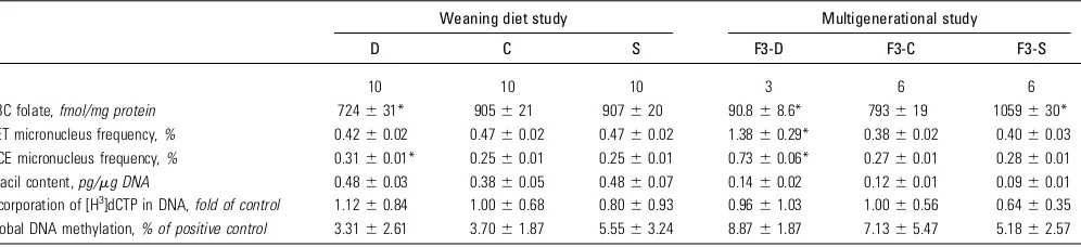
TABLE 1 RBC folate, micronucleus frequencies, uracil content in nuclear DNA, and global DNA methylation in male mice exposed to various levels of folic acid in the weaning diet study and in the multigenerational study
Weaning diet study Multigenerational study
D C S F3-D F3-C F3-S
n 10 10 10 3 6 6
RBC folate,fmol/mg protein 724631* 905621 907620 90.868.6* 793619 1059630*
RET micronucleus frequency,% 0.4260.02 0.4760.02 0.4760.02 1.3860.29* 0.3860.02 0.4060.03 NCE micronucleus frequency,% 0.3160.01* 0.2560.01 0.2560.01 0.7360.06* 0.2760.01 0.2860.01
Uracil content,pg/mg DNA 0.4860.03 0.3860.05 0.4860.07 0.1460.02 0.1260.01 0.0960.01 Incorporation of [H3]dCTP in DNA,fold of control 1.12
60.84 1.0060.68 0.8060.93 0.9661.03 1.0060.56 0.6460.35 Global DNA methylation,% of positive control 3.3162.61 3.7061.87 5.5563.24 8.8761.87 7.1365.47 5.1862.57
1Values are means
6SE. *Different from corresponding control,P,0.05. C, control diet; D, folic acid–deficient diet; dCTP, deoxycytidine triphosphate; NCE, normochromatic erythrocyte; RET, reticulocyte; S, folic acid–supplemented diet.
by guest on July 8, 2013
jn.nutrition.org
in NCE in mice weaned to the D diet but no effect on MN-RET. RET are immature RBC with a life span of ~15 h that differentiate into NCE, which are mature RBC (22,33). NCE have estimated life spans of 30–40 d (34,35). The micronucleus frequency in the NCE reflects NCE produced over the previous month, in addition to those produced in the last 3 wk. The 3-wk period during which mice were fed the C diet would have a much smaller impact on the measured micronucleus frequency in the NCE cellular population. Thus, the experimental design allowed us to measure the transient effects of dietary changes in RET, as well as the persistent effects of the diet on the NCE population. The absence of an increase in micronuclei in RET after 3 wk of consuming the C diet indicates that the DNA damage was diet-dependent and transient. This finding is supported by other studies that showed that folic acid supplementation in humans rescues the chromosomal damage induced by deficiency (2,3). We hypothesized that micronuclei result from uracil incorpora-tion into DNA or perturbaincorpora-tions in DNA methylaincorpora-tion from the D diet. However, there were no differences in either of these measures in bone marrow cells in the weaning study, suggesting that they are either not involved in the process of micronucleus formation or, alternatively, that these DNA modifications are also transient. We speculate that it is the latter, because bone marrow cells undergo a significant rate of cell turnover and can therefore quickly clear cells with these characteristics. Together, our data show that folic acid deficiency, but not supplementa-tion, leads to transient chromosome damage in RBC, and that the reintroduction of folic acid reverses the effect.
The multigenerational study was conducted to determine the cumulative effects on DNA damage of a D or an S diet over multiple generations and for a prolonged period of time. There was a significant 260% and 174% increase in RET and MN-NCE in the D line after 3 generations and 32 wk exposure. Given that exposure to a relatively high dose of the mutagen ethyl-nitrosourea (used as a positive control in this study) produced an 11-fold greater micronucleus frequency, the 2-fold increase caused by folate deficiency is quite substantial (36). Global DNA methylation did not differ among the dietary groups. However, there was a trend (P= 0.10) toward increased DNA uracil content in bone marrow cells with declining dietary folic acid (40% higher incorporation of uracil in the DNA of D lines relative to that of S) and a significant correlation between DNA uracil content and micronuclei, supporting the hypothesis that increased uracil content in nuclear DNA leads to chromosome damage.
There are 2 possible explanations for the more robust response observed in the multigenerational F3-D mice compared with mice placed on D diets at weaning for 12 wk. First, F3 male mice were fed the D diet for a prolonged period (32 wk) before being killed, which may have led to an accumulation of micronuclei. Alternatively, folic acid deficiency over 3 genera-tions may have resulted in a transgenerational cumulative effect on chromosome damage. If the D diet results in chromosome changes or hypomethylation that is transmitted through germ cells, genetic instability could manifest in the next generation and be propagated over multiple generations (37). Although there were no significant differences in global DNA methylation, direct quantification of DNA sequence–specific methylation changes in germ cells within and across generations and diets could provide mechanistic insight into the observed response. Given this interesting finding, future studies on the transgenera-tional effects of folic acid deficiency should wean the final generation of mice to the C diet. Analysis of micronucleus frequency in these mice could delineate the immediate effects of the diet from the inherited transgenerational effects.
Character-ization of DNA damage, chromatin structure, and DNA methylation in parental gametes among the dietary groups could also shed light on this outcome. One final consideration is that 3 generations of folic acid deficiency may have selected for offspring that are genetically tolerant of chromosomal instability or that exhibit enhanced DNA repair, which could affect the observed results. For example, offspring selected for enhanced uracil repair would have less uracil content and reduced chromosome damage. A difference in absolute DNA uracil content between the 2 studies suggests that differences in age or exposure paradigm affect uracil incorporation. Gene expression analysis will aid in determining whether this is the case.
As found in the weaning study, there was no change in micronucleus frequency in mice fed the S diet for 3 generations and 32 wk. There was a trend for reduction in bone marrow DNA uracil content compared with that in the C diet (P= 0.11), suggesting that there may be a lower limit to the relationship between uracil-mediated genome instability and micronucleus formation. Folic acid supplementation did not affect global DNA methylation measures.
It has been suggested that the amount of folic acid used in fortified foods should be doubled to ensure that women of childbearing age attain optimal folic acid intake to prevent neural tube defects (38). However, potential adverse effects of high folic acid intake are unclear, and work is needed to establish whether negative health outcomes are related to high folic acid intake in the general population. We found no effect of folic acid supplementation on the induction of micronuclei in RBC in male mice, even after they were fed the S diet for 32 wk and over multiple generations. The data indicate that supplemental folic acid at the level used in this study, which is consistent with combined folic acid consumption from fortified foods and vitamin supplements, was not detrimental, nor did it provide additional benefit, such as a decrease in micronucleus frequency, compared with the C diet. Thus, chronic exposure to excess folic acid at this level does not affect chromosome damage in RBC, an important finding when the high folate status of a large proportion of the Canadian population is considered (16).
In the present study we specifically quantified chromosomal effects of folate supplementation in Balb/c male mice, which are deficient in DNA-PK and hence are compromised in the repair of DNA double-strand breaks (39). It would be useful to evaluate these effects in other compromised models, such as inbred mice deficient in folate metabolism or uracil repair. Methylated CpG dinucleotides can also be spontaneously deaminated to thymi-dine (40). Therefore, an analysis of DNA sequence mutation rates in response to folic acid deficiency or supplementation should be performed. Finally, adverse effects of folic acid supplementation, such as delayed embryonic development in mice or increased progression of cancer in individuals with preneoplastic lesions in the colon, have been shown and should therefore continue to be a focus of future research (41,42).
In conclusion, folic acid deficiency clearly leads to chromo-some damage, which is consistent with previous reports. Manda-tory fortification of cereal products with folic acid in North America has essentially eliminated folate deficiency and therefore may protect this population from chromosome damage and related pathologies such as cancer that would result from folate deficiency. Our data do not support an association of high folic acid intake with genome instability, as indicated by RBC micronucleus frequency. Our findings suggest that excess folic acid at this level is likely not detrimental nor does it provide additional protection against chromosome damage in comparison to adequate folic acid intake.
by guest on July 8, 2013
jn.nutrition.org
Acknowledgments
The authors thank Ms. Cheryll Perry and Ms. Julie Todd for technical assistance and Dr. Francesco Marchetti and Dr. Alfred Aziz for helpful comments on the manuscript. A.J.M. and C.L.Y. designed the research; B.G.S., N.A.B., C.L.Y, and A.J.M. conducted the research; B.G.S., A.W., P.J.S, C.L.Y., and A.J.M. analyzed data and wrote the manuscript; and A.J.M. had primary responsibility for final content. All authors read and approved the final manuscript.
Literature Cited
1. Stover PJ. Physiology of folate and vitamin B12 in health and disease. Nutr Rev. 2004;62:S3–12; discussion S3.
2. Everson RB, Wehr CM, Erexson GL, MacGregor JT. Association of marginal folate depletion with increased human chromosomal damage in vivo: demonstration by analysis of micronucleated erythrocytes. J Natl Cancer Inst. 1988;80:525–9.
3. Blount BC, Mack MM, Wehr CM, MacGregor JT, Hiatt RA, Wang G, Wickramasinghe SN, Everson RB, Ames BN. Folate deficiency causes uracil misincorporation into human DNA and chromosome breakage: implications for cancer and neuronal damage. Proc Natl Acad Sci USA. 1997;94:3290–5. 4. Duthie SJ, Hawdon A. DNA instability (strand breakage, uracil misincorporation, and defective repair) is increased by folic acid depletion in human lymphocytes in vitro. FASEB J. 1998;12:1491–7. 5. Goulian M, Bleile B, Tseng BY. Methotrexate-induced misincorporation
of uracil into DNA. Proc Natl Acad Sci USA. 1980;77:1956–60. 6. Macfarlane AJ, Perry CA, McEntee MF, Lin DM, Stover PJ. Shmt1
heterozygosity impairs folate-dependent thymidylate synthesis capacity and modifies risk of apc(min)-mediated intestinal cancer risk. Cancer Res. 2011;71:2098–107.
7. Reidy JA. Folate- and deoxyuridine-sensitive chromatid breakage may result from DNA repair during G2. Mutat Res. 1987;192:217–9. 8. Fenech M, Kirsch-Volders M, Natarajan AT, Surralles J, Crott JW, Parry J,
Norppa H, Eastmond DA, Tucker JD, Thomas P. Molecular mechanisms of micronucleus, nucleoplasmic bridge and nuclear bud formation in mammalian and human cells. Mutagenesis. 2011;26:125–32.
9. Sauer J, Mason JB, Choi SW. Too much folate: a risk factor for cancer and cardiovascular disease? Curr Opin Clin Nutr Metab Care. 2009;12:30–6. 10. Kelly P, McPartlin J, Goggins M, Weir DG, Scott JM. Unmetabolized folic acid in serum: acute studies in subjects consuming fortified food and supplements. Am J Clin Nutr. 1997;65:1790–5.
11. Bailey RL, Mills JL, Yetley EA, Gahche JJ, Pfeiffer CM, Dwyer JT, Dodd KW, Sempos CT, Betz JM, Picciano MF. Unmetabolized serum folic acid and its relation to folic acid intake from diet and supplements in a nationally representative sample of adults aged$60 y in the United States. Am J Clin Nutr. 2010;92:383–9.
12. Matthews RG, Daubner SC. Modulation of methylenetetrahydrofolate reductase activity by S-adenosylmethionine and by dihydrofolate and its polyglutamate analogues. Adv Enzyme Regul. 1982;20:123–31. 13. Baram J, Chabner BA, Drake JC, Fitzhugh AL, Sholar PW, Allegra CJ.
Identification and biochemical properties of 10-formyldihydrofolate, a novel folate found in methotrexate-treated cells. J Biol Chem. 1988;263:7105–11. 14. Canada Gazette Part II. Regulatory impact analysis statement,
SOR/98-550. 1998;132:3029–33.
15. Colapinto CK, O’Connor DL, Tremblay MS. Folate status of the population in the Canadian Health Measures Survey. CMAJ. 2011;183:E100–6. 16. MacFarlane AJ, Greene-Fineston LS, Shi Y. Vitamin B-12 and
homo-cysteine status in a folate-replete population: results from the Canadian Health Measures Survey. Am J Clin Nutr. 2011;94:1079–87. 17. Shakur YA, Garriguet D, Corey P, O’Connor DL. Folic acid fortification
above mandated levels results in a low prevalence of folate inadequacy among Canadians. Am J Clin Nutr. 2010;92:818–25.
18. Yang Q, Cogswell ME, Hamner HC, Carriquiry A, Bailey LB, Pfeiffer CM, Berry RJ. Folic acid source, usual intake, and folate and vitamin B-12 status in US adults: National Health and Nutrition Examination Survey (NHANES) 2003–2006. Am J Clin Nutr. 2010;91:64–72. 19. Olfert ED, Cross BM, McWilliam AA, editors. Guide to the care and use
of experimental animals. 2nd ed. Ottawa (Canada): Canadian Council on Animal Care; 1993.
20. Reeves PG, Nielsen FH, Fahey GC Jr. AIN-93 purified diets for laboratory rodents: final report of the American Institute of Nutrition ad hoc writing committee on the reformulation of the AIN-76A rodent diet. J Nutr. 1993;123:1939–51.
21. Herbig K, Chiang EP, Lee LR, Hills J, Shane B, Stover PJ. Cytoplasmic serine hydroxymethyltransferase mediates competition between folate-dependent deoxyribonucleotide and S-adenosylmethionine biosynthe-ses. J Biol Chem. 2002;277:38381–9.
22. Dertinger SD, Camphausen K, Macgregor JT, Bishop ME, Torous DK, Avlasevich S, Cairns S, Tometsko CR, Menard C, Muanza T, et al. Three-color labeling method for flow cytometric measurement of cytogenetic damage in rodent and human blood. Environ Mol Mutagen. 2004;44:427–35. 23. MacFarlane AJ, Liu X, Perry CA, Flodby P, Allen RH, Stabler SP, Stover
PJ. Cytoplasmic serine hydroxymethyltransferase regulates the meta-bolic partitioning of methylenetetrahydrofolate but is not essential in mice. J Biol Chem. 2008;283:25846–53.
24. Pogribny I, Yi P, James SJ. A sensitive new method for rapid detection of abnormal methylation patterns in global DNA and within CpG islands. Biochem Biophys Res Commun. 1999;262:624–8.
25. Higgins JJ. An introduction to modern nonparametric statistics. Pacific Grove (CA): Brooks/Cole; 2003.
26. Efron B, Tibshirani R. An introduction to the bootstrap. Boca Raton (FL): Chapman & Hall/CRC; 1993.
27. R Development Core Team. R: a language and environment for statistical computing. R Foundation for Statistical Computing; 2009 [cited Feb 15, 2011]. Available from: http://www.R-project.org. 28. Liang KY, Zeger SL. Longitudinal data analysis using generalized linear
models. Biometrika. 1986;73:13–22.
29. Prentice RL, Zhao LP. Estimating equations for parameters in means and covariances of multivariate discrete and continuous responses. Biometrics. 1991;47:825–39.
30. Yan J, Fine JP. Estimating equations for association structures. Stat Med. 2004;23:859–74.
31. Yan J. geepack: yet another package for generalized estimating equations. R-News. 2002;2:12–4.
32. Fenech M. Cytokinesis-block micronucleus cytome assay. Nat Protoc. 2007;2:1084–104.
33. Grawe´ J, Biko J, Lorenz R, Reiners C, Stopper H, Vershenya S, Vukicevic V, Hempel K. Evaluation of the reticulocyte micronucleus assay in patients treated with radioiodine for thyroid cancer. Mutat Res. 2005;583:12–25.
34. The Laboratory Mouse. Germany: Elsevier Academic Press; 2004. 35. Horky´ J, Va´cha J, Znojil V. Comparison of life span of erythrocytes in
some inbred strains of mouse using 14C-labelled glycine. Physiol Bohemoslov. 1978;27:209–17.
36. Witt KL, Livanos E, Kissling GE, Torous DK, Caspary W, Tice RR, Recio L. Comparison of flow cytometry- and microscopy-based methods for measuring micronucleated reticulocyte frequencies in rodents treated with nongenotoxic and genotoxic chemicals. Mutat Res. 2008;649:101–13. 37. Filkowski JN, Ilnytskyy Y, Tamminga J, Koturbash I, Golubov A,
Bagnyukova T, Pogribny IP, Kovalchuk O. Hypomethylation and genome instability in the germline of exposed parents and their progeny is associated with altered miRNA expression. Carcinogenesis. 2010;31:1110–5. 38. Wilson RD, Johnson JA, Wyatt P, Allen V, Gagnon A, Langlois S,
Blight C, Audibert F, Desilets V, Brock JA, et al. Pre-conceptional vitamin/folic acid supplementation 2007: the use of folic acid in combination with a multivitamin supplement for the prevention of neural tube defects and other congenital anomalies. J Obstet Gynaecol Can. 2007;29:1003–26.
39. Okayasu R, Suetomi K, Yu Y, Silver A, Bedford JS, Cox R, Ullrich RL. A deficiency in DNA repair and DNA-PKcs expression in the radiosen-sitive BALB/c mouse. Cancer Res. 2000;60:4342–5.
40. Cooper DN, Krawczak M. Cytosine methylation and the fate of CpG dinucleotides in vertebrate genomes. Hum Genet. 1989;83:181–8. 41. Pickell L, Brown K, Li D, Wang XL, Deng L, Wu Q, Selhub J, Luo L,
Jerome-Majewska L, Rozen R. High intake of folic acid disrupts em-bryonic development in mice. Birth Defects Res A Clin Mol Teratol. 2011;91:8–19.
42. Cole BF, Baron JA, Sandler RS, Haile RW, Ahnen DJ, Bresalier RS, McKeown-Eyssen G, Summers RW, Rothstein RI, Burke CA, et al. Folic acid for the prevention of colorectal adenomas: a randomized clinical trial. JAMA. 2007;297:2351–9.
by guest on July 8, 2013
jn.nutrition.org