CHAPTER 2
LITERATURE REVIEW
2.1. FXTAS: THE FMR1 GENE AND CLINICAL ASPECTS
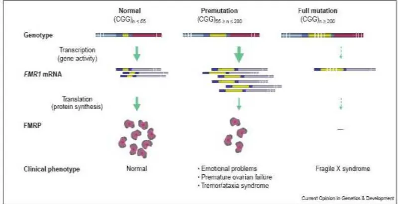
The fragile X mental retardation 1 gene (FMR1) consists of a 38 kB region with 17 exons and is located at Xq27.3 (33). This gene is responsible for producing fragile X mental retardation protein (FMRP) which is an RNA binding protein that associates with polyribosomes (34). FMRP is highly present in adult human brain, especially in neurons (35). FMRP is involved in the transport and regulation of local mRNA translation at synaptic sites, which is important for neuronal function, including modulation of synaptic plasticity (36, 37). FMRP binds to specific mRNAs and mediates the targeting of these transcripts into the dendrite. Therefore, FMRP might have a role in transport and/or translational efficiency of specific mRNAs at the synapse (20).
FMR1 with a variation in
and is transcriptionally silenced, hence no FMRP is produced. This condition results in a mental retardation disease called fragile X syndrome. The prevalence of Caucasian individuals with fragile x syndrome is estimated about 1 in 4000 (4). In Indonesia Faradz et al. found that 5 of 262 male students (1.9%) with developmental disability in special schools were positive for fragile x syndrome (5).
Expansion to full mutation usually occurs from premutation and is restricted upon maternal transmission. In other words, mothers with premutation CGG size have a high risk to have children affected with fragile X syndrome (39). Sperm of full mutation males contains only premutation alleles, which explains why daughters of full mutation males are never affected with the fragile X syndrome (40). AGG interruption commonly appears every 9-11 CGG repeats of normal individuals, but it is absent in mostly premutation individuals. The presence of AGG is considered to be able to stabilize the repeat. Premutation females with an AGG interruption transmit the CGG repeat to the children more stable than without AGG interruption (38, 39). The presence of AGG in expanded premutation CGG does not influence either transcription or translation, according
to in vitro and cell culture study (41). Study in general population of North
America has shown that one in 813 males and one in 259 are premutation carriers (38, 42).
premutation females manifest premature ovarian failure (POF), in which the menstruation cycle stops before the age of 40 (43). Moreover the premutation males have a chance to develop a disease called fragile X-associated tremor/ataxia syndrome (FXTAS). More than a third of premutation males older than 50 years develop FXTAS. The penetrance increases with age, becoming 50% at the age of 70-90. It has been suggested that the longer the CGG repeats the more increase in penetrance of FXTAS (9, 10). Allen et al. defined CGG repeats with more than 70 trinucleotides as a risk allele, while lower than 70 as low risk allele (44).
Patients with FXTAS develop some clinical features. They generally have cerebellar gait ataxia and intention tremor (8). Other features common seen are Parkinsonism, lower extremity neuropathy, autonomic dysfunction, and cognitive decline. The cognitive decline varies from mild frontal executive and memory deficits to global dementia (7, 8, 11). Besides the clinical features, individuals with FXTAS also display some psychological features. Psychiatric features present are anxiety, disinhibition, depression, and apathy (13).
Figure 1. CGG length, FMR1 expression and the clinical outcomes (48).
2.2. NEUROPATHOLOGICAL OF FXTAS
Neuroanatomical studies have shown Purkinje cell loss and Bergmann gliosis in premutation carriers. Global brain atrophy; white matter disease in subcortical, middle cerebellar peduncle (MCP), and periventricular region; and dilated ventricles are seen as MRI features of FXTAS (8, 49, 50). The MCP sign is a diagnostic criterion, since it is observed in 60% of premutation carriers with tremor and/or ataxia. Reduction in cerebellar volume, increased ventricular and white matter volume were also observed in a study of male premutation carriers (50).
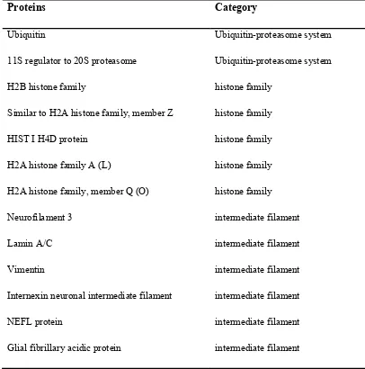
Purkinje cells, although these cells are dropped out in FXTAS patient (15). Many proteins now have been identified to co-localize in the inclusions. Those proteins can be divided into eight categories: Ubiquitin-proteasome system, histone family, intermediate filament, microtubule, myelin associated protein, RNA-binding protein, stress-related protein, and chaperone (16, 17) (see table for detail). Recently another protein, sam68, was just identified to be present in human FXTAS brain (51).
Table 1. Proteins found in the human brain inclusions (16)
Beta5-tubulin
2.3. MOLECULAR PATHOGENESIS OF FXTAS
A toxic RNA gain of function mechanism has been proposed as the mechanism underlying FXTAS (21, 22). The fact that premutation carriers have elevated levels of FMR1 RNA as much as 8 fold over normal allele gives a clue to the proposed mechanism (6). The finding of FMR1 RNA in the inclusions of premutation carriers also supports the toxic RNA gain of function mechanism. Another clue drives to this proposed mechanism is the fact that older full mutation carriers (which is not producing FMR1 RNA) have totally different outcomes with FXTAS (18, 19).
Premutation carriers exhibit elevated levels of FMR1 message with reduced levels of FMRP (6, 53). A feedback mechanism proposes that the elevated FMR1 transcript levels in premutation carrier are caused by the increased transcription as an effort of the cells to compensate the low levels of FMRP (54). Based on study of Chen et al., the elevated levels of FMR1 mRNA is likely because of the CGG itself, in which the more CGG repeats will result in more open promoter leading to enhanced access for transcription factor (20, 55). While reduced levels of FMRP are caused by reduction of translational efficiency in alleles with long CGG tracts, in which this long CGG in the RNA will result in secondary structures, hairpins, causing an impediment of ribosome to effectively doing its work (41, 55, 56).
non-coding
myotonic dystrophy protein kinase (DMPK) gene or by expanded CCTG in intron 1 of ZNF9 gene (57). The expansions in DM produce nuclear foci similar to intranuclear inclusion seen in FXTAS. The nuclear foci contain the expanded repeat RNA as well as sequestered proteins especially RNA-binding protein such as MBNL1. Studies in DM conclude the toxic RNA gain of function as the mechanism leading to the development of this disease (57).
The toxic effect of expanded premutation CGG RNA was also found when human neural cells were transfected with FMR1 CGGs and a GFP reporter. Expression of this expanded premutation CGG repeat reduced cell viability. Inclusions containing alpha-beta crystalline were observed, as well as an alteration in morphological and nuclear lamin structure. The cytotoxicity was indeed caused by the CGG RNA instead of the DNA, because cells transfected with expanded premutation CGG with deleted promoter, which means no (58). One study using microarray has shown the upregulation of genes required for apoptosis in cells transfected with construct expressing premutation CGG (59).
Expressing the expanded premutation CGG as a transcript in drosophila
melanogaster flies displayed deterioration of neurons in the fly eyes. It also
COS7 cells transfected with expanded 60 and 100 CGGs formed CGG RNA nuclear aggregates, according to studies using cell culture with fluorescence in situ hybridization (FISH) technique. The CGG RNA aggregates expanded over time and finally formed giant inclusions. The cells exhibited nuclear lamin A/C architecture disruption and cell death 72-96 hours after transfection. Cells transfected with only 20 CGGs did not exhibit those aggregates and disruptions, while transfection of 40 CGGs resulted in intermediate situation with rare and small intranuclear aggregates. Observation to the cells 24 and 72 hours after transfection demonstrated the presence of MBNL1 and hnRNP-G which are hardly seen at 24 hours, but increased after longer incubation. While sam68 was clearly seen starting from 24 hours, suggesting sam68 is an early inclusions marker (51).
aggregates. This is based on an observation if sam68 is not present in the inclusions, other proteins such as MBNL1 and hnRNP-G are absent as well (51).
2.4. MOUSE MODEL OF FXTAS
A KI mouse model was generated to study repeat instability by homologous recombination technique replacing the endogenous murine (CGG)8 with human (CGG)98. Cloning was performed with minimal changes made on the sequence. All known regulatory elements on the promoter were maintained. Although this long repeat is enough to produce repeat instability like in human case, these mice showed only minor instability of the CGG repeat and no methylation was observed even if the mice reach more than 200 repeats (24, 26, 27). After FXTAS was recognized and described, these mice then were subjected for neurohistology, biochemistry and molecular aspects of FXTAS (29). Elevated RNA levels were detected 3.5 fold compared with control mice (27).
mice compared to human FXTAS are the absence of neuronal loss, gliosis and
behavioral phenotype as seen in human patients. Only decreased performance in neuromotor tasks and mild age-dependent learning disturbances were seen in these mice (25).
Another KI mouse was generated by the Usdin group. These mice had an original size 118 of CGGs. They exhibited elevated levels of mRNA and showed positive correlation between the mRNA levels and CGG length. In some cases, large expansion to full mutation range was observed in these mice, however no methylation has been found. These KI mice exhibited similar genetic and pathophysiological performance to human FXTAS patients. These mice showed Purkinje cell loss with swollen axonal torpedos, which were not seen in the other KI mice (23).
motor-learning abilities in accelerating rotarod studies. This model has proven the toxicity of expanded premutation CGG RNA in mammals (61).
There is one sophisticated strategy to have a good mouse model for FXTAS. It is by using the tet-on-regulated inducible system and the specific driver promoter reverse tetracycline transactivator (rtTA). Inducible expression of an expanded premutation CGG repeat together with the use of specific driver promoters should give a good model to mimic the human FXTAS situation. The tet-on-regulated inducible system can control the expression by the induction of tetracycline (tet) or an analog such as doxycycline (dox) (30). This ability to control the expanded premutation CGG expression would be a great tool to study the reversibility of FXTAS. The specific reverse tetracycline transactivator (rtTA) drivers make it possible to restrict the place of toxic RNA transgene expression. The suitable driver promoters for FXTAS mouse model are PrP-rtTA which allows expression in all cell types in the brain except for the Purkinje cells (31); and GFA2-rtTA which restricts the expressions only in Bergmann glia and astrocyte (32). This restriction is a powerful tool to study the sufficiency and necessity of the cells in the development of FXTAS and allow us to know which cell types (neurons, Bergmann glia, astrocytes) contribute to FXTAS neuropathology.