Analysis of the postulated interaction between the angiotensin II
sub-type 1 receptor gene A1166C polymorphism and the
insertion
/
deletion polymorphism of the angiotensin converting
enzyme gene on risk of myocardial infarction
Richard P. Steeds
a, Anthony Wardle
b, Pamela D. Smith
b, Daniel Martin
b,
Kevin S. Channer
a, Nilesh J. Samani
b,*
aDepartment of Cardiology,Royal Hallamshire Hospital,Sheffield,UK bDepartment of Cardiology,Uni6ersity of Leicester,Leicester,UK
Received 24 August 1999; received in revised form 31 January 2000; accepted 18 February 2000
Abstract
A synergistic interaction between the insertion/deletion (I/D) polymorphism within the angiotensin-converting enzyme (ACE) gene and an A/C transversion at nucleotide position 1166 within the angiotensin II sub-type 1 receptor (AT1R) gene on risk of myocardial infarction has been reported. The risk associated with the ACE DD genotype increased with the number of AT1R C alleles present. To investigate this further, ACE I/D and AT1R A1166C genotypes were determined in 541 cases recruited at the time of infarction and 507 population-based controls. There was no difference in either the genotype distribution or allele frequencies between cases and controls for either the ACE polymorphism (P=0.48 and 0.35 respectively) or the AT1R polymorphism (P=0.35 and 0.21 respectively). Odds ratios for risk of MI associated with the ACE DD and AT1R CC genotypes were 1.09 (95% CI, 0.82 – 1.45) and 1.06 (0.67 – 1.68) respectively. 3.1% of cases versus 3.6% of controls were homozygous for both the D and C alleles (P=0.71). There was no increase in risk associated with the DD genotype in the presence of either one or two AT1R C alleles in the whole cohorts (OR 0.99, 95% CI 0.65 – 1.51 and 0.76, 95% CI 0.30 – 1.88, respectively) nor in sub-groups defined by specific risk factors. In conclusion, no evidence was found to support any interaction between the ACE gene I/D polymorphism and the ATIR gene A1166C transversion in determining the risk of myocardial infarction in the population studied. © 2001 Elsevier Science Ireland Ltd. All rights reserved.
Keywords:Gene polymorphism; Angiotensin receptors; Angiotensin converting enzyme; Myocardial infarction
www.elsevier.com/locate/atherosclerosis
1. Introduction
Epidemiological studies have identified several im-portant risk factors for ischaemic heart disease, al-though these only partly explain the occurrence of acute coronary syndromes [1]. A positive family history is a strong indicator of risk, part of which accrues from a shared genetic inheritance and part from experiencing a shared early environment [2]. Genetic factors exert their influence on susceptibility to atherosclerosis and acute thrombosis by the modulation of plasma and
tissue levels of their biologically active products. Envi-ronmental factors may subsequently modify this influ-ence in such a way as to either increase or decrease the associated risk. Acute myocardial infarction is a com-plex clinical event which, is the culmination of a multi-plicity of interacting pathologic processes. In contrast to rare monogenetic diseases, risk of ischaemic heart disease is likely to be carried by several genes, capable of multifaceted interactions with each other and with environmental factors [3].
The best example of a gene – gene interaction influ-encing the risk of MI reported to date is the purported interaction between the A1166C polymorphism in the angiotensin II sub-type 1 receptor (AT1R) gene and the insertion/deletion (I/D) polymorphism in the an-* Corresponding author. Present address: Department of
Cardiol-ogy, Clinical Sciences Wing, Glenfield Hospital, Groby Road, Leices-ter, LE3 9QP UK. Tel.: +44-116-2563038; fax: +44-116-2875792.
giotensin converting enzyme (ACE) gene. In Cau-casian male subjects recruited in four European cen-tres 3 – 9 months following their MI compared with unaffected controls, Tiret et al. [4] found that the risk associated with the ACE DD genotype compared with the ACE II/ID genotypes increased from 1.05 (95% CI 0.75 – 1.49) in those bearing the AT1R AA geno-type, to 3.95 (95% CI 1.26 – 12.4) in those ho-mozygous for the AT1R CC genotype with an intermediate risk (1.52) (1.06 – 2.18) in AC het-erozygotes. The risk was particularly increased in sub-jects considered at low risk of MI on the basis of a low apolipoprotein B level and body mass index [4]. If such an interaction could be confirmed it would represent an important advance in our understanding of the genetic basis of acute coronary syndromes and could provide a valuable tool for risk stratification. Therefore, the aim of this study was to further exam-ine the postulated interaction between the ACE I/D and AT1R A/C gene polymorphisms in a cohort of subjects with acute myocardial infarction and popula-tion-based controls.
2. Methods
2.1. Subjects
The method of recruitment of subjects has been described previously [5]. In brief, cases were recruited from patients admitted to the coronary care units (CCU) of the Leicester Royal Infirmary, Leicester (July 1993 – April 1994) and the Royal Hallamshire Hospital, Sheffield (November 1995 – March 1997). Pa-tients were recruited who satisfied the World Health Organisation criteria for acute myocardial infarction in terms of symptoms, elevations in cardiac enzymes or electrocardiographic changes [6]. Recruitment crite-ria were identical in the two centres and more than 95% of eligible subjects were enlisted. Control subjects were recruited in each hospital from adult visitors to patients with non-cardiovascular disease on general medical, surgical, orthopaedic and obstetric wards to provide subjects likely to be representative of the source population from which the subjects originated. Subjects who reported a history of coronary heart disease were excluded. Again, \95% controls ap-proached agreed to take part. Data on the ACE gene I/D polymorphism from the current Leicester cohort and a previous Sheffield cohort have been reported [7], but a new cohort of cases and controls was re-cruited for this study in Sheffield to optimise the matching of cases and controls and to improve the data available on classical risk factors for MI.
After informed consent was obtained, cases and controls filled in a standard questionnaire about their
personal histories, had height and weight measured, and provided blood samples for genotype analysis and measurement of serum total cholesterol. For the pur-pose of this study, genotype analysis was restricted to Caucasian cases B75 years of age.
The studies in both centres were approved by the respective local clinical research ethics committees.
2.2. Genetic analysis
Deoxyribonucleic acid (DNA) was prepared from a small aliquot of whole blood collected in ethylenedi-amine tetraacetic acid by using a DNA extraction ma-trix (Instagene, Biorad, Hemel Hempstead, Hertfordshire, England) and following standard tech-niques [8]. The extracted DNA was stored at −70oC until analysis. The ACE gene D and I alleles were identified on the basis of polymerase chain reaction (PCR) amplification of the respective fragments from intron 16 of the gene, as previously described [9]. In order to reduce mistyping of ID heterozygotes as DD homozygotes, 5% dimethylsulfoxide was included in the reaction mixture [10]. Amplified fragments were analysed on ethidium-bromide agarose gels as previ-ously described [7].
The AT1R A1166-C polymorphism was also as-sayed by PCR amplification with a slight modification of the method described by Bonnardeaux et al. [11]. Briefly, 100 ng of DNA was amplified with 100 pmol/ l of the sense (5% GCT TTG TCT TGT TGC AAA AGG 3%) and anti-sense (5%CCC ACT CAA ACC TTT CAA CA 3%) oligonucleotide primers in a final volume of 100 ml. The DNA was amplified for 35
cycles with denaturation at 94°C for 1 min, annealing at 52°C for 1 min, and extension at 72°C for 1 min. 10 ml of PCR product was digested overnight at 37°C
with 1 unit of DdeI in a final volume of 20 ml
con-taining 5 mg bovine serum albumin according to the manufacturers’ instructions (New England Biolabs). This reaction yielded fragments of 143, 106 and 47 base pairs in the presence of the C allele, and 249 and 47 base pairs in the presence of the A allele. After the addition of 2 ml of sucrose-based loading buffer, the
reaction product was loaded onto a 6% polyacry-lamide gel (polyacrypolyacry-lamide to bisacrypolyacry-lamide ratio of 40:1). Electrophoresis was performed at 200 v for 30 min at room temperature. The DNA fragments were visualised by ultraviolet transillumination after stain-ing with ethidium bromide.
Table 1
Characteristics of cases and controls according to centre
Leicester Sheffield
Controls
Cases P Cases Controls P
6199 0.28
Age (years) 6299 6299 55912 B0.001
62 0.49 72
64 61
Male (%) 0.01
2694
Body mass index (kg/m2) 2694 0.99 2694 2594 0.29
6.091.2
Total cholesterol (mmol/l) 5.991.0 0.22 5.791.2 5.691.0 0.49
18 B0.001 34
29 17
Hypertension (%) B0.001
2
Diabetes (%) 8 B0.001 9 4 0.06
15 B0.001 41 20 B0.001
41 Current smokers (%)
2.3. Biochemical measurements
Serum total cholesterol was measured using a Kodak Ektachem E700 CXR Automatic Analyser in Leicester and an Olympus AU 5223 machine in Sheffield in national quality-controlled hospital biochemistry labo-ratories. For cases, the first blood sample taken follow-ing admission was used for analysis.
2.4. Statistical analysis
The distribution of the ACE I/D and AT1R A/C genotypes and qualitative risk factors for patients and control subjects were compared using the chi-square test. Quantitative sample means were compared by analysis of variance. Odds ratios and 95% confidence intervals (CI) estimating the relative risk of myocardial infarction associated with the DD/CC genotype were calculated using the Mantel – Haenszel method with stratification, where described, for age, sex and centre.
3. Results
In total, 1048 subjects (541 cases of acute myocardial infarction and 507 control subjects) were analysed from the two centres. Table 1 summarises the characteristics of the cases and controls in each centre. The Sheffield cohorts were well matched for age and sex. In Leicester, the cases were significantly older than controls (average 7 years) and there was a slight male preponderance in the cases. In both centres, approximately one-third of cases had suffered with pre-existing ischaemic heart disease. As would be expected, several classical risk factors (hypertension, diabetes, and smoking lence) for acute myocardial infarction were more preva-lent in the cases than controls in both centres. However, body mass index and total cholesterol level were similar in cases and controls.
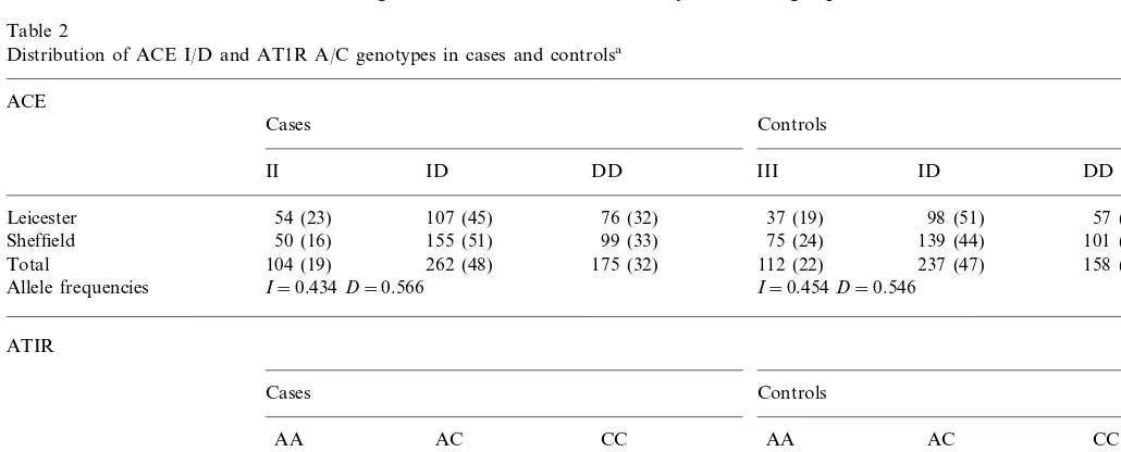
The distributions of the ACE I/D and the ATIR A/C alleles and genotypes in cases and controls are shown in Table 2. For both polymorphisms, all cohorts were in Hardy – Weinberg equilibrium. There was no difference
Table 2
Distribution of ACE I/D and AT1R A/C genotypes in cases and controlsa
ACE
Cases Controls
II ID DD III ID DD
54 (23) 107 (45) 76 (32) 37 (19) 98 (51)
Leicester 57 (30)
50 (16) 155 (51) 99 (33)
Sheffield 75 (24) 139 (44) 101 (33)
Total 104 (19) 262 (48) 175 (32) 112 (22) 237 (47) 158 (31)
Allele frequencies I=0.434D=0.566 I=0.454D=0.546
ATIR
Cases Controls
AA AC
AC CC
AA CC
121 (51) 97 (41) 19 (8)
Leicester 102 (53) 71 (37) 19 (10)
27 (9) 120 (38)
168 (53) 33 (11)
Sheffield 143 (47) 128 (42)
264 (49) 225 (42) 52 (10)
Total 270 (53) 191 (38) 46 (9)
Allele frequencies A=0.696C=0.304 A=0.721C=0.279
Table 3
Combined distributions of ACE I/D and AT1R A/C genotypes in cases and controlsa
Cases Controls
aNumbers in parentheses are% of total.
in genotype distributions between the Sheffield and Leicester cohorts. For the ACE I/D polymorphism, there was no significant difference in either the geno-type distribution (P=0.48) or the allele frequencies (P=0.35) between cases and controls. The age, sex and centre-stratified odds ratio (OR) for risk of MI associ-ated with the DD genotype compared with the com-bined ID/II genotypes was 1.09 (95% CI 0.82 – 1.45, P=0.57). Likewise, there was no significant difference in the genotype distribution (P=0.35) or allele fre-quencies (P=0.21) for the AT1R A/C polymorphism (Table 2). The age, sex and centre-stratified OR for risk of MI associated with the CC genotype compared with the combined AA/AC genotypes was 1.15 (95% CI 0.89 – 1.50,P=0.31), and the OR for the AC/CC com-bined genotypes compared with AA genotype was 1.06 (95% CI 0.67 – 1.68, P=0.86). For both polymor-phisms, there was no increase in risk in sub-groups stratified by other risk factors (body mass index, total cholesterol, smoking status, hypertension or diabetes) (data not shown).
Table 3 shows the relative distribution of ACE I/D and ATIR A/C genotypes in cases and controls. There was no increase in the number of DD/CC homozygotes in cases compared with controls (3.1 vs 3.6%,P=0.71). No interaction was observed between the two polymor-phisms on risk of MI. Specifically, there was no graded increase in risk associated with the DD genotype in the presence of an increasing number of AT1R C alleles. Thus, the OR associated with the DD genotype in subjects with the AA genotype was 1.19 (0.81 – 1.73, P=0.36), in subjects with the AC genotype 0.99 (0.65 –
1.51, P=0.97) and in subjects with the CC genotype 0.76 (0.30 – 1.88,P=0.51). The risk associated with the combined genotypes remained non-significant when subjects were stratified by either age, sex, body mass index or total cholesterol (Table 4).
4. Discussion
Genetic interactions influencing clinical phenotypes are much more plausible when the genes concerned influence the same or related biochemical pathway or cellular process. In this regard, the reported interaction between the ACE gene I/D polymorphism and the AT1R A/C polymorphism is attractive. There is consid-erable evidence implicating the renin-angiotensin sys-tem in vascular biology [12]. Further the ACE I/D polymorphism has a major influence on plasma and tissue ACE levels [13,14] which in turn could affect angiotensin II generation [15], the direct effector molecule for the ATI receptor. Although, a direct func-tional effect of the ATIR gene A/C polymorphism, which is located in the 3% untranslated region of its mRNA, has not been identified, the polymorphism has been associated with increased vascular reactivity [16] and arterial stiffness [17], suggesting a biological effect perhaps through linkage disequilibrium with another polymorphism.
There is an on-going debate about the importance of the ACE I/D polymorphism in influencing the risk of MI. [18,19]. Further, most studies that have investi-gated the AT1R A/C polymorphism have not observed
Table 4
Odds ratios of risk of myocardial infarction associated with the ACE DD genotype in subjects grouped by the AT1R A/C genotype for the whole cohort and in specified sub-groupsa
CC AC
AA
0.76 (0.30–1.88) 0.51 1.19 (0.81–1.73) 0.36 0.99 (0.65–1.51) 0.97
Overall
0.71 (0.16–3.12) 0.65 1.66 (0.82–3.36) 0.16 0.59 (0.24–1.42) 0.23
AgeB55
0.77 (0.29–2.09) 0.61 1.15 (0.67–1.98) 0.62
Male 1.06 (0.67–1.67) 0.79
BMIBmedianb 1.25 (0.73–2.14) 0.41 1.42 (0.77–2.62) 0.26 1.40 (0.35–5.63) 0.64
0.33 (0.08–1.34) 0.11 1.18 (0.65–2.15) 0.59
1.19 (0.69–2.06) 0.53 CholesterolBmedianb
Non-smokers 1.07 (0.69–1.66) 0.77 1.11 (0.67–1.85) 0.68 0.58 (0.21–1.54) 0.27
an independent effect on MI risk [4,20 – 22]. Despite this, an important epistatic interaction between the two polymorphisms could still exist. However, in a study involving 541 cases of acute myocardial infarction and 507 control subjects we did not find any evidence to support this possibility. Our findings are in contrast with those of Tiret et al. [4] but in accord with more recent studies [20 – 23]. Small study size could obscure any association. However, our study had \99% power at a significance of 0.01% to detect the 3.95 fold in-crease in risk observed by Tiret et al. [4] in subjects carrying the combined DD and CC genotypes.
There are several differences in design between our study and that of Tiret et al. [4]. Their cases were all male below the age of 65 and recruited between 3 and 9 months following MI. However, we did not find any association of the genotypes with risk in younger sub-jects or when stratified by sex (Table 4). Further re-cruitment at the time of the incident event as done by us, should have enhanced the association with MI, unless the genotypes influenced survival rather than MI risk. However, no data exists to suggest such a possibil-ity. It is also noteworthy that in the study by Tiret et al. [4], the interaction of the two genotypes arose not because of a relative increase in the number of cases with the ACE DD genotype in those bearing the AT1R CC genotype, but instead because of a marked reduc-tion in the relative proporreduc-tion of ACE DD subjects in controls bearing the AT1R CC genotype compared with the AT1R AA genotype (from 30 to 10%). An association based on this finding is only possible if one postulates that controls bearing the combined DD/CC genotypes die prematurely. However, our findings do not support this contention, since we found no differ-ence in the proportion of controls with the ACE DD genotype bearing the AT1R CC versus AA genotypes (30 vs 39%).
A further observation in the study of Tiret et al. [4] was that the effect of the interaction between the ACE and ATIR polymorphisms was greater in subjects deemed to be at low risk on the basis of plasma apolipoprotein B level and body mass index. In this sub-group the risk in DD/CC subjects was 13.3 (0.79 – 707) fold higher. Although we were not able to classify subjects in precisely the same manner, our sub-group analyses (Table 4), based on stratification by relevant individual risk factors, do not provide any evidence to support this hypothesis. In this regard, our data also agree with the findings in more recent studies [23].
In summary, in a two centre study of patients with myocardial infarction recruited at the time of infarc-tion, no significant interaction was found between the ACE I/D polymorphism and AT1R A/C polymorphism in determining risk of myocardial infarction, either in the whole population or in sub-groups stratified by the presence or absence of other coronary risk factors.
Taken together with other recent data, our findings suggest that genotyping for these polymorphisms is unlikely to be of widespread clinical utility in assessing the risk of myocardial infarction.
References
[1] Wang XL, Tam C, McCredie RM, Wilcken DEL. Determinants of severity of coronary artery disease in Australian men and women. Circulation 1994;89:1974 – 81.
[2] Marenberg ME, Risch N, Berkman LF, Floderus B, deFaire U. Genetic susceptibility to death from coronary heart disease in a study of twins. N Eng J Med 1994;330:1041 – 6.
[3] Marion AJ. Genetic risk factors for myocardial infarction. Curr Opinion Cardiol 1998;13:171 – 8.
[4] Tiret L, Bonnardeaux A, Poirier O, et al. Synergistic effects of angiotensin-converting enzyme and angiotensin-II type 1 recep-tor gene polymorphisms on risk of myocardial infarction. Lancet 1994;344:910 – 3.
[5] Steeds R, Adams M, Smith P, Channer K, Samani NJ. Distribu-tion of tissue plasminogen activator inserDistribu-tion/deletion polymor-phism in myocardial infarction and control subjects. Thromb Haemostas 1998;79:980 – 4.
[6] WHO. (Report of the Joint International Society and Federation of Cardiology/World Health Organisation Task Force on stan-dardisation of clinical nomenclature). Nomenclature and criteria for diagnosis of ischaemic heart disease. Circulation 1979;59:607 – 9.
[7] Samani NJ, O’Toole L, Martin D, et al. Insertion/deletion polymorphism in the angiotensin-converting enzyme gene and risk of and prognosis after myocardial infarction. J Am Coll Cardiol 1996;28(2):338 – 44.
[8] Sambrook J, Fritsch EF, Maniatis T. Molecular Cloning: A Laboratory Manual. Cold Spring Harbor: Cold Spring Harbor Laboratory Press, 1989.
[9] Rigat B, Hubert C, Corvol P, Soubrier F. PCR detection of the insertion/deletion polymorphism of the human angiotensin I converting enzyme gene (dipeptidyl carboxypeptidase 1). Nuc Acid Res 1992;20:1433.
[10] Shanmugam V, Sell KW, Saha BK. Mistyping ACE het-erozygotes. PCR Meth Applic 1993;3:120 – 1.
[11] Bonnardeux A, Davies E, Jeunemaitre X, et al. Angiotensin II type 1 receptor gene polymorphisms in human essential hyper-tension. Hypertension 1994;24:63 – 9.
[12] Dzau VJ. Implications of local angiotensin production in cardio-vascular physiology and pharmacology. Am J Cardiol 1987;59:59A – 65A.
[13] Rigat B, Hubert C, Alhenc-Gelas F, Cambien F, Corvol P, Soubrier F. An insertion/deletion polymorphism in the an-giotensin 1-converting enzyme gene accounting for half of the variance of serum enzyme levels. J Clin Invest 1990;86:1343 – 6. [14] Danser AHJ, Schalekamp MADH, Bax WA. Angiotensin-con-verting enzyme in the human heart: effect of the insertion/ dele-tion polymorphism. Circuladele-tion 1995;92:1387 – 8.
[15] Ueda S, Elliott HL, Morton JJ, Connell JM. Enhanced pressor response to AI in normotensive men with the deletion genotype (DD) for angiotensin-converting enzyme. Hypertension 1995;25:1266 – 9.
[16] Henrion D, Amant C, Benessiano J, et al. Angiotensin II type 1 receptor gene polymorphism is associated with an increased vascular reactivity in the human mammary artery in vitro. J Vasc Res 1998;35:356 – 62.
[18] Samani NJ, Thompson JR, O’Toole L, Channer K, Woods K. A meta-analysis of the association of the deletion allele of the angiotensin-converting enzyme gene with myocardial infarction. Circulation 1996;94:708 – 12.
[19] Staessen JA, Wang JG, Ginocchio G, et al. The deletion/ inser-tion polymorphism of the angiotensin converting enzyme gene and cardiovascular-renal risk. J Hypertens 1995;15:1579 – 92. [20] Katsuya T, Koike G, Yee TW, et al. Association of
angiotensi-nogen gene T235 variant with increased risk of coronary heart disease. Lancet 1995;345:1600 – 3.
[21] Nakauchi Y, Suehiro T, Yamamoto M, et al. Significance of the
angiotensin I-converting enzyme and angiotensin II type 1 recep-tor gene polymorphisms as risk facrecep-tors for coronary heart dis-ease. Atherosclerosis 1996;125:161 – 9.
[22] Berge KE, Bakken A, Bohn M, Erikssen J, Berg K. A DNA polymorphism at the angiotensin II type 1 receptor (AT1R) locus and myocardial infarction. Clin Genet 1997;52:71 – 6. [23] Gardemann A, Nguyen QD, Humme J, et al. Absence of an
association with the risk of coronary artery disease and myocar-dialinfarction and of a synergistic effect with angiotensin-con-verting enzyme gene polymorphism on the risk of these disease. Eur Heart J 1998;19:1657 – 65.