www.elsevier.comrlocateranireprosci
Department of Large Animal Medicine and Surgery, College of Veterinary Medicine, Texas A&M UniÕersity,
College Station, TX 77843-4475, USA
b
Department of Physiology and Pharmacology, College of Veterinary Medicine, Texas A&M UniÕersity,
College Station, TX 77843-4475, USA
c
Department of Veterinary Anatomy and Public Health, College of Veterinary Medicine, Texas A&M UniÕersity, College Station, TX 77843-4475, USA
Abstract
Numerous techniques may be used for evaluation of the different reproductive disorders of the stallion. Approaches may vary from real-time ultrasonography and biopsy for evaluating testicular tumors to use of special assays for evaluating sperm or plasma for presence of antisperm antibodies.This communication addresses techniques used to evaluate five relatively uncommon,
Ž . Ž .
but perplexing, disorders of breeding stallions: 1 seminal vesiculitis, 2 hemospermia associated
Ž . Ž .
with idiopathic urethral defects, 3 acrosomal dysfunction, 4 abnormal spermatozoal chromatin,
Ž .
and 5 azoospermia.q2000 Published by Elsevier Science B.V. All rights reserved.
Keywords: Horse; Stallion; Reproductive disorders; Semen
1. Seminal vesiculitis
Ž .
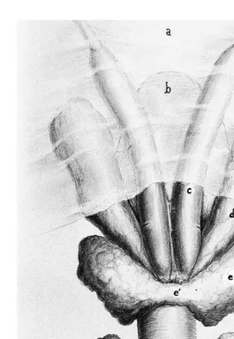
The stallion has four distinct accessory genital glands: 1 the paired seminal vesicles
Ž .
or vesicular glands, 2 the paired ampullae, which constitute a specialized portion of the
Ž . Ž .
terminal segment of each deferent duct, 3 the bilobed prostate gland, 4 and the paired
Ž .
bulbourethral glands Fig. 1 . The vesicular glands are slightly pyriform sacs which are situated on either side of the bladder neck, and extend craniolaterally into the posterior abdomen. Each gland cones into a singular excretory duct which communicates with the
)Corresponding author.
0378-4320r00r$ - see front matterq2000 Published by Elsevier Science B.V. All rights reserved. Ž .
Ž . Ž . Ž . Ž .
Fig. 1. Illustration of the accessory genital glands of the stallion dorsal view . a Genital fold, b bladder, c
Ž . Ž . Ž . Ž . Ž .
ampulla, d vesicular gland seminal vesicle , e lobe of prostate gland, e isthmus of prostate gland, f´
Ž . Ž . Ž .
lumen of the proximal urethra via a protuberance on its dorsal surface, the colliculus
Ž .
seminalis Fig. 2 .
Seminal vesiculitis is quite uncommon in stallions; however, it is a noteworthy disorder because of its persistent nature and interference with fertility. Reported bacterial
Ž . Ž .
Fig. 2. Illustration of pelvic urethra, as viewed by urethral videoendoscopy. a Bladder orifice, b colliculus
Ž . Ž .
seminalis, bI ejaculatory duct common opening for ductus deferens and excretory duct of vesicular gland ,
Ž .c openings of ducts of prostate gland, d opening of ducts of urethral glands, e openings of ducts ofŽ . Ž .
etiologic agents include Pseudomonas aeruginosa, Klebsiella pneumoniae,
Streptococ-cus spp., StaphylococStreptococ-cus spp., and Brucella abortus. We recently identified a stallion
with unilateral erosive seminal vesiculitis associated with Acinetobacter calcoaceticus. Characteristically, stallions with seminal vesiculitis do not manifest overt clinical signs. In general, affected horses tend to be afebrile and do not exhibit signs of pain at the time of urination, defecation, or breeding. Presumptive diagnosis of seminal vesiculi-tis is usually made through gross and microscopic analysis of semen. Upon cytological examination, variable numbers of neutrophils may be present. Grossly, the semen sample may be blood-tinged and flocculent, with clumps of purulent material. Large numbers of bacteria may also be detectable microscopically. Spermatozoal quality may appear unaffected when examined immediately post-ejaculation, but spermatozoal longevity is usually reduced.
It can be difficult to determine the site of inflammation if neutrophils are detected in semen, because they may arise from lesions involving the exterior of the penis, urethra, upper urinary tract, deferent ducts, epididymides, testes, or any of the accessory genital glands. An internal source of infection is suspected if no lesions are observed on the penile integument or urethral process.
A comparison of bacterial types and numbers recovered from the exterior of the penis, urethra, and semen can be used to help localize an infectious process. Culture swabs of the unwashed penis and preputial cavity are first obtained to determine the resident bacterial flora in these areas. This step is important because many cases of suspected ‘‘seminal vesiculitis’’ have an external source of infection by potentially pathogenic bacteria that reside on the skin of the penis and prepuce. In preparation for further cultures, the penis is washed with a bactericidal soap and thoroughly rinsed and dried. Pre- and post-ejaculate swabs are taken from the distal urethra. Culture swabs of the semen are also procured. Semen is collected using an artificial vagina that has been sterilized prior to use to prevent possible contamination. A heavy pure growth of bacteria in the post-ejaculate urethra and semen is consistent with an internal genital infection.
Both culture and cytologic evaluation of expressed secretions of the vesicular glands aid in diagnosis of seminal vesiculitis. To fill the vesicular gland lumina with secretions,
Ž .
the stallion is first aroused sexually teased for approximately 15 min by exposure to a mare in estrus. The distal penis is washed, rinsed and dried. A sterile 100-cm rubber catheter with an inflatable cuff is then passed into the urethra until its tip is immediately caudal to the colliculus seminalis, where the ducts of the vesicular glands enter the
Ž .
Transrectal ultrasonography has been used to evaluate the accessory genital glands of
Ž .
normal stallions Little and Woods, 1987 . This procedure may not detect seminal
Ž
vesiculitis, but can be used to support a diagnosis made by traditional means Malmgren
.
and Sussemilch, 1992 .
We have used a flexible endoscope for definitive diagnosis of seminal vesiculitis in
Ž .
four stallions. A small-diameter 7–9 mm fiberoptic or video endoscope can be passed directly into the vesicular gland lumina, via the duct openings at the seminal colliculus
ŽFig. 2 . Care should be taken to prevent undue trauma to the duct openings when the.
endoscope is inserted into each vesicular gland. The gland lumina can be directly visualized to facilitate sampling for bacterial culture and cytology analysis. Culture instruments or catheters designed for endoscopes can be passed into position via the biopsy channel for acquisition of samples. Using this urethral endoscopic approach, the character of fluid expressed manually per rectum from the vesicular glands, prostate gland, and bulbourethral glands can also be visualized.
2. Hemospermia associated with urethral defects
Hemospermia refers to contamination of ejaculates with blood. Blood-laden ejacu-lates are pink to reddish in hue on gross examination. Cytological examination may be
required to identify less obvious blood contamination. Hemospermia can result from a variety of conditions. Trauma to the penis may result in injuries such as bruising, lacerations, and hemorrhage, and most commonly involves the urethral process. Lacera-tions or contusions of the proximal penis, either prescrotal or in the area of the ischial arch, can lead to hemospermia. The application of stallion rings or brushes to prevent
masturbation may lead to penilerurethral bruising, lacerations, pressure necrosis, and
subepithelial vessel prolapse through splits in the urethral epithelium. Tumors of the penis may also result in hemospermia.
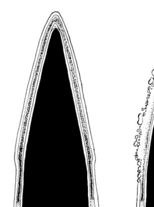
Profuse arterial hemorrhage in ejaculates usually results from an idiopathic defect in
Ž
the urethra that communicates with the surrounding corpus spongiosum penis
Schu-.
macher et al., 1995 . The lesion is typically longitudinal, and is almost always located on the posterior surface of the urethra near the pelvic ischium. Diagnosis of the urethral defect requires urethral endoscopy. The urethral defect can be identified near the pelvic
Ž .
ischium Fig. 3 . The defect can be small and difficult to identify. Endoscopy immedi-ately following ejaculation may aid the examiner in locating the defect because blood can often be detected within the defect at this time. Bleeding is profuse because the lesion communicates with the underlying corpus spongiosum penis. As such, bleeding only occurs when blood pressure within the corpus spongiosum penis increases with erection or micturition. The cause of the lesion has not been determined.
3. Acrosomal dysfunction
In some cases, results of a routine breeding soundness examinations are unable to provide a primary etiology for a stallion’s reduced fertility. Despite proper mare management, and normal semen evaluations, some stallions are still unable to impreg-nate an acceptable proportion of mares bred. Determining the cause for subfertility in stallions with normal semen parameters can be frustrating. For such stallions, additional tests are required to help explain their condition. These may include assays for sperm
Ž .
chromatin structure described below , antisperm antibodies or the acrosome reaction. Subfertile stallions have a higher incidence of elevated antisperm antibody titers than
Ž .
fertile stallions Kenney et al., 1998; Love, 1997 . In human males with unexplained infertility, the presence of antisperm antibodies has been shown to interfere with the
Ž .
induction of the acrosome reaction by the zona pellucida Francavilla et al., 1997 . A direct correlation has been demonstrated between the acrosome reaction and fertilization rate in human males. This parameter may be a better predictor of IVF success than the
Ž .
more traditionally evaluated semen parameters. Calvo et al., 1994a,b .
The acrosome is an membrane-bound organelle that covers the anterior portion of the
Ž .
mammalian spermatozoal nucleus Fig. 4 . The term, acrosome reaction, refers to the
Ž .
fusion and vesiculation of the outer acrosomal and plasma membranes Fig. 5 . During the acrosome reaction, hydrolytic enzymes are released from this organelle which facilitate penetration of the investments of the ovum. Whether or not the functions of these enzymes go beyond aiding in development of the acrosome reaction, penetration of the cumulus oophorus and binding of the zona pellucida to actually lysing a path
Ž .
Ž .
Fig. 4. Illustration of transmission electron microscopic view left and cut-away view of an equine
Ž . Ž . Ž . Ž .
spermatozoan. a Plasma membrane, b acrosome, c outer acrosomal membrane, d inner acrosomal
Ž . Ž . Ž . Ž . Ž .
membrane, e nuclear membrane, f post-acrosomal lamina, g nucleus, h capitulum, i proximal centriole,
Ž .j segmented column, k dense fiber, l and m doublets of axoneme, n mitochondria.Ž . Ž . Ž . Ž .
Fig. 5. Illustration of transmission electron microscopic view of an equine spermatozoan. Left side represents an intact acrosome. Right side represents an acrosome reaction, depicted by fusion and vesiculation of plasma and outer acrosomal membranes with resultant pore formation.
The acrosome reaction in equine spermatozoa can be induced by exposing capacitated
Ž .
spermatozoa to progesterone Meyers et al., 1995 or calcium ionophores such as
Ž .
physio-logic, it is also more time consuming. Incubation in a capacitating medium for up to 5 h may be required prior to exposing spermatozoa to progesterone. Results of one study suggested spermatozoa from fertile stallions may be more responsive to
progesterone-in-Ž .
duced acrosome reactions than those of subfertile stallions Meyers et al., 1995 .
Similarly, lower levels of both spontaneous and progesterone-stimulated acrosome reactions have been reported to occur in semen from infertile men with severe
Ž .
teratozoospermia Oehninger et al., 1994 .
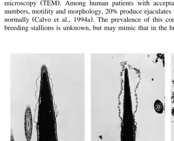
Our laboratory has identified four stallions with previously unexplained infertility, whose spermatozoa do not respond to A23187 to the same degree as fertile stallions.
Exposure of fertile stallion semen to the calcium ionophore resulted in )80% of the
spermatozoa becoming acrosome reacted, whereas -20% of spermatozoa from the
stallions with unexplained subfertility or infertility underwent the acrosome reaction under the same conditions. Acrosomal status was evaluated by transmission electron
Ž .
microscopy TEM . Among human patients with acceptable levels of spermatozoal numbers, motility and morphology, 20% produce ejaculates which fail to acrosome react
Ž .
normally Calvo et al., 1994a . The prevalence of this condition in the population of breeding stallions is unknown, but may mimic that in the human population.
Ž . Ž .
Fig. 6. Transmission electron micrographs of stallion spermatozoa, demonstrating a intact acrosome, b
Ž .
Briefly, the procedure we use for induction and assessment of the acrosome reaction is as follows: Semen is collected in a routine manner using an artificial vagina. Aliquots
of gel-free semen are exposed to 10 mM A23187 in a milk-based extender for 2 h at
Ž .
378C, then fixed and processed for TEM Varner et al., 1992 . TEM is expensive, time
consuming and requires sophisticated laboratory facilities. Consequently, alternative methods of assessing the acrosome reaction are being investigated. The use of fluores-cent probes is the most commonly employed alternative to TEM for evaluating the acrosome reaction. Chlortetracycline can be used to assess both capacitation and the acrosome reaction. Unfortunately, it does not appear to be reliable when semen has been
Ž
diluted with milk-based extenders. Conjugated lectins such as FITC-PNA Arachis
. Ž .
hypogea and FITC-PSA Pisium satiÕum are commonly used fluorescent acrosomal probes. These probes can be used with milk-based extenders. Initial studies in our laboratory revealed that the fluorescent lectin, PSA, produced a high incidence of false acrosome reactions when used with a stallion exhibiting failure of the acrosome reaction following spermatozoal exposure to A23187. In this stallion, fragmentation of acrosomal and plasma membranes following exposure to A23187 created a fluorescent pattern on
Ž .
the spermatozoa which simulated that of a true acrosome reaction Fig. 6 . At this time, TEM remains the ‘‘gold standard’’ for evaluating acrosomal integrity.
4. Abnormal spermatozoal chromatin
Sperm chromatin implies sperm DNA and associated nucleoproteins. The relationship between sperm chromatin stability and male fertility has been reported for several species. Repeated abortions in women impregnated by men with large numbers of easily denatured sperm is an example of how abnormal sperm chromatin can result in
Ž .
subfertility Ibrahim and Pedersen, 1988 . Recently, the relationship between sperm
Ž
chromatin stability and fertility in stallions had been described Kenney et al., 1995;
.
Love, 1993, Love and Kenney, 1998; .
All stallions have some level of abnormal spermatozoal chromatin. However, the degree of the abnormality is what determines the severity and how it relates to the fertility status of the stallion. Therefore, the higher the percentage of abnormal chro-matin, the greater the reduction in the fertility of the stallion that can be expected.
While there are many potential causes for disruption of sperm chromatin structure, clinically there appear to be two manifestations in the stallion. The first is a result of a traumatic or stressful incident to the testes, such as a kick or any event that would result in fluid accumulation around the testes. This has the effect of insulating the testes, resulting in a heat stress condition. In this case, changes in spermatozoal motility and
Ž
morphology can be seen, as well and changes in sperm chromatin structure Love,
.
1993 . Depending on the severity of the heat stress, the morphology and motility will be affected first, while the chromatin appears more resistant to mild forms of heat stress. As the adverse effects resolve, the chromatin returns to normal. Therefore, it appears that chromatin disruption is reversible when the inciting cause is removed.
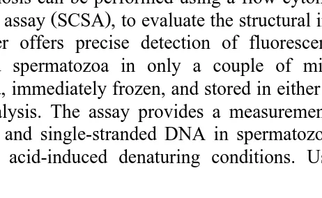
Fig. 7. Illustrated scattergram of the distribution of spermatozoa, based on green and red fluorescence. Percent COMP represents the percentage of spermatozoa that lie outside the main population.
Sperm chromatin may deteriorate progressively as stallions age, or may be manifest as an inherent trait at the onset of their breeding career.
Diagnosis can be performed using a flow cytometric procedure, the sperm chromatin
Ž .
structure assay SCSA , to evaluate the structural integrity of sperm chromatin. The flow cytometer offers precise detection of fluorescent labels and assessment of several thousand spermatozoa in only a couple of minutes. Raw semen samples can be
collected, immediately frozen, and stored in either liquid nitrogen or in ay208C freezer
until analysis. The assay provides a measurement of the relative amounts of double-stranded and single-double-stranded DNA in spermatozoal populations following exposure to heat- or acid-induced denaturing conditions. Using the metachromatic DNA stain,
acridine orange, double-stranded DNA appears green and single-stranded DNA appears red under special fluorescing conditions. Quantitation of DNA denaturation is
deter-mined by the term, alpha-t, which is defined as the ratio of redrredqgreen
fluores-Ž .
cence, and is measured for each spermatozoan represented in a scatter plot Fig. 7 or
Ž .
histogram Fig. 8 . Several parameters can be created using this scatter plot or histogram
Ž .
to describe the susceptibility of the DNA to denaturation. The mean of alpha-t at
reflects the degree of DNA denaturation of the whole spermatozoa population. The SD
at indicates the extent of denaturation or how far individual spermatozoa may deviate
from the main population. The percentage COMPat is the percentage of cells outside
the main population, and it is determined by selecting those cells to the right of the main
Ž .
Fig. 9. Sperm chromatin structure assay SCSA scattergrams representing varying levels of fertility among
Ž . Ž .
stallions from highest A to lowest D .
Ž . Ž .
Stallion Seasonal pregnancy rate % First-cycle pregnancy rate % Meanat SDat COMPat
A 94 80 222 79 11
B 81 46 266 124 28
C 67 40 279 99 68
population and represents the number of cells outside the main population as a
Ž .
percentage of the total number of spermatozoa evaluated Figs. 7 and 8 . Fig. 9
demonstrates the progressive decline in fertility of stallions as deterioration of chromatin
Ž .
increases. Stallions of high fertility stallion A have low values for all SCSA variables, indicative of chromatin that is structurally sound. As chromatin deteriorates, the SCSA values increase, as indicated for stallions B–D. Stallion B is of lesser fertility than
Ž .
stallion A seasonal pregnancy rate of 81 vs. 94% and this is represented by a higher
Ž .
mean, SD and COMP of at. Stallion C has a very high COMPat 68% and a lower
seasonal pregnancy rate than stallions A and B. Stallion D is an extreme example of a highly subfertile stallion, in which the entire spermatozoal population has shifted off the
‘‘normal’’ axis, resulting in essentially 100% COMPat. Stallions exhibiting this type of
chromatin can be easily misdiagnosed unless the axial shift is recognized.
5. Azoospermia
Azoospermia implies a complete absence of spermatozoa in ejaculates. Causes include spermatogenic failure, obstruction of extracurrent ducts, failure of emission
Žrelease of sperm and accessory gland fluids into the pelvic urethra or ejaculation.
Žforceful expulsion of the combined fluids from the urethra , and retrograde ejaculation..
Severe testicular hypoplasia or degeneration can cause azoospermia due to failure of spermatozoal production by the seminiferous epithelium. Complete obstruction of the epididymal or deferent ducts also results in azoospermia, as sperm produced by the testes cannot pass through the excurrent duct system to enter the urethra for ejaculation.
Spermatozoa occasionally become impacted within the epididymalrdeferent duct lumina
to the point that they are not released by the strong muscular contractions of the ducts at the time of ejaculation. The condition is often referred to as ‘‘plugged ampullae’’ when
Ž .
ampullary lumina become obstructed Love et al., 1992 . The size and glandular nature of the ampullae of the stallion provides a potential for excessive accumulation of spermatozoa, occasionally leading to inspissation and blockage of the lumina. With complete bilateral blockage, azoospermia occurs. Blockage can be sporadic, with affected stallions having poor spermatozoal morphology and motility due to a high percentage of tailless heads when being bred infrequently. Densely packed spermatozoal casts may also be present in the semen. Diagnosis is based on and ultrasonic physical
Ž .
examination and characteristics of semen quality Love et al., 1992 .Azoospermic
stallions with this underlying etiology will likely have negligible alkaline phosphatase
Ž .
concentrations in semen Turner, 1996 . Palpation per rectum may reveal enlarged, tense ampullae, and occasionally narrowing or loss of the echolucent lumen that is characteristic of a patent ampulla when examined by ultrasonography.
A major cause of azoospermia in stallions is anejaculation due to an ejaculatory disorder. In this instance, little to copious amounts of pre-ejaculatory fluid can be produced during the act of coitus, but sperm are not present. Many of these cases are
Ž .
psychogenic in nature McDonnell, 1992a,b, 1986 , but physical disorders can also
Ž
cause ejaculatory failure. Examples include musculoskeletal disorders e.g., back pain,
. Ž
. Ž
protozoal myeloencephalitis or nerve root injury , vascular disorders e.g., aorto-iliac
. Ž
thrombosis , gastrointestinal problems e.g., abdominal adhesions or integument scarring
.
secondary to colic surgery, or inguinal herniation or lesions specific to the reproductive
Ž
tract e.g., spermatic cord torsion, orchitisrepididymitis, seminal vesiculitis, or penile
. Ž .
injury Martin et al., 1998; McDonnell, 1992a,b, 1999; Varner et al., 1991 . Retrograde
Ž .
ejaculation i.e., ejaculation into the bladder has also been documented in a stallion. Azoospermia can be a diagnostic dilemma, as the most common cause of this
Ž
condition is failure to ejaculate i.e., that associated with psychogenic or physical
.
ailments . Diagnostic efforts are directed first toward determining if the stallion is actually ejaculating. In the stallion with normal libido and breeding behavior, indirect evidence for ejaculation includes engorgement of the glans penis, urethral pulsations, flagging of the tail, and the presence of gel in the sample collected. After removal of the gel portion, the gel-free liquid is examined, using a phase-contrast microscope at
200–400=magnification, for the presence of spermatozoa or premature germ cells. If
no spermatozoa are present, assay of the gel-free liquid for alkaline phosphatase is
Ž .
performed to confirm presence of epididymalrtesticular fluids Turner, 1996 . High
alkaline phosphatase values confirm the presence of epididymalrtesticular secretions in
the seminal fluid. If necessary, a testicular biopsy can be procured to determine if
Ž . Ž
spermatogenesis is proceeding to completion Fig. 10 . Both incisional DelVento et al.,
. Ž
1992; Threlfall and Lopate, 1993 and needle Faber and Roser, 1999; Hillman et al.,
.
1994; Threlfall and Lopate, 1993 biopsy techniques have been reported recently for use in stallions. Although detrimental effects have been associated with testicular biopsy, these studies suggest that it is a relatively safe procedure. The technique is used
Ž
routinely in subfertile men Carpi et al., 1999; Chen, 1997; Craft et al., 1997; Ezeh et
.
al., 1998; Morey et al., 1994 .
To illustrate the diagnostic approach for azoospermia, we present the data from three stallions. A 5-year-old stallion with slightly small testes appeared to ejaculate normally
Ž .
with gel being present in his ejaculates, but the semen contained only premature round
Ž .
germ cells 0.5 millionrml . Assay of seminal plasma from this stallion yielded 1130
Url alkaline phosphatase, indicating testicularrepididymal contribution to the ejaculate.
Ž
Testicular biopsy revealed spermatogenesis was not proceeding to completion seminif-erous tubules of smaller than normal diameter with no elongating or elongated
sper-.
matids, and only a few round spermatids were present . Another 7-year-old stallion with
Ž
small testes ejaculated gel and gel-free semen containing only premature germ cells 6.5
.
millionrml . This horse was castrated and histomorphometric analyzes of fixed
testicu-Ž .
lar parenchyma Johnson et al., 1991 revealed disruption of spermatogenesis between
Ž
meiosis and spermiogenesis counts of late primary spermatocyte counts yielded a daily
sperm production estimate of 30 millionrg, while greatly reduced numbers of round
.
spermatids yielded an estimate for daily sperm production of less than 2 millionrg .
Only occasional elongating spermatids were seen in the seminiferous tubules of this horse. Finally, a 3-year-old stallion with slightly small testes was examined that ejaculated gel and gel-free semen with no sperm or germ cells in the ejaculate. Assay of
seminal plasma from this stallion yielded only 43 Url alkaline phosphatase, confirming
that testicularrepididymal secretions were absent from the ejaculate. The epididymides
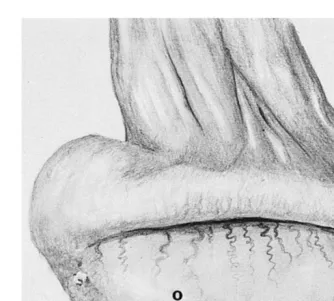
Ž . Ž
Fig. 10. Illustration of lateral aspect of testis, demonstrating vascular pattern on tunica albuginea, and
Ž .
recommended site for acquisition of punch biopsy `.
. Ž
epididymis , and were uniformly hyperechoic i.e., without the ‘‘Swiss cheese’’
.
appearance attributed to the convoluted, fluid-containing epididymal duct on ultrasound
Ž .
examination Love, 1992 . Palpation per rectum revealed normal size and location of the paired vas deferens and ampullae, seminal vesicles and lobes of the prostate. A testicular
Ž .
biopsy was taken Hillman et al., 1994 and revealed seminiferous tubules of normal appearance. The finding that spermatogenesis proceeded to completion, in conjunction with the azoospermia and low concentration of alkaline phosphatase in the gel-free semen, suggested obstruction of the epididymal ducts preventing sperm from entering the ejaculates. The presence of the extremely small epididymides in a young stallion
Ž
likewise suggested epididymal hypoplasia failure of epididymides to develop to normal
.
size .
References
Calvo, L., Dennison-Lagos, L., Banks, S.M., Sherins, R.J., 1994a. Characterization and frequency distribution of sperm acrosome reaction among normal and infertile men. Hum. Reprod. 9, 1875–1879.
Calvo, L., Dennison-Lagos, L., Banks, S.M., Dorfmann, A., Thorsell, L.P., Bustillo, M., Schulman, J.D., Sherins, R.J., 1994b. Acrosome reaction inducibility predicts fertilization success at in-vitro fertilization. Hum. Reprod. 9, 1880–1886.
Carpi, A., Fabris, F.M., Gorini, I., Gaeta, P., Romani, R., Marchetti, A., 1999. A percutaneous large-needle aspiration biopsy technique for histologic examination of the testis in infertile patients. Fertil. Steril. 71, 756–760.
Chen, C.S., 1997. Percutaneous testis biopsy: an alternative to open testicular biopsy in the evaluation of the subfertile man. J. Urol. 158, 1527.
Cooper, W.L., 1979. Methods of determining the site of bacterial injections in the stallion reproductive tract. In: Proc. Ann. Meet. Soc. Theriogenology. pp. 1–4.
Craft, I., Tsirigotis, M., Courtauld, E., Farrer-Brown, G., 1997. Testicular needle aspiration as an alternative to biopsy for the assessment of spermatogenesis. Hum. Reprod. 12, 1483–1487.
DelVento, V.R., Amann, R.P., Trotter, G.W., RaoVeeramachaneni, D.N., Squires, E.L., 1992. Ultrasono-graphic and quantitative histologic assessment of sequelae to testicular biopsy in stallions. Am. J. Vet. Res. 53, 2094–2101.
Ezeh, U.I.O., Moore, H.D.M., Cooke, I.D., 1998. A prospective study of multiple needle biopsies versus a single open biopsy for testicular sperm extraction in men with non-obstructive azoospermia. Hum. Reprod. 13, 3075–3080.
Faber, N.F., Roser, J.F., 1999. Testicular biopsy in stallions: effects on prospective fertility and diagnostic potentials. In: Proc.7th Int. Symp. Equine Reprod.. pp. 7–8.
Francavilla, F., Romano, R., Santucci, R., Marrone, V., Properzi, G., Ruvolo, G., 1997. Interference of
Ž .
antisperm antibodies with the induction of the acrosome reaction by zona pellucida ZP and its relationship with the inhibition of ZP binding. Fertil. Steril. 67, 1128–1133.
Hillman, R.B., Casey, P.J., Kennedy, P.C., Hughes, J.P., Liu, I.K.M., 1994. Testicular biopsy in the stallion. In: Proc. 6th Int. Symp. Equine Reprod.. pp. 145–146.
Ibrahim, M.E., Pedersen, H., 1988. Acridine orange fluorescence as male fertility test. Arch. Androl. 20, 125–129.
Johnson, L., Varner, D.D., Thompson, D.L. Jr., 1991. Effect of age and season on the establishment of spermatogenesis in the horse. J. Reprod. Fertil., Suppl. 44, 87–97.
Kenney, R.M., Evenson, D.P., Garcia, M.C., Love, C.C., 1995. Relationships between sperm chromatin structure, motility, and morphology of ejaculated sperm, and seasonal pregnancy rate. Biol. Reprod. Monogr. 1, 647–653.
Kenney, R.M., Cummings, M.R., Teusscher, C., Love, C.C., 1998. Possible role of autoimmunity to spermatozoa in idiopathic infertility in stallions. In: Proc.7th Int. Symp. Equine Reprod.. pp. 5–6. Little, T.V., Woods, G.L., 1987. Ultrasonography of accessory sex glands in the stallion. J. Reprod. Fertil.,
Suppl. 35, 87–94.
Love, C.C., 1992. Ultrasonographic evaluation of the testis, epididymis, and spermatic cord of the stallion. In:
Ž . Ž
Blanchard, T.L., Varner, D.D. Eds. , Veterinary Clinics of North America — Equine Practice Stallion
.
Management . Saunders, Philadelphia, PA, pp. 167–182.
Love, C.C. Relationship of protamine levels and disulfide bonding to sperm chromatin structure and fertility in the stallion. Dissertation, University of Pennsylvania, 1993, pp. 54–176.
Love, C.C., 1997. Examination of the male reproductive tract: evaluation of potential breeding soundness. In:
Ž .
Youngquist, R.S. Ed. , Current Therapy in Large Animal Theriogenology. Saunders, Philadelphia, PA, pp. 12–15.
Love, C.C., Kenney, R.M., 1998. The relationship of increased susceptibility of sperm DNA to denaturation and fertility in the stallion. Theriogenology 50, 955–972.
Ž .
Love, C.C., Riera, F.L., Oristaglio-Turner, R.M., Kenney, R.M., 1992. Sperm occluded plugged ampullae in the stallion. In: Proc. Ann. Meet. Soc. Theriogenology. pp. 117–127.
Malmgren, L., Sussemilch, B., 1992. Ultrasonography as a diagnostic tool in a stallion with seminal vesiculitis: a case report. Theriogenology 37, 935–938.
McDonnell, S.M., 1986. Stallion sexual behavior dysfunction: experimental models and clinical considera-tions. In: Proc. Ann. Meet. Soc. Theriogenology. pp. 1–12.
Ž .
McDonnell, S.M., 1992a. Ejaculation: physiology and dysfunction. In: Blanchard, T.L., Varner, D.D. Eds. ,
Ž .
Veterinary Clinics of North America — Equine Practice Stallion Management . Saunders, Philadelphia, PA, pp. 57–70.
Ž .
McDonnell, S.M, 1992b. Sexual behavior dysfunction in stallions. In: Robinson, N.R. Ed. , Current Therapy in Equine Medicine. Saunders, Philadelphia, PA, pp. 668–671.
McDonnell, S.M., 1999. Libido, erection, and ejaculatory dysfunction in stallions. Comp. Contin. Educ. Pract. Vet. 21, 263–266.
Meyers, S.A., Overstreet, J.W., Liu, I.K.M., Drobnis, E.Z., 1995. Capacitation in vitro of stallion spermatozoa: comparison of progesterone-induced acrosome reactions in fertile and subfertile males. J. Androl. 16, 47–54.
Morey, A.F., Plymyer, M., Rozanski, T.A., Deshon, G.E., Myers, J.B., Dresner, M.L., 1994. Biopty gun testis needle biopsy: a preliminary clinical experience. Br. J. Urol. 74, 366–369.
Oehninger, S., Blackmore, P., Morshedi, M., Sueldo, C., Acosta, A.A., Alexander, N.J., 1994. Defective
Ž .
calcium influx and acrosome reaction spontaneous and progesterone-induced in spermatozoa of infertile men with severe teratozoospermia. Fertil. Steril. 61, 349–354.
Schumacher, J., Varner, D.D., Schmitz, D.G., Blanchard, T.L., 1995. Urethral defects in geldings with hematuria and stallions with hemospermia. Vet. Surg. 24, 250–254.
Ž .
Threlfall, W.R., Lopate, C., 1993. Testicular biopsy. In: McKinnon, A.O., Voss, J.L. Eds. , Equine Reproduction. Lea & Febiger, Philadelphia, PA, pp. 943–949.
Turner, R.M., 1996. Alkaline phosphatase activity in stallion semen: characterization and clinical implications. In: Proc. Ann. Meet. Soc. Theriogenology. pp. 284–293.
Varner, D.D., Ward, C., Storey, B.T., Kenney, R.M., 1987. Induction and characterization of acrosome reaction in equine spermatozoa. Am. J. Vet. Res. 48, 1383–1389.
Ž .
Varner, D.D., Schumacher, J., Blanchard, T.L., Johnson, L. Eds. , Diseases and Management of Breeding Stallions. American Veterinary Publications, Goleta, CA.
Varner, D.D., Bowen, J.A., Johnson, L., 1992. Capacitation and acrosome reaction of equine spermatozoa by heparin. Mol. Androl. 4, 81–100.