Production of chitooligosaccharides from
Rhizopus oligosporus
NRRL2710 cells by chitosanase digestion
Maria Mahata
b,, Shoko Shinya
a,, Eiko Masaki
a, Takashi Yamamoto
a, Takayuki Ohnuma
a,
Ryszard Brzezinski
c, Tapan K. Mazumder
d, Kazuhiko Yamashita
d, Kazue Narihiro
d, Tamo Fukamizo
a,⇑aDepartment of Advanced Bioscience, Kinki University, 3327-204 Nakamachi, Nara 631-8505, Japan bUniversity of Andalas, Padang, West Sumatera, Indonesia
cDépartement de Biologie, Centre d’Étude et de Valorisation de la Diversité Microbienne, Université de Sherbrooke, Canada dYaegaki Bio-industry, Inc., 681 Mukudani, Hayashida-cho, Himeji 678-4298, Japan
a r t i c l e
i n f o
Article history:
Received 24 April 2013
Received in revised form 1 June 2013 Accepted 4 June 2013
The intact cells ofRhizopus oligosporusNRRL2710, whose cell walls are abundant source ofN -acetylglu-cosamine (GlcNAc) and glu-acetylglu-cosamine (GlcN), were digested with three chitinolytic enzymes, a GH-46 chitosanase fromStreptomycessp. N174 (CsnN174), a chitinase fromPyrococcus furiosus, and a chitinase fromTrichoderma viride, respectively. Solubilization of the intact cells by CsnN174 was found to be the most efficient from solid state CP/MAS13C NMR spectroscopy. Chitosanase products fromRhizopuscells
were purified by cation exchange chromatography on CM-Sephadex C-25 and gel-filtration on Cellulofine Gcl-25 m. NMR and MALDI-TOF-MS analyses of the purified products revealed that GlcN–GlcNAc, (GlcN)2–GlcNAc, and (GlcN)2were produced by the enzymatic digestion of the intact cells. The
chitosan-ase digestion ofRhizopuscells was found to be an excellent system for the conversion of fungal biomass without any environmental impact.
Ó2013 Elsevier Ltd. All rights reserved.
1. Introduction
Chitin, a water-insoluble polymer of b-1,4-linkedN -acetylglu-cosamine (GlcNAc), is the most abundant biomass next to cellu-lose, and widely distributed in organisms, such as crustacean shells, insect cuticles, and fungal cell wall.1Commercial chitin is
mainly extracted by demineralization and deproteinization of the shells of crustaceans such as shrimps, crabs, lobsters, and krill, which are supplied in large quantities by shellfish processing.2
Chitosan, ab-1,4-linked polymer of glucosamine (GlcN), has been produced on an industrial scale by the N-deacetylation of chitin using sodium hydroxide. The enzymatic hydrolysis of chitin and chitosan has been conducted to produce corresponding oligosaccharides,3which have been drawing attention because of
their presumed biological functions, such as the inhibition of tumor angiogenesis,4 control of cell growth, differentiation, and
development in vertebrates,5,6antioxidative effect,7and elicitation
of defensive actions in plants.8,9However, the extraction process of
chitin from crustacean shells is well known to produce a significant impact on the environment. In this context, it is highly desirable to establish a strategy for directly producing chitin and chitosan oligosaccharides by the enzymatic digestion of raw materials containing chitin and chitosan.
Chitin is associated with proteins and minerals in crustacean shells,2 resulting in a sclerotized and recalcitrant structure, and
further protecting the polysaccharide chain from enzymatic diges-tion. However, fungal cell wall consists of chitin, branchedb -1,3-glucan, and b-1,6-glucan, which are cross-linked to each other. Chitosan is also found in the fungal cell wall,10and makes the cell
wall structure more pliable. Therefore, the chitinous components of the fungal cell wall appear to be degradable by enzymatic ac-tions. In fact, the digestion of the cell wall fraction ofFusarium oxy-sporum by chitinase and chitosanase was found to produce oligosaccharides composed of GlcNAc and GlcN.11,12 However,
most studies on the chitinous components of fungal cell wall thus far have been devoted to efficient utilization of fungi as the source of polysaccharide chitin/chitosan13or GlcN monomer.14,15
Experi-mental data on the oligosaccharide production from the fungal cell wall have not been fully accumulated. In order to utilize the chi-tooligosaccharide products obtained from the fungal cell wall as
0008-6215/$ - see front matterÓ2013 Elsevier Ltd. All rights reserved.
http://dx.doi.org/10.1016/j.carres.2013.06.002
Abbreviations: GlcNAc, 2-acetamido-2-deoxy-b-D-glucopyranose; GlcN, 2-amino-2-deoxy-b-D-glucopyranose; (GlcN)n,b-1,4-linked oligosaccharide of GlcN with a polymerization degree ofn; MALDI-TOF-MS, matrix assisted laser desorp-tion/ionization time-of-flight mass spectrometry; NMR, nuclear magnetic resonance.
⇑ Corresponding author. Tel.: +81 742 43 8237; fax: +81 742 43 8976.
E-mail address:fukamizo@nara.kindai.ac.jp(T. Fukamizo). These authors contributed equally to this work.
Contents lists available atScienceDirect
Carbohydrate Research
food additives or pharmaceutics, fungi as the source materials should be harmless; therefore, pathogenic fungi such as the Fusar-iumspecies could not be used for this purpose.
SomeRhizopusspecies have been used in the fermentation food industry in Asia, and their products have been generally recognized as safe.16Thus we determined the chitin/chitosan content in sev-eralRhizopusspecies, and the content inR. oligosporuswas found to be the highest (32%, Yamashita et al., unpublished). In this study, the intact cells ofR. oligosporuswere digested by chitinolytic en-zymes, and the products were analyzed by NMR spectroscopy and MALDI-TOF-MS. We found that the chitosanase digestion of Rhizopuscells is an excellent strategy for chitooligosaccharide pro-duction without any impact on the environment.
2. Results and discussion
2.1. Enzymatic digestibility ofRhizopus oligosporuscells
To evaluate the enzymatic digestibility ofRhizopuscells, three enzymes were tested, an endo-splitting chitosanase from Strepto-mycessp. N174 (CsnN174), a thermophilic chitinase from Pyrococ-cus furiosus (PfChit), and a chitinase from Trichoderma viride (TvChit). These two chitinases belong to the GH18 family and hydrolyze the internal b-1,4-glycosidic linkages (endo-splitting mode) through substrate-assisted mechanism.17 The production
systems of these enzymes were highly efficient and available in our laboratory. Individual digestions of the cells were conducted for 24 h. We confirmed that the release of reducing sugars into the supernatant fraction completely ceased after 24 h incubation. The amounts of the insoluble materials remaining after the enzy-matic digestion were determined and compared with the starting amounts of the cells. The results are listed inTable 1. The amount of insoluble materials remaining was the lowest in the digestion with the chitosanase, CsnN174, indicating that the chitosanase has the highest digestibility (67%). The digestibilities of the chiti-nase preparations (PfChit and TvChit) were comparable to each other (35–36%), and much lower than those of CsnN174. Since the chitinase (PfChit) used in this experiment was thermophilic, the enzymatic reaction was also conducted at a higher tempera-ture, 70°C. However, solubilization was not enhanced by elevating
the temperature. The accessibility of the enzymes to the chitin polysaccharide chain might have been restricted in theRhizopus cell wall probably due to the ordered polysaccharide structure. When the control experiments were conducted in the absence of enzymes, reducing sugars did not significantly increase in the reac-tion mixture. It is evident that the cell wall polysaccharides were digested by the exogenous enzymes, but not by the endogenous ones.
2.2. Solid-state CP/MAS13C NMR analysis ofRhizopuscells
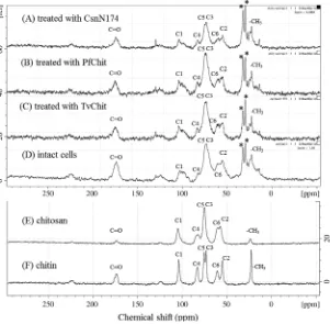
The insoluble materials remaining after the enzymatic digestion ofRhizopuscells were used for solid-state CP/MAS13C NMR
analy-sis. The NMR spectra are shown inFigure 1. We initially compared the spectrum of intactRhizopuscells (Fig. 1D) with those of authen-tic chitin and chitosan preparations (Fig. 1E and F) to roughly
estimate the overall structure of the cell wall. Based on the previous assignment data,18 the resonances for polysaccharides
were assigned as labeled in the figure. The resonances designated by asterisks and some portion of the carbonyl resonance were supposed to be derived from the protein and lipid components of Rhizopuscells. Since carbonyl and methyl carbon resonances were clearly observed, theRhizopus cell wall was found to contain a significant portion of chitin. Line broadening of the resonances for the pyranose-ring carbons (C1–C6) indicated that a major fraction of the cell wall is a mixture of polysaccharides, such as chitin/chitosan andb-1,3- andb-1,6-glucans. However, the shape of the C6 and C2 resonances of Rhizopus cells appeared to be similar to that of chitosan. Rhizopus cells are likely to contain chitin/chitosan abundantly in their cell walls.
WhenRhizopuscells were digested by TvChit or PfChit, the res-onance intensities of the methyl and carbonyl carbons were re-duced by the enzymatic digestion (Fig. 1B and C), indicating the significant digestion of chitin polysaccharides. However, the rela-tive intensities of C1–C6 in the spectra appeared to be similar to those of the intact cells. The major polysaccharide components were not markedly disrupted by the treatment with TvChit or PfChit. On the other hand, the insoluble fraction obtained after digestion ofRhizopuscells by CsnN174 exhibited lower intensities of C1–C6 (Fig. 1A). Major polysaccharide components were inten-sively disrupted by CsnN174. These results indicated again that solubilization ofRhizopuscells by CsnN174 was the most efficient. In the intact fungal cells, it appears that the polysaccharide struc-ture of chitosan region is flexible, but the chitin region is rigid. This situation might have made the chitosanase enzyme more accessi-ble to the polysaccharide chain, resulting in the more efficient deg-radation by CsnN174.
2.3. Purification of the oligosaccharide products obtained by CsnN174 digestion
The soluble fraction (1.38 g) obtained after the chitosanase digestion ofRhizopus cells was applied onto a charcoal column, and the adsorbed fraction was further employed for cation-ex-change chromatography. The profile is shown inFigure 2. After the elution of a void-volume fraction, several reducing-sugar frac-tions were obtained, and the three major fracfrac-tions I, II, and III were further purified by gel-filtration. The three purified fractions were dialyzed against distilled water, lyophilized, and employed for solution NMR spectroscopy and MALDI-TOF-MS. The recoveries of fractions I, II, and III were 21, 8, and 25 mg, respectively.
2.4. Structure analysis of the oligosaccharide products
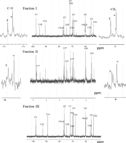
Figure 3shows the13C NMR spectra of fractions I, II, and III in
solution. Two major resonances were observed in the carbonyl and methyl carbon regions of fractions I and II, and assigned to the
a
- andb-anomers of the reducing end GlcNAc residue based on their chemical shifts. In the C1 carbon region (90–100 ppm) of fraction I, one resonance (C10) was found at 97.8 ppm in additionto the resonances corresponding to the
a
- andb-anomers of the reducing end GlcNAc (C1a
and C1b), and assigned to thenon-Table 1
Amounts of the insoluble and soluble products obtained from the enzymatic digestion ofRhizopuscells
Enzyme Intact cells*(g) Insoluble products after enzymatic digestion*(g) Soluble products (g) Hydrolysis %
CnsN174 2.06 ± 0.04 0.68 ± 0.06 1.38 67
PfChit 2.10 ± 0.05 1.34 ± 0.04 0.76 36
TvChit 2.05 ± 0.02 1.34 ± 0.06 0.71 35
*Mean values of three independent reactions with ± SD (standard deviations).
reducing end GlcN. Thus, fraction I was identified as GlcN–GlcNAc. The chemical shift values of the resonances for other pyranose ring carbons were consistent with the values reported for GlcN–Glc-NAc.19No resonances were observed in the carbonyl and methyl
carbon regions for fraction III. Chemical shifts of the resonances for the pyranose ring carbons were completely identical to those
reported for (GlcN)2.20,21Thus, fraction III was identified as (GlcN)2.
Although most resonances for fraction II were identical to those observed for fraction I, six additional resonances designated by C1⁄–C6⁄were found in the region of the pyranose-ring carbons.
The chemical shifts of the six resonances corresponded to those of the internal GlcN residues.21Although fraction II contained some Figure 1.Solid state CP/MAS13C NMR spectra of intact cells ofRhizopus oligosprusNRRL2710 (D) and insoluble fractions obtained after enzyme digestion (A, B, and C). Authentic preparations of chitin (F) and chitosan (E) were used as reference standards. NMR experiments were performed on a Bruker DSX-400WT spectrometer. Experimental conditions are described in the text. Resonance assignments were performed based on the chemical shift values reported previously.14
Figure 2.Cation-exchange chromatography of the products obtained from digestion ofRhizopuscells by CsnN174. The oligosaccharide products were put on a column of CM-Sephadex C-25 (1.075 cm) previously equilibrated with 0.01 M sodium acetate buffer, pH 5.0. After elution with the buffer, oligosaccharides adsorbed to CM-Sephadex resin were eluted with a linear gradient of NaCl from 0 to 1.0 M in the same buffer. The reducing sugars were determined for individual tubes according to the method of Imoto and Yagishita.27
impurities, a major portion of this fraction was identified as (GlcN)2–GlcNAc.
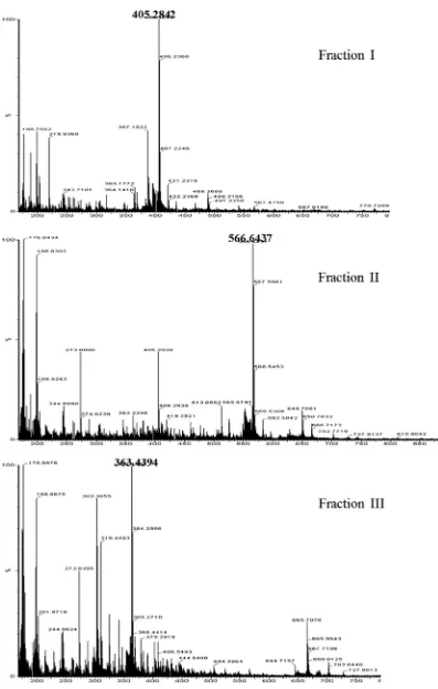
Figure 4shows the MALDI-TOF-MS profiles of the three frac-tions. The major mass signals of 405.2842, 566.6437, and 363.4394 for the individual fractions corresponded to the sodium adducts of the compounds GlcN–GlcNAc, (GlcN)2–GlcNAc, and
(GlcN)2, respectively. The mass spectra confirmed the structures
of the oligosaccharides obtained from the chitosanase digestion ofRhizopuscells.
The structure and function of CsnN174 have been intensively studied.22,23 Splitting specificity of the enzyme was determined
using the soluble substrate, partially N-acetylated chitosan, and the enzyme was shown to hydrolyze theb-1,4-glycosidic linkage
of the two sequences, GlcNAc–GlcN and GlcN–GlcN. Thus, the reducing-end residue of the oligosaccharides produced by this en-zyme is GlcNAc or GlcN, while the nonreducing-end residue is exclusively GlcN.24 The enzymatic digestion of theRhizopuscells
produced the oligosaccharides GlcN–GlcNAc, (GlcN)2–GlcNAc,
and (GlcN)2, indicating that the enzyme acts towardRhizopuscells
with the same splitting specificity as that toward the soluble sub-strate. It is likely that the chitosan polysaccharide chain inRhizopus cells is sufficiently flexible such that the chitosanase directly inter-acts with the cell wall polysaccharide in a similar manner to that with the soluble substrate. The yields of 21 mg (1.1%), 8 mg (0.4%), and 25 mg (1.3%) for the individual products, respectively, from 2 g of the intact cells appear to be very low. However, in
Figure 3.13
C NMR spectra of fractions I, II, and III in solution. Resonance assignments were performed as designated in the spectra based on the chemical shift values reported previously.15–17
C10, C1⁄, C1a, and C1brepresent the13
C-resonances of the pyranose-ring C1 carbons from the nonreducing-end residue, internal residue,a-anomer of the reducing-end residue, andb-anomer of the reducing-end residue, respectively. Similar symbols are used for the other pyranose-ring carbons (C2–C6).
the purification steps including charcoal column fractionation and cation exchange chromatography (Fig. 2), a number of the other
oligosaccharide products, which were not structurally character-ized, were removed. By means of other purification strategies,
useful oligosaccharide products might be found in the products obtained from the same digestion system.
2.5. Conclusion
IntactR. oligosporuscells were directly hydrolyzed by chitino-lytic enzymes. Among the enzymes tested, CsnN174 was found to act most efficiently toward the intact cells, producing GlcN–Glc-NAc, (GlcN)2–GlcNAc, and (GlcN)2. Since the chitosanase digestion
of intactRhizopuscells does not produce any environmental pollu-tion, this strategy is excellent for the conversion of fungal biomass.
3. Experimental
3.1. Materials
Rhizopus oligosporus NRRL2710, of which the chitin/chitosan content was determined to be 32% (Yamashita et al., unpublished), was cultivated in a medium containing 5% dextrin, 1% yeast ex-tract, and 0.5% ammonium sulfate (pH 5.5) at 30°C for 4 days. Cells
were harvested, centrifuged, and washed with distilled water three times. After lyophilization, we obtained 7.3 g of the cells from one liter culture. A chitosanase fromStreptomycessp. N174 (CsnN174) was produced by the expression system usingStreptomyces lividans TK-24,25and gave a single band on SDS–PAGE. A thermophilic
chi-tinase fromPyrococcus furiosus(PfChit) was kindly donated from Dr. Kazuhiko Ishikawa, Advanced Industrial Science and Technol-ogy (AIST), Japan,26 and also gave a single band on SDS–PAGE.
The chitinase preparation obtained from the culture supernatant ofTrichoderma viride(TvChit) was the product of Kyowa Chemical Co. We found that the TvChit preparation contains some impurities on SDS–PAGE and includesb-1,4-glucanase activity. Since the glu-canase activity included in the TvChit preparation was less than 1% of the chitinase activity as judged from the degradation rates of the corresponding hexamer substrates, the contamination could be disregarded with respect to the experiments conducted in this pa-per. One unit of activity was defined as the amount of enzyme releasing 1
l
mol of the corresponding monomer per min at 37°C.3.2. Enzymatic digestion ofRhizopuscells
Three enzymes, CsnN174, PfChit, and TvChit, were tested for their ability to digest the intact cells. Dried Rhizopus cells (2 g) were suspended in 60 ml of sodium acetate buffer at pH 5.0. At this pH, the amino groups of chitosan polysaccharide are protonated and positively charged. Since the state of ionization of the cell wall appears to affect the accessibility of enzymes, the pH value was fixed in all enzymatic digestions. Individual enzymes (50 units) and 0.5 ml of 2% NaN3 were added to the cell wall suspension,
and the reaction mixture was incubated for 24 h at 40°C. For the
thermophilic enzyme, PfChit, the reaction was also conducted at 70°C, while the other reaction conditions were fixed. The
enzy-matic reaction was terminated by incubation in boiling water for 5 min. After centrifugation of the reaction mixture, the precipitate was washed with distilled water three times, lyophilized, and weighed to calculate the solubilization percentage. Using the dried Rhizopus cells from an identical batch, each enzymatic digestion was repeated three times under the identical conditions.
3.3. Solid state CP/MAS13C NMR spectroscopy
Rhizopusdried cells, the insoluble fraction remaining after enzy-matic digestion, and other reference compounds including chitin and chitosan were packed into a sample rotor (zirconium dioxide, 7 mm). The solid state13C NMR experiments were performed on a
Bruker DSX-400WT spectrometer operating at a1H frequency of
400 MHz using the combined techniques of proton dipolar decou-pling (DD), magic angle spinning (MAS), and crosspolarization (CP). The contact time was 2 ms, sweep width 35,211 Hz, and recycle delay 5 s. Typically, 764 scans were acquired for each spectrum. Chemical shifts were externally referred by setting the carbonyl resonance of glycine to 176.03 ppm. The spinning speed was set to 5000 Hz for all samples.
3.4. Purification of the products obtained from the chitosanase digestion ofRhizopusdried cells
After digestion with CsnN174, the supernatant was applied to a column of charcoal (3.510 cm), and the salts and unadsorbed
fractions were removed by eluting with distilled water. The ad-sorbed oligosaccharides were recovered by eluting with 60% etha-nol and evaporated under reduced pressure. Dried oligosaccharides were dissolved in 0.01 M sodium acetate buffer, pH 5.0 (buffer A), and put on a column of CM-Sephadex C-25 (1.075 cm) previ-ously equilibrated with buffer A. After the elution with buffer A, oligosaccharides adsorbed to CM-Sephadex resin were eluted with a linear gradient of NaCl from 0 to 1.0 M in buffer A. The reducing sugars were determined for individual tubes according to the mod-ified Schales’ method,27and the fractions containing reducing
sug-ars were pooled, evaporated under reduced pressure, and then applied to a gel-filtration column of Cellulofine Gcl25-m (2.0180 cm) using 0.05 M NaCl as an eluent. The purified
oligo-saccharide fractions were pooled, dialyzed against distilled water using an electric dialyzer, Micro Acilyzer G1 (AsahiKasei Kogyo), and then lyophilized. The lyophilized oligosaccharide products were used for structural characterization.
3.5. Solution NMR spectroscopy
Several milligrams of the purified oligosaccharide product were lyophilized three times from2H
2O, and then dissolved in 0.5 ml of 2H
2O. The pH value of each sample was determined by a direct
me-ter reading without a correction for the isotope effect, and the sam-ple was adjusted to approximately pH 4.0 by the addition of concentrated2HCl or NaO2H.1H and13C NMR spectra were
ob-tained using a 5 mm probe on a Bruker AV400 N instrument.
3.6. Mass spectrometry
The oligosaccharide products were also analyzed by MALDI-TOF-MS. A portion of the oligosaccharide solution was mixed with an equal volume of 2,5-dihydroxy benzoic acid (2,5-DHB) and placed onto a plate in a MALDI micro MX (Waters).
Acknowledgments
This work was supported by ‘Strategic Project to Support the Formation of Research Bases at Private Universities: Matching Fund Subsidy from MEXT (Ministry of Education, Culture, Sports, Science and Technology), 2011-2015 (S1101035). We are grateful to Dr. Kazuhiko Ishikawa, AIST, Japan, for kindly providing us the thermophilic chitinase (PfChit) preparation.
References
1. Muzzarelli, R. A. A. InChitin and Chitinases; EXS, 1999; 87, pp. 1–6. 2. Hayes, M.; Carney, B.; Slater, J.; Bruck, W.Biotechnol. J.2008,3, 871–877. 3. Eijsink, V.; Hoell, I.; Vaaje-Kolstade, G.Biotechnol. Genet. Eng. Rev.2010,27,
331–366.
4. Wang, Z.; Zheng, L.; Yang, S.; Niu, R.; Chu, E.; Lin, X.Biochem. Biophys. Res. Commun.2007,357, 26–31.
5. Snaar-Jagalska, B. E.; Krens, S. F.; Robina, I.; Wang, L. X.; Spaink, H. P.
Glycobiology2003,13, 725–732.
6. Semino, C. E.; Allende, M. L.Int. J. Dev. Biol.2000,44, 183–193.
7. Ngoa, D. N.; Kimb, M. M.; Kim, S. K.Carbohydr. Polymer.2008,74, 228–234. 8. Kaku, H.; Nishizawa, Y.; Ishii-Minami, N.; Akimoto-Tomiyama, C.; Dohmae, N.;
Takio, K.; Minami, E.; Shibuya, N.Proc. Natl. Acad. Sci. U.S.A.2006,103, 11086– 11191.
9. Miya, A.; Albert, P.; Shinya, T.; Desaki, Y.; Ichimura, K.; Shirasu, K.; Narusaka, Y.; Kawakami, N.; Kaku, H.; Shibuya, N.Proc. Natl. Acad. Sci. U.S.A. 2007,104, 19613–19618.
10. Baker, L. G.; Specht, C. A.; Donlin, M. J.; Lodge, J. K.Eukaryot. Cell2007,6, 855– 867.
11. Fukamizo, T.; Ohkawa, T.; Sonoda, K.; Toyoda, H.; Nishiguchi, T.; Ouchi, S.; Goto, S.Biosci., Biotechnol., Biochem.1992,56, 1632–1636.
12. Fukamizo, T.; Honda, Y.; Toyoda, H.; Ouchi, S.; Goto, S.Biosci., Biotechnol., Biochem.1996,60, 1705–1708.
13. Tan, S. C.; Tan, T. K.; Wong, S. M.; Khor, E.Carbohydr. Polymer.1996,30, 239– 242.
14. Sitanggang, A. B.; Sophia, L.; Wu, H. S.Int. Food Res. J.2012,19, 393–404. 15. Hsieh, J. W.; Wu, H. S.; Wei, Y. H.Biotechnol. Prog.2007,23, 1009–1016.
16. Zamora, R. G.; Venum, T. L.J. Nutrit.1979,109, 1333–1339.
17. van Aalten, D. M.; Komander, D.; Synstad, B.; Gåseidnes, S.; Peter, M. G.; Eijsink, V. G.Proc. Natl. Acad. Sci. U.S.A.2001,98, 8979–8984.
18. Heux, L.; Brugnerotto, J.; Desbrieres, J.; Versali, M.-F.; Rinaudo, M.
Biomacromolecules2000,1, 746–751.
19. Fukamizo, T.; Kramer, K. J.; Mueller, D. D.; Schaefer, J.; Garbow, J.; Jacob, G. S.
Arch. Biochem. Biophys.1986,249, 15–26.
20. Tsukada, S.; Inoue, Y.Carbohydr. Res.1981,88, 19–38.
21. Fukamizo, T.; Ohtakara, A.; Mitsutomi, M.; Goto, S.Agric. Biol. Chem.1991,55, 2653–2655.
22. Marcotte, E. M.; Monzingo, A. F.; Ernst, S. R.; Brzezinski, R.; Robertus, J. D.Nat. Struct. Biol.1996,3, 155–162.
23. Fukamizo, T.; Brzezinski, R.Biochem. Cell Biol.1997,75, 687–696.
24. Fukamizo, T.; Honda, Y.; Goto, S.; Boucher, I.; Brzezinski, R.Biochem. J.1995,
311, 377–383.
25. Boucher, I.; Fukamizo, T.; Honda, Y.; Willick, G. E.; Neugebauer, W. A.; Brzezinski, R.J. Biol. Chem.1995,270, 31077–31082.