www.elsevier.com/locate/ibmb
Isolation of juvenile hormone esterase and its partial cDNA clone
from the beetle, Tenebrio molitor
Beth Ann Thomas, Andrew C. Hinton, Haim Moskowitz, Tonya F. Severson, Bruce D.
Hammock
*Department of Entomology, One Shields Avenue, University of California, Davis, CA 956 16, USA
Received 12 July 1999; received in revised form 13 January 2000; accepted 18 January 2000
Abstract
Juvenile hormone esterase (JHE) plays an essential role in insect development. It is partially responsible for the clearance of juvenile hormone (JH) which regulates various aspects of insect development and reproduction. Because of its role in regulating JH titer, this enzyme has been targeted for development of biologically-based insecticides. JHE was partially purified from the beetle, Tenebrio molitor, using a transition state analog as the affinity ligand. Two forms of JHE were characterized by activity analysis, isoelectric focusing, two-dimensional SDS–PAGE and N-terminal sequence analysis. The esterase is associated with two proteins of sizes 71 and 150 kDa, both of which are active on JH III. A partial cDNA clone for the enzyme was isolated based on the sequence of N-terminal and internal peptides. Its sequence indicates that JHE from T. molitor and Heliothis virescens may have a common origin.2000 Published by Elsevier Science Ltd. All rights reserved.
Keywords: Esterase; Affinity chromatography; Juvenile hormones; Insects; Hormones; Tenebrio molitor
1. Introduction
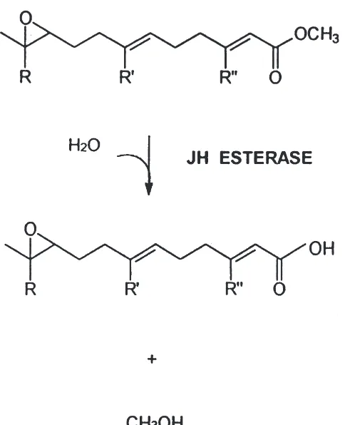
Juvenile hormones (JHs), such as JH III (methyl(2E, 6E)-[10R]-10, 11-epoxy-3, 7, 11-trimethyl-2, 6-dodecanoate) (Fig. 1), are insect hormones that are involved in the regulation of the development of insects. These hormones are also required in the adult form of many insects for the regulation of reproductive processes such as oogenesis (Sehnal, 1985). In order for metamor-phosis to occur in holometabolous insects, the JH titer must decrease during the last larval stage, resulting in pupation. JH titers must remain low during metamor-phosis of the pupa in butterflies and moths (Lepidoptera). This decrease in JH is modulated, in part, by juvenile hormone esterase (JHE), which hydrolyzes the methyl ester of JH to the corresponding carboxylic acid (Fig. 1). There is now indication that the JH acid metabolite also may have intrinsic biological activity (Ismail et al., 1998).
* Corresponding author. Tel.:+1-530-752-7519; fax:+ 1-530-752-1537.
E-mail address: [email protected] (B.D. Hammock).
0965-1748/00/$ - see front matter2000 Published by Elsevier Science Ltd. All rights reserved. PII: S 0 9 6 5 - 1 7 4 8 ( 0 0 ) 0 0 0 2 0 - 5
Fig. 1. Enzymatic reaction catalyzed by JHE. Note that the JH ester is in a chemically stableα,β-conjugated system. JH 0: R=R9=R0=ethyl; JH I: R=R9=ethyl; R0=methyl; JH II: R=ethyl, R9=R0=methyl; JH III: R=R9=R0=methyl.
gradient PAGE and the cloning of two JHE-related genes from this insect (Vermunt et al., 1998a). The sequence of this protein is quite different from the JHEs reported from H. virescens; it does not contain the characteristic GQSAG sequence which contains the active site serine (underlined and bold). This raises the question of whether JHE evolved from a monophyletic or polyphy-letic origin in beetles and moths.
JHE from H. virescens has been particularly well characterized due to interest in exploitation of this enzyme for use as a biopesticide (Bonning and Ham-mock, 1996). Three cDNA clones have been isolated, and the recombinant form of the enzyme expressed in a nuclear polyhedrosis virus has been shown to reduce caterpillar feeding and increase the speed of kill over wild type virus (Bonning et al., 1995). Both the natural and recombinant JHE cause anti-JH effects upon injec-tion into the caterpillar, Manduca sexta (Hammock et al., 1988; Philpott and Hammock, 1990). Modified forms of JHE recombinantly produced in baculovirus have been shown to be more highly insecticidal than the wild-type recombinant JHE in T. ni and H. virescens (Bonning et al., 1997).
Our goal is to characterize JHEs from insects that are evolutionarily distant from H. virescens and other Lepi-doptera. It is our hope that one could design
biopestic-ides based on JHE by exploiting unusual or otherwise different properties of JHEs found in diverse organisms. Herein, we describe partial purification by affinity chro-matography, subsequent characterization of a putative dimeric form of JHE, and partial cloning of the gene.
2. Materials and methods
Chemicals: C-10[3
H]-JH III was purchased from New England Nuclear Research (Boston, MA) at a specific radioactivity of approximately 16.4 Ci/mmol and was mixed with unlabeled JH III (5×1024M), which was pur-chased from Calbiochem (San Diego). 3-Octylthio-1,1,1-trifluoropropan-2-one (OTFP, C8H17SCH2C(O)CF3) and 3-pentylthio-1,1,1-trifluoro-2-propanone (PTFP, C4H9SCH2C(O)CF3) were used as inhibitors to elute JHE from the affinity column and were synthesized as
described in Hammock et al. (1984).
3-(4-Mercaptobutylthio)1,1,1-trifluoropropan-2-one (MBTFP, HSC4H8SCH2C(O)CF3) was used as a ligand for the affinity gel (Abdel-Aal and Hammock, 1986).
2.1. Insects
Larvae of T. molitor were purchased from Carolina Biological Supply (Burlington, NC) and were reared at 28°C on a 18L:6D photoperiod and fed a diet of Cheeriosand potatoes. Purification was performed on pupae that were 0–48 h post-pupation and frozen at
220°C until further use. Enzyme activity was stable under these storage conditions. Repeated freeze–thaw was found to result in no detectable loss of JHE activity.
2.2. JHE purification
Sepharose. This was done by combining the crude extract with the gel in a polypropylene tube and then rotating on a wheel at 4°C. At every 30 min the tube was set upright and the gel was allowed to settle. An aliquot of the supernatant was assayed for JHE activity. This method was repeated until more than 80% of the JHE activity had bound to the gel. This typically took about 6 h. The affinity gel varied in epoxide activation from approx. 6–9µmol/g gel. A typical purification pro-cedure started with an extract from approximately 200 pupae. The resulting soluble fraction was diluted three-fold and was loaded onto |3 ml of affinity gel. Affinity purification of JHE was accomplished similarly to the method of Abdel-Aal et al., 1988 (Abdel-Aal and Ham-mock, 1986). 1-Octyl-β-D-glucoside (OG) (0.1% (w/v)) was used to encourage elution of the protein by minimiz-ing hydrophobic interaction with the gel. This concen-tration provided the greatest yield of JHE elution from the column in the presence of the trifluoroketone inhibi-tor, OTFP, at a final concentration of 1 mM (Abdel-Aal and Hammock, 1986).
The enzyme was eluted batchwise in several steps in the presence of several milliliters of fresh buffer and inhibitor; the elutions were carried out for 4–8 h with shaking at 4°C. Eluates were concentrated by using Cen-tricon concentrators (Amicon, Beverly, MA). Eluates were reactivated by dialysis in purification buffer at 4°C for 96 h in the presence of exogenously added ovalbumin as a carrier at a final concentration of 200µg/ml. Further dialysis resulted in some additional reactivation (approx. 22%) and the activity was stable to further dialysis for up to 7 days.
2.3. Electrophoresis
SDS–PAGE and native Tris–glycine gels were run using either 10% polyacrylamide gels or prepared 8– 16% gradient gels (Novex, San Diego). The following standards were run on the native Tris–glycine gels: oval-bumin (43 kDa), bovine serum aloval-bumin (67 and 134 kDa) and catalase (232 kDa). Wide-range protein stan-dards (Novex) were run on SDS–PAGE. Isoelectric focusing (IEF) was performed using precast slab gels (Pharmacia) in a range of pH 3–7 and 3–10. IEF Wide Range Standards (Pharmacia) were used to calibrate the gels. JHE activity from both crude fractions and in 2 mm gel slices from IEF was verified by both partition assay (Hammock and Sparks, 1977) and TLC (Casas et al., 1991; Stauffer et al., 1997). Gels were stained with Coomassie Brilliant Blue according to the method of Sambrook et al. (1989).
2.4. Renaturation of JHE from SDS–PAGE
p71 and p150 forms of JHE were renatured following SDS–PAGE on 8–16% gradient gels (Novex, San Diego)
as described above. The gel was stained with Nile Red (Sigma) according to the procedure of Daban et al. (1996). The gel was agitated for 5 min in a solution of 40µg/ml Nile Red in distilled water. The stained bands were promptly excised from the gel under UV illumi-nation and immediately placed in 500µl 12% (v/v) Tri-ton X-100, phosphate buffered saline (PBS) for 2 h at room temperature with shaking for renaturation. Enzy-matic activity on JH III was measured by a partition assay with radiolabeled JH III (Hammock and Sparks, 1977).
2.5. Sequencing and amino acid analysis
For N-terminal sequencing, the samples were elec-trophoresed on precast 8–16% SDS–PAGE (Novex). The gel was transferred to Problot (Applied Biosystems), stained with Coomassie Blue R-250 according to Biorad instructions and submitted to the Protein Structure Lab (University of California, Davis) for amino acid analysis or sequence analysis on a Beckman 6300 Analyzer or a Applied Biosystems 470A Sequencer, respectively. For in-gel Lys C digestion the samples were electrophoresed on SDS–PAGE, stained with Coomassie R-250; in-gel proteolysis was performed according to the methodology of Y.M. Lee (personal communication) using Lys C endoprotease from Achromobacter lyticus (Wako). The peptides were separated on a 1×150 mm, 5 µm, 300 A˚ Michrom reverse-phase C18 Reliasil column running on a Michrom Ultrafast Protein Analyzer. A gradient of 5– 70% B (A=2% acetonitrile, 0.075% (v/v) TFA, B=100% acetonitrile, 0.1% (v/v) TFA) in 70 min at a flow rate of 50 µl/min with detection at 210 nm was used. Peak fractions were collected and submitted for analysis to the Protein Structure Lab (University of California, Davis).
2.6. Cloning of JHE
PolyA RNA was isolated from fat bodies of three 0– 48 h old pupae of T. molitor. Approximately 350 mg of wet tissue was used, and yielded 6.65 µg of poly A RNA. mRNA was isolated using the Poly(A) Pure kit from Ambion per manufacturer’s instructions. This mRNA was used as template for Reverse Tran-scriptase—PCR. The degenerate oligonucleotides,
TmGPA (59–TAY–ACN–AAR–TAY–GGI–CCI–GC–
39), TmAIG (59–AAR–TAY–GGI–CCI–GCI–ATH–
sequence, TAANPNPFEK. For the first strand of cDNA synthesis, 500 ng of mRNA was hybridized with 10 pmol of the oligonucleotide, TmFEK in TE buffer (10 mM Tris–HCl, 1 mM EDTA, pH 8.0) in a total volume of 5 µl. The mixture was incubated at 70°C for 5 min, cooled slowly to 46°C and then placed on ice. The RNA-:oligonucleotide hybrid mixture was then mixed in a 20
µl reaction containing 20 U/µl Superscript Reverse Tran-scriptase 2 (Gibco Life Technologies), 2 U/µl of RNasin ribonuclease inhibitor (Promega), and Reverse Tran-scriptase buffer (50 mM Tris–HCl (pH 8.3), 75 mM KCl, 3 mM MgCl2, 2 µM DTT) (Gibco Life Technologies) and 0.25 mM dNTP (TaKaRa). The reverse transcriptase reaction was carried out at 42°C for 50 min followed by 50°C for 20 min. An aliquot (0.5 µl) of this first strand reaction was used for the first round of PCR amplifi-cation in a 100µl reaction containing 100 pmol each of the primers, TmFEK and TmGPA, 2.5 U of Taq DNA Polymerase (TaKaRa), 0.2 mM of each dNTP (TaKaRa), and Taq polymerase buffer (10 mM Tris–HCl (pH 8.3), 50 mM KCl, 1.5 mM MgCl2) (TaKaRa). The thermocy-cling was performed in a PTC-100 thermocycler from MJ Research, Inc. The reaction was carried out with an initial 2 min denaturing step at 94°C followed by 4 cycles of 30 set at 94°C 30 set at 61°C and 2 min at 72°C; then 6 rounds of 30 s at 94°C, 30 s at 59°C 2 min at 72°C; then 8 cycles of 30 s at 94°C, 30 s at 57°C, 2 min at 72°C; and finally 19 rounds of 30 s at 94°C, 30 s at 55°C, and 2 min at 72°C. A second round of nested PCR was performed using 2µl of the first reaction with the same conditions as above except that the primers TmAIG and TmPNP were used. The second amplifi-cation resulted in an 800 base-pair PCR product, which was then cloned into pPCR-Script Amp SK(+) plasmid according to manufacturer’s instructions using the PCR-Script Amp Cloning Kit (Stratagene). The recombinant plasmid, pPNP, was amplified in Epicurian coli XL1 Blue MRF’ cells (Stratagene). pPNP DNA was pur-ified from the recombinant bacteria using published tech-niques (Sambrook et al., 1989). The subcloned PCR pro-duct was sequenced from pPNP DNA using an ABI sequencing machine Model #377 (Perkin Elmer).
3. Results
3.1. JH metabolites in crude homogenates
All experiments in this paper utilized 0–48 h old pupa, in part because larvae are difficult to stage. Previous reports by J.L. Connat (1983) and Sparks and Hammock (1980) have demonstrated that during development of T. molitor, JHE activity is high in early pupae. Whole body homogenate was used in this case because T. molitor consumes little water and thus, inherently has relatively
low quantities of hemolymph where most JHE is typi-cally located in other species.
Quantitative experiments indicate that approximately 98% of the total esterase activity on JH III is found in the soluble crude pupal fractions following 10,000g spins and filtering. Of this soluble activity 80% is reco-vered in the 100,000g supernatant and approx. 1.0% is recovered in the 100,000g pellet fraction. Thus, 19% of activity is lost during ultracentrifugation. Similar results were obtained in a second independent experiment.
Crude 100,000g supernatants and pellets of insect pupae were analyzed for JH-esterase and JH epoxide hydrolase activities using TLC to separate metabolites of radiolabeled JH. JH-acid was found to be the major metabolite present in the 100,000g supernatant; in a 30 min incubation 87% of the radioactivity was present as acid, the balance of the activity was present as JH-diol and JH-JH-diol-acid. No parent JH was detected at this time point. The 100,000g membrane fraction, incubated with substrate under the same conditions, yielded 74 and 26% JH-acid and JH-acid-diol, respectively. A second independent experiment yielded similar results. These results indicate the presence of a low amount of epoxide hydrolase activity, and for this reason we assayed the subsequent affinity eluate for epoxide hydrolase activity.
3.2. Purification by affinity chromatography
JHE from T. molitor was purified by an affinity chro-matography method designed for JHEs from extracts of other insects. This method relies on the use of a trifluo-roketone-based transition state analog bound to Sepharose CL-6B to selectively bind JHE with sub-sequent elution using a stronger inhibitor. This technique typically yields homogeneous JHE from crude prep-arations of many different insects in a single step (Abdel-Aal and Hammock, 1986).
Table 1
Purification table for JHE from T. molitor
Total protein (mg) Specific activity Fold purification Recovery activity (%) (nmol/min mg)
100,000g supernatant 1243 13.9 1 100
Affinity stepa 2.02 N.D.a N.D.a N.D.a
Reactivationb 1.11 286.1 20.1 1.8
aSample was inhibited with the trifluoroketone, OTFP.
b Sample was dialyzed in the presence of 1.6 g/ml ovalbumin for 3 days.
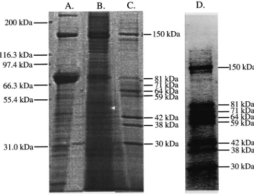
observed recoveries are close to a previous purification in T. molitor (49-fold purification and 1.56% yield of activity) that was reported by Stauffer et al. (1997). Fig. 2 shows SDS–PAGE analysis of the affinity eluate using OTFP which reveals that eight major bands were purified by this procedure. SDS–PAGE analysis of purification
Fig. 2. SDS–PAGE analysis of 100,000g supernatant (A), insoluble precipitate from affinity purification (B) and affinity eluate (C) from T. molitor. An 8–16% SDS–PAGE was run in which 12 and 3.1µg total protein of the crude supernatant and affinity eluate, respectively, and 30 µl of a SDS-solubulized suspension were loaded onto the gel. The resulting gel was stained with Coomassie Blue R-250. Numbers on the left-hand side show positions of molecular weight markers and on the right-hand side show estimated molecular weights of bands discussed in the text. Lane D contains the same affinity eluate as in lane C, but in this lane the resolution is much improved such that each band can clearly be visualized.
exchange, hydrophobic interaction and lectin chromato-graphy.
We observed during the affinity purification of JHE from T. molitor that a large amount of protein precipi-tates out of solution during incubation with the trifluo-roketone inhibitor used for elution. In a single experi-ment 50% of the total protein following an elution was present as an insoluble material as measured by Biorad protein assay. SDS–PAGE analysis showed that a sub-stantial amount of proteins ranging from 71 to 150 kDa appeared to be present in this fraction with a much greater proportion of p150 relative to the amount seen in nonprecipitated affinity eluate (Fig. 2).
Solubilization of the pellet was attempted with both the non-ionic and ionic detergents, Triton X-100 and sodium dodecyl sulfate (SDS), respectively. Of the reagents tried, only SDS was capable of fully solubiliz-ing the pellet. Thus a major factor in the apparent poor yield may be due to aggregation of JHE that sub-sequently precipitates out of solution.
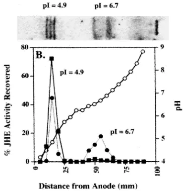
3.3. Identification of JHE on isoelectric focusing
JHE from the affinity eluate of T. molitor was ident-ified and characterized by isoelectric focusing (pH 3– 10), two-dimensional SDS–PAGE and activity analysis. Fig. 3 shows the IEF for the affinity eluate and corre-sponding activity profile as measured by the radiometric substrate, [3H]-JH III, across an IEF gel for both the 100,000g supernatant and the purified affinity eluate. Two major areas of activity were identified at a pI=4.9 and 6.7, which correspond to the Coomassie Blue stained areas on the IEF gel (Fig. 3). All of the enzyme activity that was loaded onto the gel was recovered in these two peaks in a ratio of 46 and 54% for the pI 6.7 and 4.9 forms, respectively, for the crude extract and 5% and 95% for the pI 6.7 and 4.9 forms, respectively, for the affinity eluate.
A TLC analysis of products formed by incubation of JH III with the two pI forms was accomplished to test whether the enzymatic activity of the two pI forms is due to ester hydrolysis. Both pI forms produce JH-acid as the major product on TLC analysis with no evidence of any JH-diol or JH-diol-acid that would be indicative of epoxide hydrolase activity (data not shown).
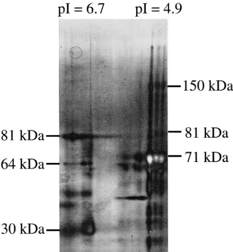
A two-dimensional SDS–PAGE of the affinity eluate after the components were separated based on their pIs is shown in Fig. 4. These data show that the pI 4.9 form appears to be the one that consists of the 71 and 150 kDa proteins. The pI 6.7 form appears to contain only proteins smaller than 81 kDa, and appears to be associa-ted with at least the majority of the 30 kDa protein that copurifies on affinity chromatography. These results sug-gest more than one of the protein bands in Fig. 2 are JHEs.
3.4. N-terminal sequence analysis
An N-terminal sequence analysis was performed on several bands of the affinity eluate. Table 2 shows the N-terminal sequence data for the bands of interest in this study. The N-terminal sequence of the 71 kDa protein and the 150 kDa protein are identical. A BLAST datab-ase search (Karlin and Altschul, 1990) using each N-terminal sequence as a query shows similarity of the 71 kDa protein and 150 kDa protein to the JHE of H. vires-cens (Hammock et al., 1988). Thus we tested the hypoth-esis that the 71 kDa band is a JHE, and that the 150 kDa protein represents a dimer of the the 71 kDa protein. Identification of homologous proteins in the BLAST dat-abase for the remaining three bands which migrate close to the 71 kDa band, namely, the 81, 64 and 59 kDa bands, was not possible with the present data.
3.5. Characterization of 150 kDa protein as a putative JHE dimer
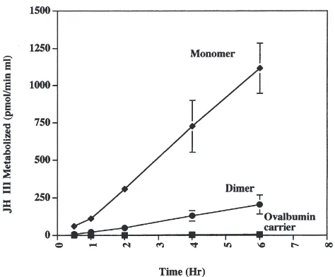
We tested the hypothesis that the 150 kDa protein is a dimer of the 71 kDa protein. Surprisingly, both the p71 and p150 proteins were detected on SDS–PAGE even under strongly denaturing conditions. We could not separate the p71/p150 couple, and the two forms appear to interconvert on the time scale of many of our experi-ments. Interconversion can be demonstrated by concen-trating a sample of JHE that eluted from an S200 gel filtration column at a molecular weight of 71 kDa in which no 150 kDa protein was initially present as exam-ined by SDS–PAGE. Concentration of this sample 12-fold (65–780µg/ml) caused partial interconversion to the p150 form as measured by SDS–PAGE (data not shown). Because of the inability to separate the p71/p150 couple under native conditions, we resorted to separation on SDS–PAGE, followed by renaturation in the presence of Triton X-100 and measurement of renat-ured bands for enzyme activity on JH III. Not surpris-ingly, renaturation after incubation in the ionic detergent, SDS, was associated with a low recovery of JHE activity and in all cases only a few percent of the total activity loaded on the gels was recovered after renaturation. Fig. 6 shows a time course of activity on JH III for the p71 and p150 bands following SDS–PAGE and renaturation in the presence of Triton X-100. The enzyme activity for both p71 and p150 is essentially linear with time over a period of 6 h (Fig. 6).
Fig. 3. Enzymatic activity analysis of JHE affinity eluate and crude 100,000g supernatant on isoelectric focusing (IEF), pH 3-10 (Pharmacia). (A) 1.1µg affinity eluate and 6µg crude material (not shown) were electrophoresed and stained with Coomassie Blue R-250. (B) Esterase activity on JH III as a function of distance in mm from the anode. (I) and (j) represent gel slices from crude homogenate and affinity eluate, respectively.
Activity is displayed as a percent of total activity initially loaded onto the gel. pH gradient is shown as a function of the distance from the anode, indicated bys(mm).
and are similar to those obtained by Vermunt et al. on the JHE of L. decemlineata (Vermunt et al., 1998b). Thus the possibility of an electrostatic interaction between the two putative monomers was tested by serial dilution of the affinity eluate up to 100-fold; this did not result in interconversion. Finally, the affinity eluate was incubated in the presence of 1 mM β-mercaptoethanol at 90°C in a time course up to 4 h. We found that not only did extensive heat and reducing agent not dissociate the 150 kDa protein into smaller proteins, but the 150 kDa protein appeared to produce higher order aggregates that did not migrate into an 8–16% SDS–PAGE. Thus the results indicate the possibility that a putative dimer associates by an uncharacteristically strong electro-static interaction.
3.6. Cloning of JHE
Ver-Fig. 4. Two-dimensional SDS–PAGE of JHE affinity eluate. 8–16% 2D SDS–PAGE (Novex) was run on the JHE affinity eluate following separation in the first dimension by IEF (pH 3–7) (Novex) in the first dimension. Approx. 2.9µg total protein was run on the first dimen-sional gel. The IEF gel was prepared for loading onto the subsequent 2D SDS–PAGE per instructions (Novex). Numbers on the left and right of the gel show estimated molecular weights of bands of the pI isoforms discussed in the text. Thus both the 150 and 71 kDa JHE bands can be detected in the band focusing at pI=4.9.
Table 2
N-terminal sequence analysis of proteins inthe affinity eluate from
T. molitor
Molecular weighta Sequence
150 FNTLSPWDKEVIYNWKA
81 HSVHSTNYAQKDV
71 FNTLSPWDKEVIYNWKA
64 YAPKSPIVY
59 PPEVTIEQGKLR
aMolecular weights of proteins sequences were determined on a 8–16% SDS–PAGE.
munt et al. (1998a). Further analysis by the CLUSTAL W alignment program (Thompson et al., 1994) for the JHEs of H. virescens, T. molitor and L. decemlineata yielded homology scores of 34 for the alignment of H. virescens and T. molitor, 4 for the alignment of T. moli-tor and L. decemlineata and 5 for the alignment of H. virescens and L. decemlineata. These results suggest that while JHE from T. molitor has significant similarity to that of JHE from H. virescens, the JHE reported from L. decemlineata does not. The clone reported here shows similarity to the other JHEs studied, and contains the characteristic GQsAG sequence around the active site
serine (underlined and bold) (Fig. 5). The other two members of the catalytic triad were not identified due to an incomplete clone. All of the peptides obtained from Lys C digestion gave sequences which exactly matched the corresponding amino acid sequence derived from the cDNA clone sequence.
4. Discussion
Several JHEs have been successfully purified to hom-ogeneity by the MBTFP–Sepharose-based affinity puri-fication method, however, these successes have been lar-gely limited to insects of the Lepidopteran order. JHEs from insects of other orders have proven more difficult to purify. This was recently indicated by Stauffer et al. (1997) and Vermunt et al. (1998b) for JHE of L. decem-lineata. In this study, we purified JHE from the coleop-teran, T. molitor, using a trifluoroketone-based affinity purification system, but failed to obtain a homogeneous preparation of JHE. Upon further study, however, we found some rather interesting biochemical characteristics of this JHE which are presented in the results.
In the application of the affinity chromatography tech-nique to whole body homogenate of T. molitor, our con-sistent result was the purification of a small number of proteins. SDS–PAGE analysis (Fig. 2) reveals the pres-ence of major protein bands at eight different molecular weights. The fact that more than one protein eluted from the affinity gel could either indicate multiple forms of JHE, other esterase enzymes, or unrelated proteins.
As the purification table (Table 1) indicates, there is a 20-fold increase in specific activity as measured by radiometric JHE assay, and a 1.8% yield in total JHE activity. Though greater than 90% of the soluble JHE activity bound to the affinity gel, elution and reactivation of the enzyme was difficult. The low yield of cata-lytically active enzyme from the affinity purification scheme, in this case, is likely due to two causes. The first reason is the apparent aggregation and precipitation of the enzyme following purification under the con-ditions presented in this paper. The precipitate was resistant to resolubilization, and the analysis of partially solubilized precipitate by SDS–PAGE revealed a sig-nificant amount of 150 kDa protein. Possibly, the smaller JHE protein(s) has a tendency to dimerize and then pre-cipitate out of solution, or the majority of JHE activity exists as a 150 kDa protein in vivo, but is unstable under the in vitro conditions that have been reported here.
Fig. 5. Protein sequence alignment of partial clone of JHE from T. molitor (upper sequence) with that of H. virescens (Hanzlik, et al, 1989) (lower sequence). Partial clone of JHE from T. molitor is aligned with that of H. virescens using the software, DNASIS. Numbering system includes amino acids and gaps. Thus, catalytic serine 203 from JHE of H. virescens is 238 in this figure. Underlined residues correspond to peptides whose sequences were obtained from in-gel Lys C digests. All of the peptides obtained by this method agree exactly with this sequence. Shaded region corresponds to the catalytic serine sequence. JHE from T. molitor and H. virescens display 42% identity in the region of the clone, which comprises the majority of the N-terminal domain. The sequence from L. decemlineata (Vermunt et al., 1998a) did not have high enough similarity to present a meaningful alignment by either DNASIS or CLUSTAL W (Thompson et al., 1994).
elution, low observed recovery may be due to incom-plete removal of the trifluoroketone inhibitor from the enzyme despite extensive dialysis. This observation has been made before with other JHEs. Trifluoroketones exhibit slow tight-binding kinetics, and OTFP has been reported to have an I50of 10210M on JHE of T. molitor (Stauffer et al., 1997).
The affinity eluate contains two groups of JH hydrolyzing proteins which can be separated based on their pIs. The two sets at different pIs are enzymatically active and both produce JH-acid as their sole reaction product. Though the majority of JHE activity of the affinity eluate migrates at pI=4.9 with IEF analysis, part of the original JHE activity is apparently present within a group of proteins with a pI of 6.7. A higher proportion
Fig. 6. Time course of activity on JH III for the putative dimer and monomer of JHE after renaturation from SDS–PAGE. Each point rep-resents the average±SD of two independent experiments in which the data were each collected in triplicate. A blank gel slice was averaged and subtracted from each datum point for each independent experi-ment.
The significance of additional forms of JHE activity remains uncertain. When both pI groups of JHE from IEF are run on a second dimension of electrophoresis using SDS–PAGE, each pI group separates into various molecular weights. There is some overlap between the 2 groups (64 and 81 kDa), but several other bands are unique to each pI group. The 71 and 150 kDa proteins seem to be limited to the pI 4.9 group of JHE activity. In the future, the thorough explanation of the roles of these different forms of JHE will likely require the pro-duction of antibodies and/or molecular cloning and expression of individual proteins within these groups. With such tools available it would be feasible to deter-mine whether these are isoforms of the same protein, or different gene products of completely independent genes. It will also be interesting to assess the roles of each form and assess their prominence throughout devel-opment.
Under the conditions used for affinity chromato-graphy, a variety of proteins were eluted. All major bands were eluted from SDS–PAGE, and the eluate was dialyzed and tested for esterase activity. Two of the pro-teins (71 and 150 kDa) are able to hydrolyze JH. One other protein (38 kDa) had catalytic activity onα -naph-thyl acetate but not JH. The remainder of proteins that co-eluted from the affinity gel have no known function, as they neither exhibited any catalytic activity nor revealed any homology to known proteins by N-terminal sequence analysis. The inability to recover JHE activity from the other eluted proteins following SDS–PAGE does not rule out the possibility that these are JH hydrolyzing proteins which are less stable or which fail to reactivate under these conditions.
Despite the fact that there is more than one protein which possibly represents an independent JHE, we were able to make a logical decision to focus upon the 71 kDa protein and the 150 kDa protein for several reasons. Firstly, both of these proteins exhibited JHE activity after elution from SDS–PAGE and dialysis. Secondly, they both are represented in the group of proteins which migrates at pI=4.9 by isoelectric focusing. This area of the IEF gel represents the majority of JHE activity in the affinity eluate. Thirdly, N-terminal analysis indicates not only that the two proteins have identical N-termini, but that this protein sequence has homology to a pre-viously cloned JHE from another species, H. virescens. Based upon these reasons, we decided to focus upon this pair of proteins.
Analysis of the 71 kDa band by partial cloning was accomplished. This clone, which encompasses the N-ter-minal domain of p71, is 42% identical to the region of H. virescens JHE to which it aligns. This sequence dem-onstrates that the isolated clone is probably that of an esterase similar to JHE based on homology to JHE of H. virescens and the signature sequence in the vicinity of the catalytic serine, GQSAG. Although this clone shows high homology to JHE sequences from T. ni, M. sexta (Hinton, A.C., unpublished), and Lymantria dispar (Nussbaumer, C., 2000), it shows low identity (21%) to the JHE-related sequences recently reported from the beetle L. decemlineata (Vermunt et al., 1998a). Com-parison of our sequence data from T. molitor with JHEs from Lepidopterans strongly suggests a monophyletic origin, at least with the homologue or paralogue described here. The observation that our sequence data are not highly similar to JHE from L. decemlineata is unexplainable at this point (Vermunt et al., 1998a). However, Vermunt et al. (1998a) report the absence of both a catalytic serine and the characteristic GQSAG sequence in the vicinity of the active site serine in their translated sequences of the two JHE-related genes in Colorado potato beetle. Furthermore, catalytic activity on JH III has not been demonstrated for the proteins purified by Vermunt et al. (1998b). This suggests that the JHE reported herein is not the same as that isolated from L. decemlineata (Vermunt et al., 1998a,b), although the JHE reported by Vermunt et al. (1998a,b) may corre-spond to a small percentage of JHE from T. molitor unidentified in this study.
identical N-terminal sequences out to 17 amino acids
(FNTLSPWDKEVIYNWKA). The N-terminal
sequences of four individual bands in the molecular weight range of approx. 153–174 kDa determined fol-lowing separation by SDS–PAGE appear to have ident-ical sequences at least out to the first five amino acids, which is as far as we sequenced in these cases. These data indicate the possibility that these bands are N-gly-cosylated isoforms of the p150 form. We attempted to ascertain whether the putative dimer is a homo-dimer or hetero-dimer. However, peptide mapping of Lys-C digests of the dimer was unsuccessful, presumably due to challenges associated with this type of analysis on large proteins. Thus, we propose the hypothesis that the JHE dimer is a homo-dimer, based on the fact that only one sequence is obtained on N-terminal sequencing of all four bands in the 150 kDa molecular weight region. However, one cannot rule out the possibility that there is actually a heterodimer with one of the sequences N-terminally blocked.
Although the N-terminal sequence data and similar pI on isoelectric focusing would strongly suggest dimeriz-ation, the inability to dissociate this putative dimer by SDS–PAGE is disconcerting. The putative dimer was stable to strongly denaturing conditions, including incu-bation with SDS, urea, reducing reagents and high tem-perature. Most noncovalent interactions and disulfide linkages would be disrupted under these conditions. Possible interpretations of this are (1) the 150 kDa tein represents a single polypeptide (2) the 150 kDa pro-tein represents a dimer which is covalently linked by some covalent linkage other than disulfide bonds (3) the 150 kDa protein represents two monomers which are bound by unusually strong electrostatic and/or hydro-phobic interactions. The latter two possibilites are con-sidered due to the observation that a small amount of the p71 form demonstrated conversion to p150 after sep-aration of the two forms on S200 gel filtration column, and then concentrating the p71 sample 12-fold. The con-centrated sample (65µg/ml) revealed a small p150 band on SDS–PAGE. Thus, there is evidence that the 150 kDa protein can arise from monomers of pure 71 kDa protein. It is now clear with the growing number of JHEs characterized outside the Lepidopteran order that JHEs can vary widely among insect species and orders. This variation comprises not only catalytic parameters but possibly also quaternary structure, as we have demon-strated here. The future cloning and comparison of JHEs from different species, as well as different forms of JHE within species, will allow discovery of mechanisms of enzyme function, degradation, and their roles in insect physiology and development.
Acknowledgements
B.A.T. was supported on USDA postdoctoral fellow-ship #95-37302-1861, T.F.S. was supported on a Univer-sity of California Biotechnology Fellowship. This work was partly funded by USDA Competitive Research Grants Program #97-35302-4406. The University of Cal-ifornia, Davis is a NIEHS Center for Environmental Health Sciences (P30 ES05707) and an EPA Center for Ecological Health Research (CR8 19658).
References
Abdel-Aal, Y.A.I., Hammock, B.D., 1988. Kinetics of binding and hydrolysis of JH II in the hemolymph of Trichoplusia ni (Hubner). Insect Biochemistry 18, 743–750.
Abdel-Aal, Y.A.I., Hammock, B.D., 1985. Apparent multiple catalytic sites involved in the ester hydrolysis of juvenile hormones by the hemolymph and by an affinity-purified esterase from Manduca
sexta Johannson (Lepidoptera: Sphingidae). Archives of
Biochem-istry and Biophysics 243, 206–219.
Abdel-Aal, Y.A.I., Hammock, B.D., 1986. Transition state analogs as ligands for affinity purification of juvenile hormone esterase. Science 233, 1073–1076.
Abdel-Aal, Y.A.I., Hanzlick, T.N., Hammock, B.D., Harshman, L.G., Prestwich, G., 1988. Juvenile hormone esterases in two heliothines: kinetic, biochemical and immunogenic characterization. Compara-tive Biochemistry and Physiology 90B, 117–124.
Baker, F., 1990. Techniques for identification and quantitation of juv-enile hormones and related compounds in arthropods. In: Gupta, A. (Ed.), Morphogenetic Hormones of Arthropods—Discoveries, Syntheses, Metabolism, Evolution, Modes of Action and Tech-niques. Rutgers University Press, New Brunswick, pp. 389–453. Bonning, B.C., Hoover, K., Booth, T.F., Duffey, S., Hammock, B.D.,
1995. Development of a recombinant baculovirus expressing a modified juvenile hormone esterase with potential for insect con-trol. Archives of Insect Biochemistry and Physiology 30, 177–194. Bonning, B.C., Hammock, B.D., 1996. Development of recombinant baculoviruses for insect control. Annual Review of Entomology 41, 191–210.
Bonning, B.C., Ward, B.K., Meet-, M.M.M., Booth, T.F., Hammock, B.D., 1997. Disruption of lysosomal targeting associated with insecticidal potency of juvenile hormone esterase. Proceedings of the National Academy of Science USA 94, 6007–6012.
Campbell, P.M., Oakeshott, J.G., Healy, M.J., 1998. Purification and characterization of juvenile hormone esterase from Drosophila
mel-anogaster. Insect Biochemistry and Molecular Biology 28, 501–
515.
Casas, J., Harshman, L.G., Hammock, B.D., 1991. Epoxide hydrolase activity on juvenile hormone in Manduca sexta. Insect Biochemis-try 21, 17–26.
Connat, J.L., 1983. Juvenile hormone esterase activity during the last larval and pupal stages of Tenebrio molitor. Journal of Insect Physi-ology 29, 515–521.
Daban, J.R., Bartolome, S., Bermudez, A., 1996. Rapid staining of proteins in polyacrylamide gels with Nile red. In: Walker, J.M. (Ed.), The Protein Protocols. Humana Press, New Jersey, pp. 179–185.
Grieneisen, M., Mok, A., Kieckbusch, T., Schooley, D., 1997. The specificity of juvenile hormone esterase revisited. Insect Biochem-istry and Molecular Biology 27, 365–376.
inhibitors of juvenile hormone esterase. Pesticide Biochemistry and Physiology 22, 209–223.
Hammock, B.D., Harshman, L.G., Philpott, M.L., Szekacs, A., Ottea, J.A., Newitt, R.A., Wroblewski, V.J., Halarnkar, P.P., Hanzlik, T.N., 1988. Strategies for the discovery of insect control agents: exploitation of biomechanisms regulating insect development. In: Steffens, G.L., Rumsey, T.S. (Eds.), Biomechanisms Regulating Growth and Development. Kluwer, Dordrecht, Netherlands, pp. 137–173.
Hammock, B.D., Roe, R.M., 1985. Analysis of juvenile hormone ester-ase activity. In:. Law, J.H., Rilling, H.C. (Eds.), Methods in Enzy-mology. Steroids and Isoprenoids, Part B, III. Academic Press, Orlando, FL, pp. 487–494.
Hammock, B.D., Sparks, T.C., 1977. A rapid assay for insect juvenile hormone esterase activity. Annals of Biochemistry 82, 573–579. Hanzlik, T.N., Hammock, B.D., 1987. Characterization of juvenile
hor-mone hydrolysis in early larval development of Trichoplusia ni. Journal of Biological Chemistry 262, 13584–13591.
Hanzlik, T.N., Abdel-Aal, Y.A.I., Harshman, L.G., Hammock, B.D., 1989. Isolation and sequencing of cDNA clones coding for juvenile hormone esterase from Heliothis virescens: evidence for a charge relay network of the serine esterases different from the serine pro-teases. Journal of Biological Chemistry 264, 12419–12425. Ismail, S.M., Satyanarayana, K., Bradfield, J.Y., Dahm, K.H.,
Bhaska-ran, G., 1998. Juvenile hormone acid: evidence for a hormonal function in induction of vitellogenin in larvae of Manduca sexta. Archives of Insect Biochemistry and Physiology 37, 305–314. Jesudason, P., Venkatesh, K., Roe, R.M., 1990. Haemolymph juvenile
hormone esterase during the life cycle of the tobacco hornworm,
Manduca sexta. Insect Biochemistry 20, 593–604.
Karlin, S., Altschul, S., 1990. Methods for assessing the statistical sig-nificance of molecular sequence features by using general scoring schemes. Proceedings of the National Academy of Science USA 87, 2264–2268.
Khlebodarova, T.M., Gruntenko, N.E., Grenback, L.G., Sukhanova, M.Z., Mazurov, M.M., Rauschenback, I.Y., Thomas, B.A., Ham-mock, B.D., 1996. A comparative analysis of juvenile hormone metabolizing enzymes in two species of Drosophila during devel-opment. Insect Biochemistry and Molecular Biology 28, 829–835. McCutchen, B.F., Uematsu, T., Sze´ka´cs, A., Huang, T.L., Shiotsuki,
T., Lucas, A., Hammock, B.D., 1993. Development of surrogate substrates for juvenile hormone esterase. Archives of Biochemistry and Biophysiology 307, 231–241.
Nussbaumer, C., Hinton, A.C., Schopf, A., Stradner, A., Hammock, B.D., 2000. Isolation and characterization of juvenile hormone esterase from hemolymph of Lymantria dispar by affinity and by anion exchange chromatography. Insect Biochemistry and Molecu-lar Biology 30, 307–314.
Philpott, M.L., Hammock, B.D., 1990. Juvenile hormone esterase is a biochemical anti-juvenile hormone agent. Insect Biochemistry 20, 451–459.
Rauschenbach, I.Y., Khlebodarova, T.M., Chentsova, N.A., Grun-tenko, N.E., Grentach, L.G., Yantsen, E.I., Filipenko, M.L., 1995. Metabolism of the juvenile hormone in Drosophila adults under normal conditions and heat stress. Journal of Insect Physiology 41, 179–189.
Sambrook, J., Fritsch, E.F., Maniatis, T., 1989. Molecular Cloning: A Laboratory Manual, 2nd ed. Cold Spring Harbor Laboratory, Cold Spring Harbor, NY.
Sehnal, F., 1985. Morphology of insect development. Annual Reviews in Entomology 30, 89–109.
Sparks, T.C., Hammock, B.D., 1980. Comparative inhibition of the juvenile hormone esterases from Trichoplusia ni, Tenebrio molitor and Musca domestica. Pesticide Biochemistry and Physiology 14, 290–302.
Stauffer, C., Shiotsuki, T., Chan, W., Hammock, B.D., 1997. Charac-terization of the esterase isozymes of Ips typographicus (Coleoptera Scolytidae). Archives of Insect Biochemistry and Physiology 34, 203–221.
Thompson, J., Higgins, D., Gibson, T., 1994. CLUSTAL W: improv-ing the sensitivity of progressive multiple sequence alignment through sequence weighting, position-specific gap penalties and weight matrix choice. Nucleic Acids Research 22, 4673–4680. Vermunt, A.M.W., Koopmanschap, A.B., Vlak, J.M., DeKort, C.A.D.,