Bipolar Disorder
Husseini K. Manji and Robert H. Lenox
Clinical studies over the years have provided evidence that monoamine signaling and hypothalamic–pituitary– adrenal axis disruption are integral to the pathophysiol-ogy of bipolar disorder. A full understanding of the pathophysiology from a molecular to a systems level must await the identification of the susceptibility and protective genes driving the underlying neurobiology of bipolar disorder. Furthermore, the complexity of the unique biol-ogy of this affective disorder, which includes the predis-position to episodic and often progressive mood distur-bance, and the dynamic nature of compensatory processes in the brain, coupled with limitations in experimental design, have hindered our progress to date. Imaging studies in patient populations have provided evidence of a role for anterior cingulate, amygdala, and prefrontal cortex in the pathophysiology of bipolar disorder. More recent research strategies designed to uncover the molec-ular mechanisms underlying our pharmacologic treat-ments and their interaction in the regulation of signal transduction as well as more advanced brain imaging studies remain promising approaches. This experimental strategy provides data derived from the physiologic re-sponse of the system in affected individuals and addresses the critical dynamic interaction with pharmacologic agents that effectively modify the clinical expression of the pathophysiology. Biol Psychiatry 2000;48:518 –530 ©2000 Society of Biological Psychiatry
Key Words: Bipolar disorder, lithium, signal transduc-tion, protein kinase C, G proteins, protein kinase A
Introduction
C
linical studies over the past 40 years have attempted to uncover the biological factors mediating the patho-physiology of bipolar disorder (BD) utilizing a variety of biochemical and neuroendocrine strategies. These studieshave for the most part rested upon the conceptual founda-tion that monoamine signaling and hypothalamic–pitu-itary–adrenal axis disruption are integral to the pathophys-iology of both depression and mania (Bowden 1997; Goodwin and Jamison 1990). Although such investiga-tions have been heuristic over the years, they have been of limited value in elucidating the unique biology of this affective disorder, which must include an understanding of the underlying basis for the predilection to episodic and often profound mood disturbance that can become pro-gressive over time. Kindling and propro-gressive sensitization to seizures, taken from the epilepsy literature, has been very useful as a model for focusing attention to the long-term course of the illness, but thus far has been less useful in explicating the biological processes leading to instability in the regulation of mood; however, recent research characterizing the contributory roles of signaling pathways in various facets of kindling and sensitization holds promise in this regard. More recent research strate-gies designed to uncover the molecular mechanisms un-derlying our pharmacologic treatments coupled with more advanced brain imaging studies remain promising ap-proaches as we enter the next millennium.
We should also keep in mind that a true understanding of the pathophysiology of BD must address its neurobiol-ogy at different physiologic levels (i.e., molecular, cellu-lar, systems, and behavioral; Figure 1). Abnormalities in gene expression undoubtedly underlie the neurobiology of the disorder at the molecular level, and this will become evident as we identify the susceptibility and protective genes for BD in the coming years. Once this has been accomplished, however, the even more difficult work of examining the impact of the faulty expression of these gene products (proteins) on integrated cell function must begin. It is at these levels that we have identified some protein candidates using the psychopharmacologic strate-gies noted above and that will be more fully elucidated below. The precise manner in which these candidate molecular and cellular targets may or may not relate to the faulty expression of susceptibility gene products is yet to be determined. The task becomes even more daunting when one considers the possibility that a major component of the pathophysiology of BD may stem from discordant From the Laboratory of Molecular Pathophysiology, Departments of Psychiatry and
Behavioral Neurosciences, and Cellular and Clinical Neurobiology Program, Wayne State University School of Medicine, Detroit, Michigan (HKM) and the Molecular Neuropsychopharmacology Program, Departments of Psychiatry, Pharmacology, and Neuroscience, University of Pennsylvania School of Medicine, Philadelphia (RHL).
Address reprint requests to Husseini K. Manji, M.D., Wayne State University School of Medicine, 4201 St. Antoine, UHC 9B-29, Detroit MI 48201. Received February 4, 2000; revised May 11, 2000; accepted May 17, 2000.
© 2000 Society of Biological Psychiatry 0006-3223/00/$20.00
biological rhythms ranging from ultradian to infradian that ultimately drive the periodic recurrent nature of the disor-der (Bunney and Bunney 2000; Ehlers et al 1988; Ikono-mov and Manji 1999; Lenox and Manji 1998; Mandell et al 1984; Wehr and Goodwin 1983). The subsequent challenge for the basic and clinical neuroscientist will be the integration of these molecular/cellular changes to the systems and ultimately to the behavioral level, wherein the clinical expression of BD becomes fully elaborated.
Before critically reviewing the data relevant to the pathophysiology of BD, it is necessary to consider the complexity of our task (Table 1). To begin with, we would suggest that altered expression of critical proteins resulting from a series of susceptibility genes predisposes to a dysregulation of signaling in regions of the brain predis-posing to periodic loss of homeostasis and clinical mani-festation of affective symptomatology (i.e., mania and/or depression; Depue et al 1987; Goodwin and Jamison 1990). Thus the biological processes underlying the risk for mood cycling may be very distinct from the biology driving the clinical symptoms of mania or depression per se (Goodwin and Jamison 1990). In this regard it should be noted that there is increasing evidence for a shared genetic risk for both bipolar and unipolar disorders (Berrettini 2000), suggesting that the underlying pathophysiologic
processes predisposing to recurrent mood disturbance may share common features. Furthermore, the clinical picture and system response are the result of a complex dynamic interaction between the dysregulated signaling systems and activation of existing physiologic feedback mecha-nisms designed to compensate for extreme changes (Post and Weiss 1999). In this way, the constellation of symp-toms including not only mood but also autonomic,
endo-Figure 1. The pathophysiology of bipolar disorder (BD). This figure highlights the fact that a complete understanding of the pathophysiology of BD must address its neurobiology at different physiologic levels (i.e., molecular, cellular, systems, and behavioral). PKC, protein kinase C; MARCKS, myristoylated alanine-rich C kinase substrate; GSK-3, glycogen synthase kinase-3; MAP kinase, mitogen-activated protein kinase; Bcl-2, B-cell leukemia/lymphoma; proteome, the population of cellular protein species and their expression level; transcriptome, the population of cellular messenger RNA species and their expression level.
Table 1. The Pathophysiology of Bipolar Disorder: Constraints for Experimental Design
● Complex disease with diagnostic heterogeneity
● Episodic nature of symptoms and distinct symptom clusters ● The biology underlying recurrence/cyclicity may be distinct from
that responsible for specific symptom clusters
● Disease progression dictates changes over the course of the illness ● Dynamic interaction between compensatory/adaptive changes in the
brain and primary neurobiology of the disorder
● Effect of treatment and treatment withdrawal on measures ● Potential circadian rhythm abnormalities suggest that single
time-point studies may be inadequate
● Relative inaccessibility of target organ and dependence on peripheral
models
● Lack of suitable animal models
● The characterization of “mood” as a quantitative trait (quantitative
crine, sleep/wake, and circadian activity determinants will reflect not only the stage and progression of illness, but also the unique individual characteristics conferring het-erogeneity in clinical presentation and diagnosis. In light of this complexity and the dynamic properties of the system, research strategies examining biochemical and endocrine variables would be expected to be subject to a high degree of inherent variability not only when using cross-sectional analyses between patients, but also when utilizing longitudinal designs over time within individual patients. Furthermore, the use of peripheral sources of tissue and postmortem brains to address biochemical/ neuroendocrine activity within patients’ brains introduces another set of variables inherent in the experimental design and most often conferring significant constraints on data interpretation.
Classical Monoaminergic Neurotransmitter
and Neuroendocrine Systems
The stimulus for the study of the biogenic amines in patients with BD was provided by the discovery of effective pharmacologic treatments for depression and mania (Goodwin and Jamison 1990). Although these pharmacologic agents have prominent effects on single episodes of depression and mania, even more pertinent to our discussion are the major effects that these agents exert on the long-term course of the illness (Goodwin and Jamison 1990; Manji and Potter 1997). Antidepressants in general, and tricyclics in particular, may increase the frequency of cycles and worsen long-term outcome (Wehr and Goodwin 1987). In addition to this compelling phar-macologic data, the biogenic amine neurotransmitter sys-tems are distributed extensively in the limbic system, which is implicated in the regulation of sleep, appetite, arousal, sexual function, endocrine function, and emo-tional states such as fear and rage. The clinical picture of BD involves disruption of behavior, circadian rhythms, neurophysiology of sleep, and neuroendocrine and bio-chemical regulation within the brain (for reviews, see Goodwin and Jamison 1990; Holsboer 1995). Thus it is not surprising that clinical research strategies over the years have identified dysregulation of noradrenergic, do-paminergic, serotonergic, and cholinergic systems as well as the hypothalamic–pituitary axis in patients with BD. The behavioral and physiologic manifestations of the illness are complex and undoubtedly mediated by a net-work of interconnected neurotransmitter pathways (Lenox 1987; Manji 1992); the monoamine neurotransmitter sys-tems are ideally placed to mediate such complex behav-ioral effects, and thus have represented attractive candi-date systems underlying the pathophysiology of BD. The multiple functions of central
neurotransmitter/neuroendo-crine systems, however, make it difficult to conceptualize, let alone demonstrate, true specificity of detected abnor-malities. Since these earlier studies have been amply reviewed elsewhere, in this perspectives article we focus upon more recent data linking signaling abnormalities with the underlying neurobiology of BD.
Signaling Networks: The Cellular
Machinery Underlying Information
Processing and Long-Term Neuroplastic
Events
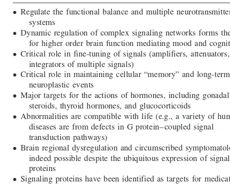
It is hardly surprising that abnormalities in multiple neurotransmitter systems and physiologic processes have been found in a disorder as complex as BD. Signal transduction pathways are in a pivotal position in the central nervous system (CNS), able to affect the functional balance between multiple neurotransmitter systems, and may therefore play a role in mediating the more “down-stream” abnormalities in multiple neurotransmitter sys-tems and physiologic processes. Moreover, recent research has clearly identified signaling pathways as therapeuti-cally relevant targets for our most effective pharmacologic treatments (Table 2). Indeed, the molecular and cellular targets underlying lithium’s ability to stabilize an under-lying dysregulation of limbic and limbic-associated func-tion strongly suggest that abnormalities in signaling path-ways may also play a critical role in the pathophysiology of BD.
Multicomponent, cellular signaling pathways interact at various levels, thereby forming complex signaling net-works that allow the cell to receive, process, and respond to information (Bhalla and Iyengar 1999; Bourne and Nicoll 1993). These networks facilitate the integration of
Table 2. Bipolar Disorder: A Putative Role for Signaling Pathways
● Regulate the functional balance and multiple neurotransmitter
systems
● Dynamic regulation of complex signaling networks forms the basis
for higher order brain function mediating mood and cognition
● Critical role in fine-tuning of signals (amplifiers, attenuators, and
integrators of multiple signals)
● Critical role in maintaining cellular “memory” and long-term
neuroplastic events
● Major targets for the actions of hormones, including gonadal
steroids, thyroid hormones, and glucocorticoids
● Abnormalities are compatible with life (e.g., a variety of human
diseases are from defects in G protein– coupled signal transduction pathways)
● Brain regional dysregulation and circumscribed symptomatology are
indeed possible despite the ubiquitous expression of signaling proteins
● Signaling proteins have been identified as targets for medications
signals across multiple time scales and the generation of distinct outputs depending on input strength and duration, and regulate intricate feed-forward and feedback loops (Weng et al 1999). These properties of signaling networks suggest that they play critical roles in cellular memory; thus, cells with different histories, and therefore express-ing different repertoires of signalexpress-ing molecules and tran-scription factors, interacting at different levels, may re-spond quite differently to the same signal over time. Given their widespread and crucial role in the integration, regu-lation, amplification, and fine-tuning of physiologic pro-cesses, it is not surprising that abnormalities in signaling pathways have now been identified in a variety of human diseases (Milligan and Wakelam 1992; Spiegel 1998; Weintraub 1995). Pertinent to the present discussion is the observation that a variety of diseases manifest relatively circumscribed symptomatology, despite the widespread, often ubiquitous expression of the affected signaling proteins.
Although complex signaling networks are likely present in all eukaryotic cells and control various metabolic, humoral, and developmental functions, they may be espe-cially important in the CNS, where they serve the critical roles of first amplifying and “weighting” numerous extra-cellularly generated neuronal signals and then transmitting these integrated signals to effectors, thereby forming the basis for a complex information processing network (Bhalla and Iyengar 1999; Bourne and Nicoll 1993; Manji 1992). The high degree of complexity generated by these signaling networks may be one mechanism by which neurons acquire the flexibility for generating the wide range of responses observed in the nervous system (Table 2). These pathways are thus undoubtedly involved in regulating such diverse vegetative functions as mood, appetite, and wakefulness and are therefore likely to be involved in the pathophysiology of BD. We now turn to a discussion of the direct and indirect evidence supporting a role for abnormalities in signaling pathways in the patho-physiology of BD.
Alterations of Transmembrane Cellular
Signaling Pathways in Animal Models of BD
The need to use caution in the appropriate application of animal models to an understanding of complex neuropsy-chiatric disorders has been well articulated (Einat et al, in press; Post and Weiss 1999), and in fact it is unlikely we will ever develop animal models that display the full range of symptomatology as clinically expressed in man. Two current models that have had reasonable heuristic value in the study of mood disorders are kindling and behavioral sensitization (Einat et al, in press; Post and Weiss 1999). In both of these paradigms, there is increased responsivity
(either behavioral or electrophysiologic) to repeated “low dose” stimulation (pharmacologic or electric) over time. Indeed, the sensitized behavioral response has been re-ported to persist for months and years after discontinuation of drug administration. Considerable evidence implicates long-term alterations in midbrain dopaminergic transmis-sion in the development of behavioral sensitization, but the cellular mechanism(s) underlying the long-term changes in excitability observed in kindled or stimulant-sensitized animals have not been fully elucidated. In recent years, an appreciation of the major role of signal transduction pathways in the regulation of neuronal excitability has led to extensive research into their involvement in kindling and behavioral sensitization, and a growing body of evidence implicates alterations in both protein kinase C (PKC; Table 3) and certain G proteins (especially Giand
Go). Studies have demonstrated that pertussis toxin (which
inactivates Giand Go) produces a significant augmentation
of psychostimulant-induced motor activity, and dopamine release in the nucleus accumbens (Chen et al 1999; Steketee and Kalivas 1991). In keeping with these studies, specific decreases in Gai and Gai levels have been
observed in response to chronic cocaine in the ventral tegmental area and nucleus accumbens (Nestler et al 1990), once again consistent with the hypothesis that regulation of G proteins represents part of the biochemical changes that underlie the effects of chronic psychostimu-lants. Similarly, injection of pertussis toxin into the amyg-dala of kindled rats markedly modifies kindled seizures, effects that appear to be mediated via an alteration in the after-discharge threshold.
Table 3. Protein Kinase C: Pathophysiology and Treatment of Bipolar Disorder
● Kindling produces dramatic increases in membrane-associated PKC
in the hippocampus and amygdala
● Amphetamine produces increases in PKC activity, and GAP-43
phosphorylation (implicated in neurotransmitter release)
● PKC inhibitors block the biochemical and behavioral responses to
amphetamine and cocaine and also block cocaine-induced sensitization
● Dexamethasone administration increases PKC activity, and increases
the levels of PKCaand PKCein the rat frontal cortex and hippocampus
● Increased membrane/cytosol PKC partitioning in platelets from
manic subjects; normalized with lithium treatment
● Increased PKC activity and translocation in bipolar disorder brains,
as compared with control subjects
● Lithium and valproate regulate PKC activity, PKCa, PKCe, and
MARCKS
● Preliminary data suggest that PKC inhibitors may have efficacy in
the treatment of acute mania
● Typical and atypical antipsychotics regulate the levels of PKCaand
PKCein areas of the rat brain
With respect to the PKC signaling system, dramatic increases in membrane-associated PKC have been ob-served in the bilateral hippocampus up to 4 weeks after the last kindled seizure and in the amygdala/pyriform cortex at 4 weeks after (Daigen et al 1991). Studies have also implicated alterations in PKC activity as mediators of long-term alterations in neuronal excitability in the brain following chronic stimulant use. Thus, both acute and chronic amphetamine produce an alteration in PKC activ-ity, its cytosol relative to membrane distribution, and the phosphorylation of a major PKC substrate, growth cone– associated protein 43 (GAP-43), which has been impli-cated in long-term alterations of neurotransmitter release (Kantor and Gnegy 1998). Furthermore, PKC inhibitors have been shown to block the acute responses to both amphetamine and cocaine (as assessed by both behavioral and in vivo microdialysis studies) as well as cocaine-induced sensitization (Kantor and Gnegy 1998).
It is indeed striking that behavioral sensitization and kindling produce robust alterations in the PKC signaling pathway in critical limbic structures, since lithium and valproate (VPA) also target the very same biochemical targets (see below). Thus, although considerable caution clearly needs to be employed when extrapolating from the findings observed in these two models, the fact that they are associated with effects on PKC signaling that are the opposite of those observed with chronic lithium or VPA is compelling indeed.
Are G Protein–Coupled Signal Transduction
Pathways Targets for Mood-Stabilizing
Agents?
The cAMP/Protein Kinase A Signaling Pathway as a Target for the Actions of Mood Stabilizers and Antidepressants
As discussed above, dysregulation of cellular signaling networks in the brain likely underlies the oscillations in behavioral states observed in mood disorders. Thus, it is not surprising that increasingly recent research on the cellular mechanisms underlying lithium’s therapeutic ef-fects has focused on the transmembrane signaling path-ways in the brain (for reviews, see Lenox and Manji 1998; Mork et al 1992; Post et al 2000).
Lithium and the AC System
It has been demonstrated that lithium exerts complex effects on the activity of adenylyl cyclase (AC), with the preponderance of the data demonstrating an elevation of basal AC activity but an attenuation of variety of receptor-mediated responses (Jope 1999a, 1999b; Manji et al 2000; Wang et al 1997). Lithium in vitro inhibits the stimulation
of AC both by Gpp(NH)p (a poorly hydrolyzable analog of guanosine triphosphate [GTP]) and by Ca11 /calmodu-lin, suggesting that lithium in vitro is directly able to inhibit the catalytic unit of AC (Mork et al 1992; Newman and Belmaker 1987). Since these inhibitory effects of lithium in vitro can be overcome by Mg11, it has been postulated that they are mediated (at least in part) by a direct competition with magnesium (whose hydrated ionic radius is similar to that of lithium) for a binding site on the catalytic unit of AC (Mork and Geisler 1989; Mork et al 1992; Newman and Belmaker 1987); however, the inhib-itory effects ofchroniclithium treatment on rat brain AC are not reversed by Mg11and still persist after washing of the membranes, but are reversed by increasing concentra-tions of GTP (Mork and Geisler 1989; Mork et al 1992; Newman and Belmaker 1987). These results suggest that the effects of chronic lithium (those that are more likely to be therapeutically relevant) may be exerted at the level of signal-transducing G proteins at a GTP-responsive step (discussed below). These two distinct actions of lithium on the AC system may explain the differences in results that have been obtained by investigators using rat membrane preparations and those obtained with the use of rat slice preparations (Lenox and Manji 1998). This has led to an investigation of lithium’s effects on the AC system in vivo, using microdialysis. These studies found that chronic lithium treatment produced a significant increase in basal and post–receptor stimulated (cholera toxin or forskolin) AC activity, while attenuating theb-adrenergic–mediated effect (Manji et al 2000; Masana et al 1992). Interestingly, chronic lithium treatment resulted in an almost absent cyclic adenosine monophosphate (cAMP) response to pertussis toxin, suggesting a lithium-induced attenuation of Gi function. It should be noted, however, that chronic
lithium has been found to increase not only cAMP levels (Wiborg et al 1999), but also the levels of AC type I and type II messenger RNA (mRNA) and protein levels in the frontal cortex (Colin et al 1991; Jensen et al 2000), suggesting that lithium’s complex effects on the system may represent the net effects of direct inhibition of AC, upregulation of AC subtypes, and effects on the stimula-tory and inhibistimula-tory G proteins. Most recently, lithium’s effects on the phosphorylation and activity of cyclic AMP response element– binding protein (CREB) have been examined in the rodent brain and in cultured human neuroblastoma cells, with somewhat conflicting results (Ozaki and Chuang 1997; Wang et al 1999).
increase platelet basal and post–receptor stimulated AC activity (Risby et al 1991), effects that are strikingly similar to those observed in the rodent brain. Consistent with a lithium-induced increase in basal cAMP and AC levels, a more recent study found that platelets obtained from lithium-treated euthymic bipolar patients exhibited enhanced basal and cAMP-stimulated phosphorylation of Rap1 (a protein kinase A [PKA] substrate), as well as another unidentified 38-kd phosphoprotein (Perez et al 2000a). Somewhat surprisingly, these investigators did not find similar effects of lithium in healthy subjects.
Carbamazepine and the AC System
The effects of carbamazepine (CBZ) on the AC system have been less extensively studied. It is noteworthy, however, that CBZ has been reported to decrease not only basal and stimulated cAMP production in rodents, but also cerebrospinal fluid levels of cAMP in manic patients (Post et al 2000). Recent studies have found that CBZ, at therapeutically relevant concentrations, inhibits both basal AC and FSK-stimulated cAMP accumulation in C6 gli-oma cells (Chen et al 1996). Furthermore, CBZ also inhibits both basal and FSK-stimulated activity of AC purified from the rat cortex, suggesting that CBZ inhibits cAMP production by acting directly on AC and/or through factor(s) that are tightly associated with and copurify with AC (Chen et al 1996). Carbamazepine’s effects on cAMP-mediated transcriptional events have also been investi-gated, and CBZ has been found to inhibit forskolin-stimulated phosphorylation of CREB (Chen et al 1996) and c-fos expression (Divish et al 1991). Together, the results suggest that CBZ brings about a dampening of the AC system; these effects may play a role in both its antiepileptic and its antimanic effects, but such a conten-tion requires extensive further validaconten-tion.
Antidepressants and the AC System
The chronic administration of antidepressants (ADs) is known to bring about a variety of biochemical effects, including an enhanced coupling between Gas and the
catalytic unit of AC (Rasenick et al 2000). Several studies have also demonstrated that the postreceptor components of the cAMP system are regulated by long-term AD treatments, including cAMP-dependent protein kinase en-zyme activity (Nestler et al 1989; Popoli et al 2000). Taken together, these results suggest that ADs, via their complex effects on G proteins and the AC signaling system, may attenuate b-adrenergic–mediated activation of Gs, while enhancing the effects of agents operating by
pathways independent of theb-adrenergic receptor (bAR). Consistent with these results, ADs have been demon-strated to activate cAMP-dependent and
calcium/calmod-ulin-dependent protein kinases, effects that are accompa-nied by increases in the endogenous phosphorylation of selected substrates (microtubule-associated protein 2 and synaptotagmin; Popoli et al 2000). Furthermore, recent data from Duman and associates (1997) have demon-strated that the chronic treatment of rats with a variety of ADs increases the levels of CREB mRNA, CREB, and CRE DNA binding activity in the hippocampus. The same laboratory has demonstrated that chronic AD treatment also increases the expression of two important genes known to be regulated by CREB—namely, brain-derived neuotrophic factor and its receptor trkB (Duman et al 1997). Preliminary postmortem human brain studies have also revealed increased CREB levels in patients treated with ADs, providing indirect support for the rodent and cell culture studies (Dowlatshahi et al 1998).
Lithium and G Proteins
Abundant experimental evidence has shown that lithium attenuates receptor-mediated second messenger generation in the absence of consistent changes in the density of the receptors themselves (Lenox and Manji 1998; Mork et al 1992). There is now considerable evidence that chronic lithium administration affects G protein function (Jope 1999a, 1999b; Lenox and Manji 1998; Wang and Fried-man 1999); however, the preponderance of data suggests that lithium, at therapeutically relevant concentrations, does not have anydirecteffects on G proteins and does not consistently alter G protein subunitlevels(Ellis and Lenox 1991; Lenox and Manji 1998). Although some studies have reported modest changes in thelevelsof G proteina
subunits, the preponderance of the data suggests that the effects of chronic lithium on signaling pathways occur in the absence of changes in the levels of G protein subunits per se (Lenox and Manji 1998); however, chronic in vivo lithium treatment has been shown to produce a significant increase in pertussis toxin– catalyzed [32P]adenosine diphosphate ([32P]ADP) ribosylation in the rat frontal cortex. Since pertussis toxin selectively ADP-ribosylates the undissociated, inactiveabgheterotrimeric form of Gi,
these results suggest that lithium attenuates Gifunction via
a stabilization of the inactive conformation. These obser-vations suggest that the removal of the “inhibitory tone” by lithium may be responsible for the elevations in basal AC and the responses to agents activating the stimulatory pathway distal to the receptor (Jope 1999b). Consistent with this hypothesis, lithium has been shown to potentiate the behavioral hyperactivity induced by intra-accumbens cholera toxin administration (which activates the stimula-tory G proteins, Gs and Golf; Kofman et al 1998).
generally observed reduced receptor/G protein coupling (Lenox and Manji 1998; Risby et al 1991). The effects of 2 weeks of lithium administration on G protein measures has also been examined in healthy volunteers. Similar to the findings in the rat brain, lithium did not affect the levels of platelet G protein a subunits, but produced a significant increase in pertussis toxin– catalyzed [32P]ADP
ribosylation, once again suggesting a stabilization of the inactive undissociated abg heterotrimeric form of Gi.
These long-term effects of chronic lithium on G protein are likely attributable to an indirect posttranslational mod-ification of the G protein(s) and a relative change in the dynamic equilibrium of the active/inactive states of pro-tein conformation. In this context, it is noteworthy that investigators have demonstrated that lithium alters the levels of endogenous ADP-ribosylation in C6 glioma cells (Young and Woods 1996) and in the rat brain (Nestler et al 1995), suggesting another novel mechanism by which chronic lithium may indirectly regulate the activity of these critical signaling proteins.
The Protein Kinase C Signaling Cascade
Over the last decade there have been major advances in our understanding of the critical role of the PKC signaling pathway as a therapeutically relevant target for the long-term actions of mood stabilizers. The “inositol depletion hypoth-esis” posited that lithium, as an uncompetitive inhibitor of inositol-1-phosphatase, produced its therapeutic effects via a depletion of neuronalmyo-inositol (mI) levels. Although this hypothesis has been of great heuristic value, numerous studies have examined the effects of lithium on receptor-mediated phosphoinositide (PI) responses, and although some report a reduction in agonist-stimulated phosphatidyl-inositol 4,5-bisphosphate (PIP2) hydrolysis in rat brain slices
following acute or chronic lithium, these findings have often been small and inconsistent, and subject to numerous meth-odological differences (Jope and Williams 1994; Lenox and Manji 1998). Most recently, a magnetic resonance spectros-copy study has demonstrated that lithium-induced mI reduc-tions are observed in the frontal cortex of BD patients after only 5 days of lithium administration, at a time when the patients’ clinical state is completely unchanged(Moore et al 1999). Consequently, these and other studies suggest that whereas inhibition of inositol monophosphatase may repre-sent an initiating lithium effect, reducing mI levels per se is not associated with therapeutic response. This led to our working hypothesis that some of theinitialactions of lithium may occur with a relative reduction of mI—this reduction of mI initiates a cascade of secondary changes in the PKC signaling pathway and gene expression in the CNS, effects that are ultimately responsible for lithium’s therapeutic efficacy.
Indeed, evidence accumulating from various laborato-ries has clearly demonstrated that lithium, at therapeuti-cally relevant concentrations, exerts major effects on the PKC signaling cascade (Table 3). Currently available data suggest that acute lithium exposure facilitates a number of PKC-mediated responses, whereas longer exposure results in an attenuation of phorbol ester–mediated responses, which is accompanied by a downregulation of specific PKC isozymes (discussed in Manji and Lenox 1999). Studies in rodents have demonstrated that chronic (but not acute) lithium produces an isozyme-selective reduction in PKCaandein the frontal cortex and hippocampus, in the absence of significant alterations in the b, g, d, or z
isozymes. Concomitant studies carried out in immortalized hippocampal cells in culture exposed to chronic lithium show a similar reduction in the expression of both the PKC
aandeisozymes in the cell as determined by immunoblot (Chen et al, in press; Manji and Lenox 1999).
A major strategy that has been utilized to investigate the downstream consequences of lithium-induced alteration in PKC isozymes is the examination of the effects of chronic lithium on endogenous PKC substrates in the brain. The most prominent substrate for PKC in the brain is an acidic protein, myristoylated alanine-rich C kinase substrate (MARCKS), which has been implicated in regulating long-term neuroplastic events. Lenox and associates (1992) provided the first evidence for the effects of chronic administration of lithium on the regulation of MARCKS in the rat brain. They demonstrated that chronic lithium administration dramatically reduced MARCKS expression in the hippocampus, effects that were not immediately reversed following lithium discontinuation. Subsequent studies carried out in immortalized hippocam-pal cells have demonstrated that this action of chronic lithium on MARCKS regulation is dependent upon both the inositol concentration and the level of receptor-medi-ated activation of PI hydrolysis (Watson and Lenox 1996). Although these effects of lithium on PKC isozymes and MARCKS are striking, a major problem inherent in neuropharmacologic research is the difficulty in attribut-ing therapeutic relevance to any observed biochemical finding. It is thus noteworthy that the structurally dissim-ilar antimanic agent VPA produces effects strikingly similar to those of lithium on PKC isozymes and on the expression of the MARCKS protein (Manji and Lenox 1999; Watson et al 1998).
Are There Abnormalities in Signaling
Pathways in BD?
The cAMP-Generating System
role in the therapeutic effects of lithium and VPA. Do these clinically effective agents, in fact,correct an under-lying abnormality in signaling pathways in BD? The future development of selective receptor and second mes-senger ligands for positron emission tomography studies may permit the direct assessment of CNS receptor and postreceptor sensitivity in humans. To date, however, studies of receptor and postreceptor function in mood disorders have been limited to indirect research strategies. The most commonly utilized strategy has been to charac-terize receptor function in readily accessible blood ele-ments, and much clinical research has focused on the activity of the cAMP-generating system in mood disor-ders. Overall, the preponderance of the evidence suggests altered receptor and/or postreceptor sensitivity of the cAMP-generating system in the absence of consistent alterations in the number of receptors themselves(for a review, see Wang et al 1997). A consistently observed decrease in leukocytebAR function in depression could reflect an inherited abnormality of thebAR/Gs/AC
com-plex, as suggested by the findings utilizing immortalized (Epstein–Barr virus–transformed) lymphocytes (Wright et al 1984). These findings need to be replicated, however, and a number of additional confounding factors need to be considered. In this context, twin studies suggest that variations in isoproterenol-stimulated cAMP production are most likely due to “environmental” effects on the number or sensitivity of bARs (Ebstein et al 1986). A recent study found higher levels of cAMP-stimulated phosphorylation of a;22-kd protein in platelets obtained from 10 treated euthymic BD patients, as compared with healthy subjects; by contrast, there was no significant difference in the basal phosphorylation between the groups (Perez et al 2000a, 2000b). Follow-up studies identified the ;22-kd protein as Rap1, and once again found higher cAMP-stimulated phosphorylation in the BD patients (Perez et al 2000a, 2000b).
Warsh and associates have undertaken the most thor-ough series of studies investigating the cAMP/PKA sys-tem in postmorsys-tem brains of BD patients. Due to the instability of cAMP in postmortem brain tissue, this research group has investigated downstream targets of cAMP. They found that the levels of PKA regulatory subunits (as assessed by [3H]cAMP binding) were
signif-icantly lower in cytosolic fractions of BD frontal, tempo-ral, occipital, and parietal cortices; cerebellum; and thala-mus, as compared with matched control subjects (Rahman et al 1997). Furthermore, preliminary findings show that the reduction of regulatory subunits of PKA in the cyto-solic fractions of the BD temporal cortex is accompanied by a higher basal kinase activity and significantly lower apparent activation constant for cAMP in the cytosolic fractions of the BD temporal cortex (Fields et al 1999).
Whether such changes result in altered endogenous cAMP-stimulated phosphorylation in the brain, similar to what has been reported in platelets from euthymic BD patients (Perez et al 2000a, 2000b), remains to be demon-strated. Nevertheless, these observed changes in PKA provide additional important evidence for a potential dysregulation of the Gas-mediated cAMP signaling
path-way in BD.
G Proteins in Mood Disorders
In view of the indirect evidence for abnormalities at postreceptor sites described above, it is not surprising that several independent laboratories have examined G pro-teins in patients with mood disorders (Chen et al 1999; Mathews et al 1997; Wang et al 1997; Warsh et al 2000). Young and associates (1993) were the first to report increased levels of Gas in BD. Compared with control
subjects matched with respect to age, postmortem interval, and brain pH, they found increased levels of Gas in the
frontal, temporal, and occipital cortices (but not in the hippocampus, thalamus, or cerebellum) in postmortem brain tissue from patients with BD. This group also found increases in forskolin-stimulated AC activity in postmor-tem brains of BD patients, compatible with enhanced Gs/AC signaling in BD (Warsh et al 2000). The findings of
elevated Gaslevels and/or function are also supported by
the recent observations of Friedman and Wang (1996), who found increased agonist-activated [35S]GTP
gS bind-ing to G protein asubunits in frontal cortical membrane preparations from BD patients. Several studies have also examined G proteins in peripheral circulating cells and have found elevated levels of Gas protein (Chen et al
1999; Wang et al 1997; Warsh et al 2000) and mRNA levels (Spleiss et al 1998), although the dependency on clinical state remains unclear. Similar to what has been observed in the CNS, one recent study has evaluated the role of platelet G proteins as “signal coincidence detec-tors,” and has found this function to be impaired in depressed patients (Mooney et al 1998). Overall, the most consistent finding to emerge is that in both peripheral cells and postmortem brain tissue from BD patients elevations are observed in thepredominantsubspecies of Gaspresent in the tissues examined. It should be emphasized, how-ever, that there is at presentno evidenceto suggest that the alterations in the levels of Gasare due to a mutation in the
Gasgene itself. Indeed, there are numerous transcriptional
they derive primarily from peripheral cell models and postmortem studies involving a small number of patients.
Abnormalities of Calcium Signaling in BD
Acting via intracellular proteins such as MARCKS and calmodulin, and enzymes such as PKC, AC, and Ca11/ calmodulin-dependent kinase, calcium ions have been shown to regulate the synthesis and release of neuro-transmitters, neuronal excitability, cytoskeletal remod-eling, and long-term neuroplastic events. Thus, it is not surprising that a large number of studies have investi-gated intracellular Ca21 in peripheral cells in BD (discussed in Dubovsky et al 1992; Emamghoreishi et al 1997; Wang et al 1997). In view of the caveats associated with studies of peripheral circulating cells, the remarkable consistency of the findings is surprising indeed. Studies have consistently revealed elevations in both resting and stimulated intracellular Ca21 levels in platelets, lymphocytes, and neutrophils of patients with BD. The calcium abnormalities have been postulated to represent state-dependent findings (Dubovsky et al 1992), but recent studies using transformed lympho-blasts from BD patients have revealed similar abnor-malities, suggesting that they may be trait dependent (Emamghoreishi et al 1997). The regulation of free intracellular Ca21 is a complex, multifaceted process involving extracellular entry, release from intracellular stores following receptor-stimulated PI hydrolysis, up-take into specific organelles, and binding to specific proteins. Thus, the abnormalities observed in BD could arise from abnormalities at a variety of levels, and recent studies suggest that the abnormality lies beyond the receptor (Hough et al 1999). Interestingly, recent studies have demonstrated that Rap1 phosphorylation state is related to platelet intracellular calcium signaling (Magnier et al 1995), suggesting a possible relationship between these two documented abnormalities. Since PKC is also known to regulate calcium signaling at multiple levels (Ozaki et al 1997; Shibata et al 1996; Si-Tahar et al 1996), more recent studies have investi-gated the putative role of PKC in mediating the calcium abnormalities in BD; preliminary analysis suggests that alterations in tonic PKC activity may play an important role in mediating the abnormal intracellular calcium responses observed in BD (H.K. Manji and R.M. Post, unpublished observations, June 1998).
Are There Abnormalities of PKC Signaling in BD?
To date, there have only been a limited number of studies directly examining PKC in BD. Friedman and associates (1993) investigated PKC activity and PKC translocation in response to serotonin in platelets
ob-tained from BD subjects before and during lithium treatment. They reported that the ratios of platelets membrane-bound to cytosolic PKC activities were ele-vated in the manic subjects. In addition, serotonin-elicited platelet PKC translocation was found to be enhanced in those subjects. With respect to brain tissue, Wang and Friedman (1996) measured PKC isozyme levels, activity, and translocation in postmortem brain tissue from BD patients; they reported increased PKC activity and translocation in BD brains, as compared with control subjects, effects that were accompanied by elevated levels of selected PKC isozymes in cortices of BD subjects.
In view of the pivotal role of the PKC signaling pathway in the regulation of neuronal excitability, neurotransmitter release, and long-term synaptic events, it was postulated that the attenuation of PKC activity may play a role in the antimanic effects of lithium and VPA. In a pilot study it was found that tamoxifen (a nonsteroidal antiestrogen known to be a PKC inhibitor at higher concentrations) may, indeed, possess anti-manic efficacy (Bebchuk et al 2000). These data further support the potential role of the PKC pathway in mediating the pathophysiology of BD. The data impli-cating the PKC signaling system in the pathophysiology of BD are particularly compelling in view of the growing body of data suggesting that this signaling pathway represents a therapeutically relevant target for both lithium and VPA (see above). The complex effects of lithium on the PKC signaling pathway represent an attractive and heuristic mechanism by which the expres-sion of various proteins involved in long-term neuronal plasticity and cellular response is modulated, thereby compensating for as yet genetically undefined physio-logic abnormalities in critical regions of the brain. It is our strong conviction that it is at the cellular and molecular level that some of the most exciting advances in our understanding of the pathophysiology of BD will take place in the coming years. Current studies of the long-term lithium-induced changes of the PKC signal-ing pathway (includsignal-ing PKC isozyme regulation, post-translational modification of key phosphoproteins, and PKC-mediated alterations in gene and protein expres-sion) are a most promising avenue for future investigation.
Concluding Remarks
only the profound changes in mood but also a constel-lation of neurovegetative features derived from dys-function in limbic-related regions such as the hip-pocampus, hypothalamus, and brain stem. The highly integrated monoamine and prominent neuropeptide pathways are known to originate and project heavily within these regions of the brain, and it is thus not surprising that abnormalities have been noted in their function across clinical studies. In fact, the contribution of these pathways to the pathophysiology of BD must be reasonably robust, given the variability that might be expected in assessing such dynamic systems under the constraints in experimental design imposed upon such research. Whereas in the past much of the research effort has been focused upon identifying which of these systems might be etiologic in nature, we suggest that we might be better served by addressing their relative contributions in the system response to the underlying neurobiology of the disease process. This will become particularly important as we begin to identify the susceptibility genes for BD in the coming years. More recently, investigators have begun to identify compo-nents of signal transduction pathways in the brain such as PKC that may not only play a role in the pathophys-iology of the disease but also represent targets for the action of our most efficacious treatments (i.e., lithium and VPA). This experimental strategy may prove to be most promising, since it provides data derived from the physiologic response of the system in affected individ-uals and addresses the critical dynamic intereraction with pharmacologic agents that effectively modify the clinical expression of the pathophysiology. It is also noteworthy that a growing body of data is demonstrat-ing significant reductions in regional CNS volume and cell numbers (both neurons and glia) in BD (Drevets et al 1997; Manji et al, in press; Rajkowska et al 1999). Furthermore, it has been increasingly recognized that for many patients there is a progressive decline in overall functioning; the potential role of cell death/ atrophy in determining long-term outcome remains to be elucidated. Nevertheless, it is noteworthy that psy-chopharmacologic research strategies have most re-cently identified long-term actions of these mood sta-bilizers in the regulation of expression of genes implicated in processes involved in neuroplasticity, neuroprotection, and even neurogenesis (Chen et al, in press; Lenox and Watson 1994; Manji et al 1999, in press; Moore et al, in press). To what extent these long-term processes in the brain are critical to the ability of these drugs to compensate for the pathophys-iology of BD as well as stabilize the clinical course and progression of BD has yet to be defined (Manji et al 1999).
The authors’ research is supported by the National Institute of Mental Health (Grants Nos. RO1-MH57743 and RO1-MH59107 [HKM] and RO1-MH6247 [RHL]), Theodore and Vada Stanley Foundation (HKM, RHL), National Alliance for Research on Schizophrenia and Depression (HKM, RHL), and Joseph Young Sr. Research grants (HKM). Outstand-ing editorial assistance was provided by Celia Knobelsdorf. Space limitations necessitate our citing of review articles in many cases. A full reference list upon which this article was based is available upon request. Aspects of this work were presented at the conference “Bipolar Disorder: From Pre-Clinical to Clinical, Facing the New Millennium,” January 19 –21, 2000, Scottsdale, Arizona. The conference was spon-sored by the Society of Biological Psychiatry through an unrestricted educational grant provided by Eli Lilly and Company.
References
Bebchuk JM, Arfken CL, Dolan-Manji S, Murphy J, Manji HK (2000): A preliminary investigation of a protein kinase C inhibitor (tamoxifen) in the treatment of acute mania.Arch Gen Psychiatry57:95–97.
Berrettini WH (2000): Are schizophrenic and bipolar disorders
related? A review of family and molecular studies. Biol Psychiatry48:531–538.
Bhalla US, Iyengar R (1999): Emergent properties of networks of biological signaling pathways.Science283(5400):381–387. Bourne HR, Nicoll R (1993): Molecular machines integrate
coincident synaptic signals.Cell72(suppl):65–75.
Bowden CL (1997): Toward an integrated biological model of bipolar disorder. In: Young LT, Joffe RT, editors.Bipolar Disorder: Biological Models and Their Clinical Application.
New York: Marcel Dekker, 235–254.
Bunney WE, Bunney BG (2000): Molecular clock genes in man and lower animals. Possible implications for circadian abnor-malities in depression. Neuropsychopharmacology 22:335– 345.
Chen G, Hasanat KA, Bebchuk JM, Moore GJ, Glitz D, Manji HK (1999): Regulation of signal transduction pathways and gene expression by mood stabilizers and antidepressants.
Psychosom Med61:599 – 617.
Chen G, Hawver D, Wright C, Potter WZ, Manji HK (1996): Attenuation of adenylyl cyclases by carbamazepine in vitro.
J Neurochem67:2079 –2086.
Chen G, Masana M, Manji HK (in press): Lithium regulates PKC-mediated intracellular cross-talk and gene expression in the CNS in vivo.Bipolar Disord.
Chen G, Rajkowska G, Du F, Seraji-Bozorgzad N, Manji HK (in press): Enhancement of hippocampal neurogenesis by lith-ium.J Neurochem.
Colin SF, Chang HC, Mollner S, Pfeuffer T, Reed RR, Duman RS, Nestler EJ (1991): Chronic lithium regulates the expres-sion of adenylate cyclase and Gi-protein alpha subunit in rat cerebral cortex.Proc Natl Acad Sci U S A88:10634 –10637. Daigen A, Akiyama K, Otsuki S (1991): Long-lasting change in the membrane-associated protein kinase C activity in the hippocampal kindled rat.Brain Res545:131–136.
Divish MM, Sheftel G, Boyle A, Kalasapudi VD, Papolos DF, Lachman HM (1991): Differential effect of lithium on fos protooncogene expression mediated by receptor and postre-ceptor activators of protein kinase C and cyclic adenosine monophosphate: model for its antimanic action.J Neurosci Res28:40 – 48.
Dowlatshahi D, MacQueen GM, Wang JF, Young LT (1998): Increased temporal cortex CREB concentrations and antide-pressant treatment in major depression. Lancet2352(9142): 1754 –1755.
Drevets WC, Price JL, Simpson JR Jr, Todd RD, Reich T, Vannier M, Raichle ME (1997): Subgenual prefrontal cortex abnormalities in mood disorders.Nature386:824 – 827. Dubovsky S, Murphy J, Christiano J, Lee C (1992): The calcium
second messenger system in bipolar disorders: Data supporting new research directions.J Neuropsychiatry Clin Neurosci4:3– 14.
Duman RS, Heninger GR, Nestler EJ (1997): A molecular and cellular theory of depression.Arch Gen Psychiatry54:597– 606. Ebstein RP, Oppenheim G, Ebstein BS, Amiri Z, Stessman J (1986): The cyclic AMP second messenger system in man: The effects of heredity, hormones, drugs, aluminum, age and disease on signal amplification.Prog Neuropsychopharmacol Biol Psychiatry10:323–353.
Ehlers CL, Frank E, Kupfer DJ (1988): Social zeitgebers and biological rhythms. A unified approach to understanding the etiology of depression.Arch Gen Psychiatry45:948 –952. Einat H, Kofman O, Belmaker RH (in press): Animal models of
bipolar disorder: From a single episode to progressive cycling models. In: Myslobodsky MS, Weiner I, editors. Contempo-rary Issues in Modeling Psychopathology.Boston: Kluwer Academic.
Ellis J, Lenox RH (1991): Receptor coupling to G proteins: Interactions not affected by lithium.Lithium2:141–147. Emamghoreishi M, Schlichter L, Li PP, Parikh S, Sen J, Kamble
A, Warsh JJ (1997): High intracellular calcium concentrations in transformed lymphoblasts from subjects with bipolar I disorder.Am J Psychiatry154:976 –982.
Fields A, Li PP, Kish SJ, Warsh JJ (1999): Increased cyclic AMP-dependent protein kinase activity in postmortem brain from patients with bipolar affective disorder.J Neurochem
73:1704 –1710.
Friedman E, Hoau YW, Levinson D, Connell TA, Singh H (1993): Altered platelet protein kinase C activity in bipolar affective disorder, manic episode.Biol Psychiatry33:520 –525. Friedman E, Wang HY (1996): Receptor-mediated activation of
G proteins is increased in postmortem brains of bipolar affective disorder subjects.J Neurochem67:1145–1152. Goodwin FK, Jamison KR (1990): Manic-Depressive Illness.
New York: Oxford University Press.
Holsboer F (1995): Neuroendocrinology of mood disorders. In: Bloom FE, Kupfer DJ, editors. Psychopharmacology: The Fourth Generation of Progress.New York: Raven, 957–969. Hough C, Lu SJ, Davis CL, Chuang DM, Post RM (1999): Elevated basal and thapsigargin stimulated intracellular cal-cium of platelets and lymphocytes from bipolar affective disorder patients measured by a fluorometric microassay.Biol Psychiatry46:247–255.
Ikonomov O, Manji HK (1999): Molecular mechanisms
under-lying mood-stabilization in manic-depressive illness: The phenotype challenge.Am J Psychiatry156:1506 –1514. Jensen JB, Mikkelsen JD, Mork A (2000): Increased adenylyl
cyclase type 1 mRNA, but not adenylyl cyclase type 2 in the rat hippocampus following antidepressant treatment. Eur Neuropsychopharmacol10:105–111.
Jope RS (1999a): Anti-bipolar therapy: Mechanism of action of lithium.Mol Psychiatry4:117–128.
Jope RS (1999b): A bimodal model of the mechanism of action of lithium.Mol Psychiatry4:21–25.
Jope RS, Williams MB (1994): Lithium and brain signal trans-duction systems.Biochem Pharmacol77:429 – 441.
Kantor L, Gnegy ME (1998): Protein kinase C inhibitors block amphetamine-mediated dopamine release in rat striatal slices.
J Pharmacol Exp Ther284:592–598.
Kofman O, Li PP, Warsh JJ (1998): Lithium, but not carbamaz-epine, potentiates hyperactivity induced by intra-accumbens cholera toxin.Pharmacol Biochem Behav59:191–200. Lenox RH (1987): Role of receptor coupling to phosphoinositide
metabolism in the therapeutic action of lithium.Adv Exp Med Biol221:515–530.
Lenox RH, Manji HK (1998): Lithium. In: Nemeroff CB, Schatzberg AF, editors.American Psychiatric Press Textbook of Psychopharmacology,2nd ed. Washington, DC: American Psychiatric Press, 379 – 430.
Lenox RH, Watson DG (1994): Lithium and the brain: A psychopharmacological strategy to a molecular basis for manic depressive illness.Clin Chem40:309 –314.
Lenox RH, Watson DG, Patel J, Ellis J (1992): Chronic lithium administration alters a prominent PKC substrate in rat hip-pocampus.Brain Res570:333–340.
Magnier C, Corvazier E, Aumont MC, Le Jemtel TH, Enouf J (1995): Relationship between Rap1 protein phosphorylation and regulation of Ca21 transport in platelets: A new ap-proach.Biochem J310:469 – 475.
Mandell AJ, Knapp S, Ehlers C, Russo PV (1984): The stability of constrained randomness: Lithium prophylaxis at several neurobiological levels. In: Post RM, Ballenger JC, editors.
Neurobiology of Mood Disorders. Baltimore: Williams & Wilkins, 744 –776.
Manji HK (1992): G proteins: Implications for psychiatry.Am J Psychiatry149:746 –760.
Manji HK, Chen G, Hsiao JK, Masana MI, Moore GJ, Potter WZ (2000): Regulation of signal transduction pathways by mood stabilizing agents: Implications for the pathophysiology and treatment of bipolar affective disorder. In: Manji HK, Bow-den CL, Belmaker RH, editors.Bipolar Medications: Mech-anisms of Action. Washington, DC: American Psychiatric Press, 129 –177.
Manji HK, Lenox RH (1999): Protein kinase C signaling in the brain: Molecular transduction of mood stabilization in the treatment of bipolar disorder.Biol Psychiatry46:1328 –1351. Manji HK, Moore GJ, Chen G (1999): Lithium at 50: Have the neuroprotective effects of this unique cation been over-looked?Biol Psychiatry46:929 –940.
Manji HK, Potter WZ (1997): Monoaminergic mechanisms. In: Joffe RT, Young LT, editors.Bipolar Disorder: Biological Models and Their Clinical Application.New York: Marcel Dekker, 1– 40.
Masana MI, Bitran JA, Hsiao JK, Potter WZ (1992): In vivo evidence that lithium inactivates Gi modulation of adenylate cyclase in brain.J Neurochem59:200 –205.
Mathews R, Li PP, Young LT, Kish SJ, Warsh JJ (1997): Increased G alpha q/11 immunoreactivity in postmortem occipital cortex from patients with bipolar affective disorder.
Biol Psychiatry41:649 – 656.
Milligan G, Wakelam M (1992):G Proteins: Signal Transduc-tion & Disease.San Diego: Academic Press.
Mooney JJ, Samson JA, McHale NL, Colodzin R, Alpert J, Koutsos M, Schildkraut JJ (1998): Signal transduction by platelet adenylate cyclase: Alterations in depressed patients may reflect impairment in the coordinated integration of cellular signals (coincidence detection). Biol Psychiatry43: 574 –583.
Moore GJ, Bebchuk JM, Parrish JK, Faulk MW, Arfken CL, Strahl-Bevacqua J, Manji HK (1999): Temporal dissociation between lithium-induced CNS myo-inositol changes and clinical response in manic-depressive illness.Am J Psychiatry
156:1902–1908.
Moore GJ, Bebchuk JM, Wilds IB, Chen G, Manji HK (in press): Pharmacologic increase in human gray matter.Lancet.
Mork A, Geisler A (1989): The effects of lithium in vitro and ex vivo on adenylate cyclase in brain are exerted by distinct mechanisms.Neuropharmacology28:307–311.
Mork A, Geisler A, Hollund P (1992): Effects of lithium on second messenger systems in the brain.Pharmacol Toxicol
71(suppl 1):4 –17.
Nestler EJ, Terwilliger RZ, Duman RS (1989): Chronic antide-pressant administration alters the subcellular distribution of cyclic AMP-dependent protein kinase in rat frontal cortex.
J Neurochem53:1644 –1647.
Nestler EJ, Terwilliger RZ, Duman RS (1995): Regulation of endogenous ADP-ribosylation by acute and chronic lithium in rat brain.J Neurochem64:2319 –2324.
Nestler EJ, Terwilliger RZ, Walker JR, Sevarino KA, Duman RS (1990): Chronic cocaine treatment decreases levels of the G protein subunits Gi alpha and Go alpha in discrete regions of rat brain.J Neurochem55:1079 –1082.
Newman ME, Belmaker RH (1987): Effects of lithium in vitro and ex vivo on components of the adenylate cyclase system in membranes from the cerebral cortex of the rat. Neurophar-macology26:211–217.
Ozaki N, Chuang D-M (1997): Effect of lithium on transcription factor binding in cultured cerebellar granule cells and the brain of rats.J Neurochem69:2336 –2344.
Ozaki N, Manji HK, Luiberman V, Lu S, Rosenthal N, Goldman D (1997): A naturally occurring amino acid substitution of the human serotonin (5-HT) 2A receptor influences amplitude and timing of intracellular calcium mobilization. J Neuro-chem68:2186 –2193.
Perez J, Tardito D, Mori S, Racagni G, Smeraldi E, Zanardi R (2000a): Abnormalities of cAMP signaling in affective dis-orders: Implications for pathophysiology and treatment. Bi-polar Disord2:27–36.
Perez J, Tardito D, Mori S, Racagni G, Smeraldi E, Zanardi R (2000b): Altered Rap1 endogenous phosphorylation and lev-els in platelets from patients with bipolar disorder.J Psychiatr Res34:99 –104.
Popoli M, Brunello N, Perez J, Racagni G (2000): Second messenger-regulated protein kinases in the brain: Their func-tional role and the action of antidepressant drugs.J Neuro-chem74:21–33.
Post RM, Chuang D-M (1991): Mechanism of action of lithium: Comparison and contrast with carbamazepine. In: Birch NJ, editor.Lithium and the Cell: Pharmacology and Biochemis-try.London: Academic Press, 199 –241.
Post RM, Weiss SRB (1999): Neurobiological models of recur-rence in mood disorder. In: Charney DS, Nester EJ, Bunney BS, editors. Neurobiology of Mental Illness. New York: Oxford University Press, 365–384.
Post RM, Weiss SRB, Clark M, Chuang DM, Hough C, He L (2000): Lithium, carbamazepine and valproate in affective illness: Biochemical and neurobiological mechanisms. In: Manji HK, Bowden CL, Belmaker RH, editors. Bipolar Medications: Mechanisms of Action.Washington, DC: Amer-ican Psychiatric Press, 219 –248.
Rahman S, Li PP, Young LT, Kofman O, Kish SJ, Warsh JJ (1997): Reduced [3H] cyclic AMP binding in postmortem brain from subjects with bipolar affective disorder.J Neuro-chem68:297–304.
Rajkowska G, Miguel-Hidalgo JJ, Wei J, Dilley G, Pittman SD, Meltzer HY, et al (1999): Morphometric evidence for neuro-nal and glial prefrontal cell pathology in major depression.
Biol Psychiatry45:1085–1098.
Rasenick MM, Chen J, Ozawa H (2000): Effects of antidepres-sant treatments on the G protein-adenylyl cyclase axis as the possible basis of therapeutic action. In: Manji HK, Bowden CL, Belmaker RH, editors. Bipolar Medications: Mecha-nisms of Action. Washington, DC: American Psychiatric Press, 87–108.
Risby ED, Hsiao JK, Manji HK, Bitran J, Moses F, Zhou DF, Potter WZ (1991): The mechanisms of action of lithium.Arch Gen Psychiatry48:513–524.
Shibata K, Morita K, Kitayama S, Okamoto H, Dohi T (1996): Ca21entry induced by calcium influx factor and its regula-tion by protein kinase C in rabbit neutrophils. Biochem Pharmacol52:167–171.
Si-Tahar M, Renesto P, Falet H, Rendu F, Chignard M (1996): The phospholipase C/protein kinase C pathway is involved in cathepsin G-induced human platelet activation: comparison with thrombin.Biochem J313:401– 408.
Spiegel AM (1998): G Proteins, Receptors, and Disease. To-towa, NJ: Humana Press.
Spleiss O, van Calker D, Scharer L, Adamovic K, Berger M, Gebicke-Haerter PJ (1998): Abnormal G protein alpha(s)-and alpha(i2)-subunit mRNA expression in bipolar affective disorder.Mol Psychiatry3:512–520.
Steketee JD, Kalivas PW (1991): Sensitization to psychostimu-lants and stress after injection of pertussis toxin into the A10 dopamine region.J Pharmacol Exp Ther259:916 –924. Wang HY, Friedman E (1996): Enhanced protein kinase C
activity and translocation in bipolar affective disorder brains.
Biol Psychiatry40:568 –575.
receptor-mediated activation of G proteins in rat brain cortical mem-branes.Neuropharmacology38:403– 414.
Wang JF, Asghari V, Rockel C, Young LT (1999): Cyclic AMP responsive element binding protein phosphorylation and DNA binding is decreased by chronic lithium but not val-proate treatment of SH-SY5Y neuroblastoma cells. Neuro-science91:771–776.
Wang JF, Young LT, Li PP, Warsh JJ (1997): Signal transduc-tion abnormalities in bipolar disorder. In: Joffe RT, Young LT, editors.Bipolar Disorder: Biological Models and Their Clinical Application.New York: Marcel Dekker, 41–79. Warsh JJ, Young LT, Li PP (2000): Guanine nucleotide binding
(G) protein disturbances in bipolar affective disorder. In: Manji HK, Bowden CL, Belmaker RH, editors. Bipolar Medications: Mechanisms of Action.Washington, DC: Amer-ican Psychiatric Press, 299 –329.
Watson DG, Lenox RH (1996): Chronic lithium-induced down-regulation of MARCKS in immortalized hippocampal cells: Potentiation by muscarinic receptor activation.J Neurochem
67:767–777.
Watson DG, Watterson JM, Lenox RH (1998): Sodium valproate down-regulates the myristoylated alanine-rich C kinase sub-strate (MARCKS) in immortalized hippocampal cells. A property of protein kinase C-mediated mood stabilizers.
J Pharmacol Exp Ther285:307–316.
Wehr TA, Goodwin FK (1983): Biological rhythms in
manic-depressive illness. In: Wehr TA, Goodwin FK editors. Cir-cadian Rhythms in Psychiatry.Pacific Grove, CA: Boxwood Press, 129 –184.
Wehr TA, Goodwin FK (1987): Can antidepressants cause mania and worsen the course of affective illness?Am J Psychiatry
144:1403–1411.
Weintraub BD (1995): Molecular Endocrinology: Basic Con-cepts and Clinical Correlations.New York: Raven. Weng G, Bhalla US, Iyengar R (1999): Complexity in biological
signaling systems.Science284(5411):92–96.
Wiborg O, Kruger T, Jakobsen SN (1999): Region-selective effects of long-term lithium and carbamazepine administra-tion on cyclic AMP levels in rat brain.Pharmacol Toxicol
84:88 –93.
Wright AF, Crichton DN, Loudon JB, Morten JE, Steel CM (1984): Beta-adrenoceptor binding defects in cell lines from families with manic-depressive disorder. Ann Hum Genet
48:201–214.
Young LT, Li PP, Kish SJ, Siu KP, Kamble A, Hornykiewicz O, Warsh JJ (1993): Cerebral cortex Gs a protein levels and forskolin-stimulated cyclic AMP formation are increased in bipolar affective disorder.J Neurochem61:890 – 898. Young LT, Woods CM (1996): Mood stabilizers have