FOURIER DOMAIN ITERATIVE APPROACH TO OPTICAL SECTIONING OF 3D
TRANSLUCENT OBJECTS FOR OPHTHALMOLOGY PURPOSES
A. V. Razgulina,∗, N. G. Iroshnikovb, A. V. Larichevb, T. E. Romanenkoa, A. S. Goncharovb
a
Lomonosov Moscow State University, Faculty of Computational Mathematics and Cybernetics, Leninskie Gory 1/52, Moscow, Russia - (razgulin, romanenko)@cs.msu.ru
bLomonosov Moscow State University, Faculty of Physics,
Leninskie Gory 1/2, Moscow, Russia - (nikita,larichev)@optics.ru, [email protected]
Commission II, WG II/10
KEY WORDS:Sectioning, 3D Deconvolution, Convolution, Iterative Regularization Method
ABSTRACT:
In this paper we deal with the problem of optical sectioning. This is a post processing step while investigating of 3D translucent medical objects based on rapid refocusing of the imaging system by the adaptive optics technique. Each image, captured in focal plane, can be represented as the sum of in-focus true section and out-of-focus images of the neighboring sections of the depth that are undesirable in the subsequent reconstruction of 3D object. The problem of optical sectioning under consideration is to elaborate a robust approach capable of obtaining a stack of cross section images purified from such distortions. For a typical sectioning statement arising in ophthalmology we propose a local iterative method in Fourier spectral plane. Compared to the non-local constant parameter selection for the whole spectral domain, the method demonstrates both improved sectioning results and a good level of scalability when implemented on multi-core CPUs.
1. INTRODUCTION
Recently, a promising tool for investigation of 3D translucent medical objects was suggested which is based on the rapid refo-cusing of the incoherent imaging system by adaptive optics tech-nique (see (Larichev et al., 2002)). By properly varying of the focal length it provides a stack of images each corresponding to different depths of cross section. In principle, having a sufficient amount of sections, one can try to reconstruct the original 3D object by known methods. However, a special step called sec-tioning is necessary for image processing of the stack obtained. Actually, images, captured in each focal plane, represent both in-focus image of true section and a sum of out-of-in-focus images of the neighboring sections of the depth. Various aberrations of an optical system, fixation fluctuations, distortions of light-sensitive sensors, etc. should be taken into account as well. Thus, the prob-lem is to elaborate a robust approach capable of obtaining a stack of cross section images that are purified from such distortions.
In this paper, we elaborate a 3D model of the incoherent imag-ing system which takes into account defocus between sections depending on their depth and high order phase aberrations. The problem consists in solving a 3D convolution equation. Direct deconvolution is an ill-posed problem. Analysis of contempo-rary 3D deconvolution methods (Wu et al., 2008) shows that it-erative methods, compared with direct ones, are more accurate, but usually are accompanied by an increase in the complexity of computing and additional requirements for tuning of parameters.
The approach we propose utilizes the iterative algorithm in trans-versal Fourier domain, allowing parallelization of computations by modern multi core CPU/GPU software and hardware, in such a way that an increase in the complexity is not a matter of principle. In (Razgulin et al., 2015) the particular case of the approach was
∗Corresponding author
presented with a constant selection of parameters for all spectral components. It was found that for different values of the parame-ters the quality of recovery for high and low Fourier harmonics is different and can not be equally good for both cases. In this paper we utilize the effective local Fourier domain approach, based on the localization of the regularization parameter selection method. The results of computer simulations demonstrate the efficiency of the approach. The performance obtained by parallelization al-lows sectioning with transversal resolution up to4Kand accept-able time which is promising in noninvasive restoration of the 3D structure of human eye fundus in vivo for a consequent diagnosis of diseases.
2. 3D MODEL OF IMAGING SYSTEM
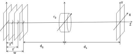
We consider the following principal scheme of imaging system for optical sectioning (see Figure 1). A thin translucent 3D object
d0 d1
X Y
Z
w z
z'
r0
Figure 1. Principal scheme of imaging system for optical sectioning
at a distanced1behind the lens. Here, for the sake of simplicity, we suppose the parametersd0,d1to be fixed and the focal length of the lensf(z)being a function of the variablez:
By alteringz from0towone can obtain, at the image plane, a stack of translucent object section images. Each cross-section image represents the true cross-cross-section superposed with blurred neighboring sections. Further we discuss this fact in de-tail.
To this end, we consider infinitely thin translucent object illu-minated by incoherent uniform plane wave and characterized by amplitude transmittance (optical density)OT(x′, y′, z′). If we
place it at the planez′ ,0≤z′
≤w, and focus imaging system at the planezwith focal lengthf(z)according to (1), then, at the image plane, we observe the following distribution
I(x, y, z, z′) =K(z, z′)
is the image of the object in geometrical optics approximation,
M(z) = d1·(d0+w−z)−1 — magnification. Point spread functionh(ξ, η, z, z′)
takes into account incoherent illumination and defocus caused by the misalignment of focal and object plane locations in the casez 6=z′
under the Fresnel scalar diffraction theory approach (Easton Jr., 2010, Gaskill, 1978):
h(ξ, η, z, z′) =
Strength of defocus is conducted by the function
ε(z, z′) = 1
Whenw≪d0, d1there is a spatially invariant approximation
ε(z, z′) = z
′ −z
(d0+w)2
. (4)
In the case of thickN-layered translucent object composed of
∆z=w/Nwidth layers placed atz′
n=n∆z,n= 0,1, . . . , N
with plane optical densityOT(x′, y′, z′n)the amplitude
transmit-tance ofn-th layer isOT(x′, y′, zn′)∆z′. In the image plane, as
mentioned in (Gu, 2000, p. 64), the total impact of all layers may be considered under linear superposition principle, which is valid for translucent thick objects where secondary diffraction is neglected. Thus the resulted distribution appears as a sum of
corresponding images of each layer:
I(x, y, z) =
Note that, under approximation (4), this formula can be easily represented in the form of 3D convolution integral, but this is not essential for us. From the relation (5) one can easily see that, due to the integration alongzaxis, the image ofz-section of 3D object contains true cross-section and defocused images of neigh-boring sections of the depth. These distortions are undesirable in the problems of 3D object reconstruction from captured cross-sections arisen in ophthalmology applications, bio-microscopy, etc. (Castleman, 2007, Wu et al., 2008). The problem is to elabo-rate robust approach capable to ”purify” stack of captured cross-section images from such distortions. This procedure is further called ”optical sectioning”.
3. DISCRETE APPROXIMATION IN FOURIER DOMAIN
Equation (5) is a 3D integral Fredholm equation of the 1st kind. The problem of its solving is an ill-posed unstable problem. In addition, from the optical point of view, this is also related to the fact that corresponding optical transfer function (OTF), a Fourier transform of PSF, is close to zero in some regions of the Fourier frequency domain. A review of the advantages and disadvantages of the available direct and iterative methods for solving similar in-verse problems of three-dimensional biomicroscopy, which differ from our statement in the form of a PSF function, is given, for ex-ample, in (Castleman, 2007, ch. 22.2) and (Wu et al., 2008, ch. 14). An analysis of these methods shows that the iteration meth-ods are more accurate in comparison with the direct ones, but are accompanied by an increase in the computational complexity. However, with the proper choice of the iterative algorithm, which allows the parallelization of computations with modern software and hardware, such an increase in computing resources is not es-sential.
Typically, the sections of a three-dimensional image of an oph-thalmic object are available for analysis and processing only in a finite set of cross-section planesz ∈ {z1, z2, . . . , zN}, equally
located at a distance∆z, where the numberN is in orders of magnitude smaller than the number of points in each horizon-tal section. As we saw above, the same situation appears for a thickN-layered translucent object composed of∆z-width lay-ers. Thus it is reasonable to turn from equation (5) to its discrete analogue in depth variablez:
On(x′, y′) =O(x′, y′, zn),m, n= 1,2, . . . , N,
hmn(x′, y′) =K(zm, zn)·h(x′, y′, zm, zn)·∆z.
Following to (Razgulin et al., 2015), we propose the iteration ap-proach allowing effective parallelization. To start with, we use compactness of support property both for imageIm(x, y)and
PSF functionh(x′, y′, z
m, zn), which allows effective sampling
of these functions and transforming continuous convolution in-tegral to discrete convolution. Using discrete Fourier transform (DFT) and discrete convolution theorem we write equations (6) in Fourier spectral domain(u, v)using notationsIbm,Obm,bhmn
for discrete Fourier coefficients of corresponding functionsIm, Om,hmn:
b
Im(u, v) = N
X
n=1 b
On(u, v)·bhmn(u, v). (7)
At each point(u, v)equations (7) form a system of linear alge-braic equations (SLAE)
HO=I (8)
for the vectorO(u, v) = (Ob1(u, v),Ob2(u, v), . . . ,ObN(u, v))of
Fourier coefficients of the sought functionOandN×Nmatrix
H(u, v) ={bhmn(u, v)}.
Direct methods are unapplicable for solving (8) sinceHis a de-generate or ill-conditioned matrix in most interesting cases. That is why we use iterated Tikhonov regularization method (Bakushin-sky and Gonchar(Bakushin-sky, 1989)
Ok+1= (E+µH∗H)−1Ok+µ(E+µH∗H)−1H∗I, (9) wherek = 0,1, . . . , K, O0 = (0,0, . . . ,0). As it is men-tioned in (Razgulin et al., 2015), in case of distortion present in observed dataI, the parameterµ >0and the number of itera-tionsKshould be chosen according to the trade-off between a rapid draft sectioning with the smallest number of iterations for a rough visualization of layers at relatively large valuesµand thor-ough restoration with fine identifying the texture due to additional iterations applied for smallµ.
4. RESULTS OF SECTIONING
4.1 Phantom three-dimensional object
We have considered a phantom translucent three-dimensional ob-ject consisting of several vessels located on different layers. The following system parameters specific to the living three-dimen-sional structure of the eye were used: the depth of the object along
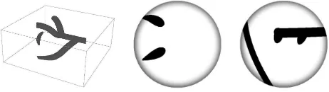
z coordinate was800microns, the number of layersN = 20. Also a small defocus was introduced to each layer as a kind of stationary aberration. The specific form of three-dimensional ob-ject and corresponding true cross-sections are shown in Figure 2.
Since Poisson noise is typical for light-sensitive sensors used in imaging system at the image plane, sectioning was performed for the case of2%Poisson noise was added to captured images, which corresponds to100,000photons per unit of pixel intensity. A typical view of noisy images for layers no.3,10,19is shown in Figure 3.
Figure 2. The image of the 3D object under consideration and of true layers no.3,10.
Figure 3. Images of noisy layers no.3,10,19.
4.2 Restoration results for a constantµ
We utilize vector frequency criterion (VFC) to quantify the qual-ity of image restoration in each cross-section focusing plane. VFC is defined by the function
A(r) = 1
m(r)
X
r2≤u2+v2≤(r+1)2
F E¯ D¯( ˜O)
F E¯ D¯(O)
(u, v)
whereO˜ – recovered image, shifted to nonnegative area,O– original object,D¯(X) =X/D(X),E¯(X) =X/E(X),D(·)– dispersion,E(·)– average,F – Fourier transform,m(r)– num-ber of points(u, v), satisfying the inequalityr2 ≤ u2+v2 ≤
(r+ 1)2. VFC allows one to control the restoration features in the spectral domain.
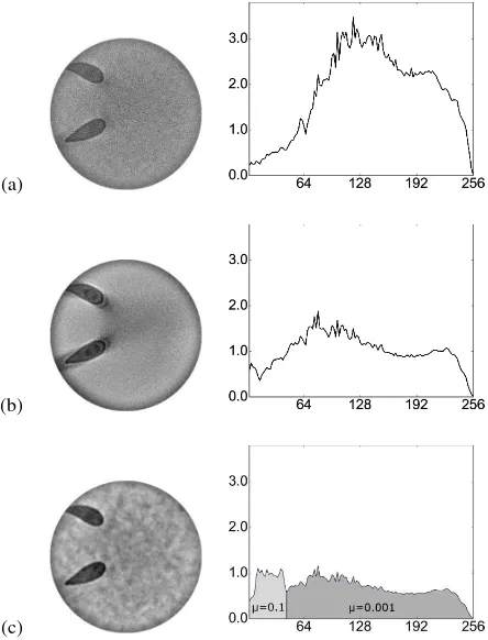
Figure 4 presents specific restoration results and graphs of the corresponding values of VFC for the section with the number of
3for different values of the parameterµfor a fixed number of iterationsK= 40of the method (9).
As demonstrated in Figure 4, the valueµ = 0.1(a) is too large for noisy images due to the high-frequency noise added to the restored image. Decrease of the parameter down to the value
0.01improves the quality of restoration insignificantly. The VFC graph still shows degradation of the restored image in the high-frequency area. Further reduction of the parameterµto the value
0.001makes it possible to significantly reduce the noise level, but leads to the sectioning of the layers being insufficient. This can also be seen on the VFC graph, where a dip in the low frequency area is noticeable, although the high-frequency graph demon-strates a sufficient quality of restoration.
4.3 Restoration results for the parameterµ depending on the harmonic number
(a)
(b)
(c)
Figure 4. Sectioning of the layer3of the image for the number of iterationsK= 40and the parameterµ= 0.1(a),µ= 0.01
(b) andµ= 0.001(c).
(a)
(b)
Figure 5. Sectioning the image layer no.3for the number of iterationsK= 40and the values of the parameterµ, depending
on the harmonic number.
was fulfilled with a larger value of the parameterµ, but with the same number of iterations.
Figure 5(a) shows the results of the reconstruction for the case of
K= 40iterations, whenµwas equal to0.1for harmonics
(u, v) :pu2+v2≤R/3, R= max (u,v)(u
2+v2),
and equal to0.001for the remaining harmonics. Despite good
sectioning, the restored image is noised by the low-frequency noise, which is confirmed by a jump in the corresponding fre-quency region of the VFC graph. By reducing the calculation area for low frequencies to(u, v) :√u2+v2 ≤R/6,a signif-icant improvement in the quality of the reconstructed image can be achieved. As we see from Figure 5(b), the reconstructed im-age is well sectioned from adjacent layers, and the VFC graph is close to unity throughout the region, including the high-frequency region.
Figure 6 compares the image restoration quality for cases the pa-rameterµ(a), (b) constant to all harmonics numbers and case (c) described above on Figure 5(b). The recovery result forµ, de-pending on the harmonic number, is characterized by VFC graph closed to unity, in contrast to the VFC graphs for constantµ, which have jumps and irregular changes in the mid and high fre-quencies. Moreover, for the case under consideration, it was pos-sible to achieve a good sectioning of the renewable layers from adjacent layers in combination with a sufficiently low level of in-put noise.
(a)
(b)
(c)
Figure 6. Comparison of restoration quality on various choices of the parameterµ: constant for all harmonics (µ= 0.1(a) and
µ= 0.001(b)) and depending on the harmonics (c).
The above results show that, by fine-tuning of the parameterµ
depending on the choice of the frequency domain, it is possible to achieve a significant improvement in the quality of the recon-struction of noisy images. If, in addition, a-priori information about the aberrations of spectral components of captured images is available then suboptimal filtration method (Sizikov, 1999) is applicable.
5. SOFTWARE IMPLEMENTATION
offi-cial release of BLAS (Basic Linear Algebra Subprograms) from Netlib for Windows within the LAPACK library.
OpenMP standard in C++ was used to parallel the implementa-tion of the iterative method for several points on selected number of processor cores. Maximum size of the RAM used by each par-allel process of the iteration method for some point(u, v)was
128×(2N2+ 3N)bytes, which allowed to increase the maxi-mum possible size of the processed data.
To measure the performance of the implemented algorithm, cal-culations for tasks with dimensions of256×256, 512×512,
1024×1024and2048×2048for20recoverable layers were per-formed on a personal computer with an Intel Core i7-4790K pro-cessor having4processor cores supporting Intel Hyper-Threading Technology,16GB of RAM under control of 64-bit operating system Windows 8.1.
With typical image sizes, as can be seen in Figure 7, the running time of the program changes little with the number of iterations and is acceptable for clinical practice when restoring medical im-age stacks.
Figure 7. Execution time forK= 10,40and100iterations.
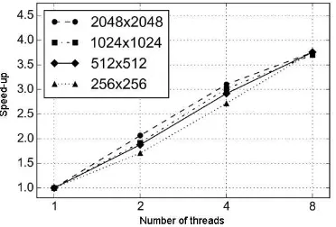
As we can see from the execution time graph shown in Figure 8 the program demonstrates good scalability for different sizes of input images on the 4-core CPU.
Figure 8. Performance acceleration graph for different image sizes depending on the number of threads.
Note that one of the important features of the implemented algo-rithm is its ability of effective parallelizing using the multi-core architecture of modern CPUs. Furthermore, it allows to transfer a part of computations to the GPU, that, taking into account the specifics of modern multi-core GPUs, will potentially lead to a significant reduction of runtime for both DFT and SLAE.
6. CONCLUSION
A local iterative method in Fourier spectral plane is considered for a typical statement of three-dimensional sectioning problem arising in ophthalmology. Compared to non-local parameter se-lection, the method demonstrates both improved sectioning re-sults and a good level of scalability when implemented on multi-core CPUs. Additional quality improvements of sectioning can be achieved while utilizing grid warping techniques that provides both denoising and sharpening of images (Krylov et al., 2016).
ACKNOWLEDGEMENTS
This work was supported by Russian Foundation for Basic Re-search grant 15-29-03896.
REFERENCES
Bakushinsky, A. and Goncharsky, A., 1989.Iterative methods for
solving ill-posed problems (In Russian). Nauka.
Castleman, K., 2007.Digital Image Processing. Pearson Educa-tion.
Easton Jr., R., 2010.Fourier Methods in Imaging. Wiley.
Gaskill, J., 1978.Linear systems, Fourier transforms, and optics. John Wiley & Sons.
Gu, M., 2000.Advanced Optical Imaging Theory, Springer series
in optical sciences. Springer.
Krylov, A., Nasonov, A., Razgulin, A. and Romanenko, T., 2016. A post-processing method for 3-d fundus image enhancement. In:
2016 IEEE 13th International Conference on Signal Processing
(ICSP), IEEE, pp. 49–52.
Larichev, A., Ivanov, P., Iroshnikov, N., Shmalhauzen, V. and Ot-ten, L., 2002. Adaptive system for eye-fundus imaging.Quantum
Electronics32, pp. 902–908.
Razgulin, A., Iroshnikov, N., Larichev, A., Pavlov, S. and Ro-manenko, T., 2015. On a problem of numerical sectioning in ophtalmology.Computer Optics39(5), pp. 777–786.
Sizikov, V., 1999. Analysis of local regularization methods and formulation of suboptimal filtration method for an equation of the first kind. Computational Mathematics and Mathematical
Physics39(5), pp. 686–701.
Wu, Q., Merchant, F. and Castleman, K., 2008. Microscope