Mouse Transgenic Model
Elisa Callegari,
1Bahaeldin K. Elamin,
1,2Ferdinando Giannone,
3Maddalena Milazzo,
3Giuseppe Altavilla,
4Francesca Fornari,
3Luciano Giacomelli,
4Lucilla D’Abundo,
1Manuela Ferracin,
1Cristian Bassi,
1Barbara Zagatti,
1Fabio Corr
a,
1Elena Miotto,
1Laura Lupini,
1Luigi Bolondi,
3Laura Gramantieri,
3Carlo M. Croce,
1,5Silvia Sabbioni,
1and Massimo Negrini
1MicroRNA-221 (miR-221) is one of the most frequently and consistently up-regulated
microRNAs (miRNAs) in human cancer. It has been hypothesized that miR-221 may act as
a tumor promoter. To demonstrate this, we developed a transgenic (TG) mouse model that
exhibits an inappropriate overexpression of miR-221 in the liver. Immunoblotting and
im-munostaining confirmed a concomitant down-regulation of miR-221 target proteins. This
TG model is characterized by the emergence of spontaneous nodular liver lesions in
approx-imately 50% of male mice and by a strong acceleration of tumor development in 100% of
mice treated with diethylnitrosamine. Similarly to human hepatocellular carcinoma, tumors
are characterized by a further increase in miR-221 expression and a concomitant inhibition
of its target protein-coding genes (i.e., cyclin-dependent kinase inhibitor [Cdkn]1b/p27,
Cdkn1c/p57, and B-cell lymphoma 2–modifying factor). To validate the tumor-promoting
effect of miR-221, we showed that
in vivo
delivery of anti-miR-221 oligonucleotides leads
to a significant reduction of the number and size of tumor nodules.
Conclusions
: This study
not only establishes that miR-221 can promote liver tumorigenicity, but it also establishes a
valuable animal model to perform preclinical investigations for the use of anti-miRNA
approaches aimed at liver cancer therapy.
(H
EPATOLOGY2012;00:000–000)
S
everal studies revealed that the expression of
microRNAs (miRNAs) is deregulated in human
hepatocellular carcinoma (HCC), in comparison
with non-neoplastic liver tissues, as reviewed recently.
1Among these, microRNA-221 (miR-221) emerged as
consistently up-regulated. In HCC, miR-221 is
up-regulated in approximately 70%-80% of cases.
2Its
up-regulation in glioblastoma, pancreatic, kidney,
bladder, colon, stomach, prostate, and thyroid cancer
strengthened its importance in tumorigenesis.
2-11The
hypothesized tumor-promoting activity was supported
by functional and molecular evidence. Forced
expres-sion of miR-221 in HCC cells could induce an
increase in growth, proliferation, migration, and
inva-sion capabilities
in vitro
.
2,10,12Conversely,
anti-miR-221 oligonucleotides could inhibit
in vitro
growth of
liver cancer cells.
13Importantly, the promotion of
tu-mor progression
in vivo
and the shortening of animal
survival was observed when miR-221 was introduced
into c-myc-immortalized P53
/liver progenitor cells,
which were implanted into irradiated nude mice.
13Surprisingly, the almost identical miR-222 miRNA,
which shares the same seed sequence of miR-221, did
not accelerate tumorigenesis in this model system.
At the molecular level, miR-221 was shown to affect
several cancer pathways by modulating multiple gene
targets, which included the cyclin-dependent kinase
inhibitors CDKN1B/p27
7,11and CDKN1C/p57,
2,10Abbreviations:a1-AT, alpha1 antitrypsin; AMOs, anti-miR oligonucleotides; Bcl2, B-cell lymphoma 2; BMF, Bcl2-modifying factor; CDKN, cyclin-dependent kinase inhibitor; PTEN, phosphatase and tensin homolog; DDIT4, DNA damage-inducible transcript 4; TIMP3, tissue inhibitor of metalloprotease 3; mTOR, mammalian target of rapamycin; DENA, diethylnitrosamine; EII, enhancer II; HBV, hepatitis B virus; HCC, hepatocellular carcinoma; IFN-c, interferon-gamma; IP, intraperitoneal; IV, intravenous; miR-221, microRNA-221; miRNA, microRNA; PCR, polymerase chain reaction; TG, transgenic; WT, wild type.
From the1Dipartimento di Medicina Sperimentale e Diagnostica, Universita di Ferrara, Ferrara, Italy;2Department of Microbiology, Faculty of Medical Laboratory Sciences, University of Khartoum, Khartoum, Sudan;3Centro di Ricerca Biomedica Applicata e Dipartimento di Medicina Interna, Policlinico S. Orsola-Malpighi e Universita di Bologna, Bologna, Italy;4Dipartimento di Scienze Medico Diagnostiche e Terapie Speciali, Universita di Padova, Padova, Italy; and5Department of Molecular Virology, Immunology and Medical Genetics, Ohio State University Medical Center, Columbus, OH.
Received November 11, 2011; accepted March 21, 2012.
This work was supported by fundings from the Associazione Italiana per la Ricerca sul Cancro and from the Italian Ministry of Research (to M.N.). E.C. was a recipient of a fellowship from the Fondazione Italiana per la Ricerca sul Cancro.
the pro-apoptotic protein B-cell lymphoma
2-modify-ing factor (BMF),
14the inhibitor of the
phosphoinosi-tide 3-kinase pathway phosphatase and tensin homolog
(PTEN),
12the DNA damage-inducible transcript 4
(DDIT4), a tumor suppressor that modulates kinase
activity of mammalian target of rapamycin (mTOR),
13the tissue inhibitor of metalloproteinase 3 (TIMP3).
12From a clinical point of view, it was shown that
higher levels of miR-221 in HCC correlated with
higher tumor stage and metastasis
15and were
associ-ated with multifocal tumors and a shorter time to
re-currence after surgical treatment.
14These pieces of evidence strongly suggested an
impor-tant role of miR-221 up-regulation in
hepatocarcinogene-sis. To prove the hypothesis and develop a more
conven-ient animal model, we produced a transgenic (TG) mouse
model that exhibits an inappropriate overexpression of
miR-221 in the liver. This TG model is characterized by
the appearance of spontaneous liver tumors in a fraction of
male mice and a strong acceleration of tumor development
in 100% of mice treated with diethylnitrosamine
(DENA). This model represents a valuable tool to perform
preclinical investigations on the use of miRNA or
anti-miRNA approaches for liver cancer therapy.
Materials and Methods
In
Vivo
Studies.
Animal experimentation was
approved by the institutional ethical committee. Mice
were maintained in a vented cabinet at 25
C with a
12-hour light-dark cycle and were provided food and water
ad libitum
. Ten-day newborn mice received one
intraper-itoneal (IP) injection of DENA (Sigma-Aldrich, St.
Louis, MO) (7.5 mg/kg body weight)
16-19and then
were sacrificed at various ages. All mice were subjected to
autopsy, and tissues were partly fixed in 10% formalin
and partly frozen in liquid nitrogen. Mice and livers
were weighed. The anti-miRNA oligonucleotide (AMO)
against miR-221 was: 5’-mG*mA*mA mAmCmC
mCmAmG
mCmAmG
mAmCmA
mAmUmG
mU*mA*mG* mC*mU-3’ (where ‘‘m’’ represents
2’-O-methyl RNA bases and asterisk [*] represents
phospho-thioate bond) and was obtained from Integrated DNA
Technologies (Leuven, Belgium). For
in vivo
evaluation
of miR-221 targeting, mice received a single intravenous
(IV) dose of 300
l
g (10 mg/kg) of anti-miR-221 diluted
in saline solution. All animals were sacrificed after 48
hours. Blood and livers were analyzed as described above.
For assessing antitumor activity of
in vivo
anti-miR
treat-ments, 10-day newborn mice received one IP injection of
DENA (7.5 mg/kg body weight), and after 2 months,
each mouse received a single IV dose of anti-miR-221 (10
mg/kg) diluted in saline solution every 15 days for a total
of three injections (approximately 1 mg total for each
mouse). Mice were sacrificed at 4 and 5 months of age.
Other reagents and methods are described in the
Supporting Materials.
Results
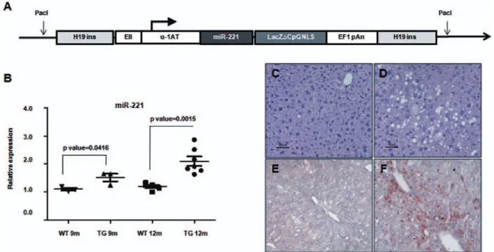
Development of a TG Mouse Model Carrying a
Liver-Deregulated miR-221.
A miR-221 expression
vector, based on the pWhere as vector backbone
(Invi-trogen, Carlsbad, CA), was developed. The pWhere
vector is characterized by the presence of two murine
H19 insulators, which protect the integrated
transcrip-tional unit from negative, as well as positive, influences
from adjacent sequences.
To specifically drive a liver-specific expression, the
pWhere vector was modified by inserting a regulatory
ele-ment that consisted of the liver-specific alpha1-antitrypsin
(
a
1-AT) promoter, coupled with the enhancer II (EII)
sequence of human hepatitis B virus (HBV). This chimeric
DNA element was previously shown to act as a potent,
steady promoter and was able to ensure a constant, high
level of gene expression in the liver.
20The tissue specificity
of this EII/
a
1-AT chimeric promoter, cloned upstream of
a luciferase reporter gene into a pGL3 plasmid, was tested
in different types of hepatic and nonhepatic cell lines,
which confirmed that the highest level of luciferase
expres-sion was detectable in hepatocytes, thereby confirming the
liver specificity of the promoter (data not shown).
A DNA segment, which included the mmu-mir-221
locus, was amplified from mouse genomic DNA and
was cloned into the pWhere/EII/
a
1-AT vector
down-stream of the EII/
a
1-AT promoter (Fig. 1A). Expression
of miR-221 from this vector was proven to be functional
in a liver-cancer–derived cell line (Supporting Fig. 1).
To generate a line of TG mice, the pWhere/EII/
a
1-AT/miR221 plasmid was linearized using the
Pac
I
Address reprint requests to: Massimo Negrini, Ph.D., Dipartimento di Medicina Sperimentale e Diagnostica, Universita di Ferrara, via Luigi Borsari 46, 44121 Ferrara, Italy. E-mail: [email protected]; fax: þ39-0532-455875 or Silvia Sabbioni, Ph.D., Dipartimento di Medicina Sperimentale e Diagnostica, Universita di Ferrara, via Luigi Borsari 46 44121, Ferrara, Italy. Email: [email protected]; fax: +39 0532 247618.
CopyrightVC2012 by the American Association for the Study of Liver Diseases.
View this article online at wileyonlinelibrary.com. DOI 10.1002/hep.25747
Potential conflict of interest: Nothing to report.
Additional Supporting Information may be found in the online version of this article.
restriction enzyme. The purified 9-kilobase fragment
containing the transgene was used to microinject
fertil-ized oocytes of a B6D2F2 mouse strain to complete their
development. After several crosses, a homozygous line of
TG mice overexpressing the miR-221 in the liver was
produced and used in all subsequent experiments.
Characterization of the Livers of miR-221 TG
Mice.
To assess miR-221 expression levels in the TG
model, livers taken from homozygous mice at different
ages were analyzed by real-time polymerase chain
reac-tion (PCR). In comparison with wild-type (WT) mice,
the analysis revealed a stable, increased expression of
miR-221 in the livers of TG animals, thereby
confirm-ing the development of homozygous TG mice
overex-pressing miR-221 in hepatic cells (Fig. 1B).
Macroscopically, livers of TG mice exhibited an
increase in volume and weigth in comparison with
controls (Supporting Fig. 2). Histologically, though
both groups displayed a conserved liver architecture,
TG livers were characterized by variable extents of
steatohepatitic changes, with hepatocyte degeneration
characterized by enlarged cells with large dysplastic
nuclei, lipidic vacuole, and focal coagulative necrosis
(Fig. 1C-F). These changes were more evident in older
TG animals and were absent among WT controls.
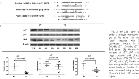
To assess whether miR-221 up-regulation could
affect the expression of its targets, we performed an
immunoblotting analysis to verify the expression of the
miR-221 target proteins, Cdkn1b/p27, Cdkn1c/p57,
and Bmf.
2,14In non-neoplastic liver tissue, we
con-firmed that Bmf and Cdkn1b/p27 were both
signifi-cantly down-regulated in TG mice. Cdkn1c/p57 was
also generally down-regulated, although it did not
reach statistical significance (Fig. 2). Immunostaining
for Cdkn1b/p27 confirmed the strong reduction of the
protein in TG animals (Supporting Fig. 3).
In addition to the above characteristics,
gene-expression profiling proved that the livers of TGs
dif-fered from WT also at a deeper molecular level
(Sup-porting Fig. 4; Sup(Sup-porting Table 1). Interaction
anal-ysis revealed that many of the identified
protein-coding genes were connected to the modulation of
the interferon-gamma (IFN-
c
) pathway (Supporting
Fig. 5).
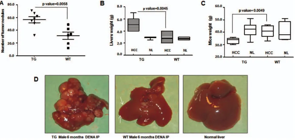
miR-221 Promotes Liver Tumorigenesis.
Because it
is well established that miR-221 is up-regulated in
human cancer, we analyzed whether the miR-221 TG
mouse model was predisposed to the development of
liver cancer. By monitoring mice at different ages (3,
6, 9, and 12 months), it emerged that a fraction of
males developed spontaneous tumors that became
visi-ble not earlier than 9 months of age. Four of eight
observed male mice (50%), at least 9 months old
(range, 9-12) showed evidence of small, but visible,
liver tumors. These tumors were characterized by a
further up-regulation of miR-221 (Supporting Fig. 6).
Females did not develop spontaneous tumors.
TG mice also exhibited an increased susceptibility to
treatment with the carcinogen, DENA. TG as well as
WT mice were injected IP with 7.5 mg/kg of DENA at
10 days of age. Animals were daily monitored and
peri-odically sacrificed at various ages. An increasing
development of tumors was observed at the different
time points in all mice, which was stronger in TG
ani-mals than in WT controls (Supporting Fig. 7). At 6
months, all male animals treated with DENA showed
evidence of multiple large tumors. TGs exhibited a
larger number of foci, which were also larger in size
than in WT control mice. Tumor burden caused a
sig-nificant increase in liver weight. Possibly because of the
presence of destructive liver tumors, TG mice exhibited
a more significant decrease in body weight than controls
(Fig. 3; Supporting Table 2). In females treated with
DENA, liver tumors were not visible at 6 months.
However, starting at 9 months of age, tumors began to
become visible in TG, but not in WT, control females
(Supporting Figs. 8 and 9).
In both miR-221 TG mice and controls, multifocal
liver nodules were detectable. Their size varied in
di-ameter from 1 mm to 1 cm. Small nodules displayed
the histopathological features of liver adenomas or
HCCs, whereas large nodules were HCC with either a
pseudoglandular or, more often, a trabecular pattern of
growth, with some clearly anaplastic HCCs
(Support-ing Fig. 10A). At 6 months of age, in DENA-treated
TG males, tumors almost completely substituted the
entire liver by confluent neoplastic nodules, which
were characterized by an infiltrative invasive front with
no demarcation from the surrounding liver
paren-chyma, presence of necrotic areas, marked angiogenesis
with slit-like sinusoids lined by endothelium, and
intravasation of tumor cells (Supporting Fig. 10).
Con-versely, DENA-treated control mice displayed tumor
nodules smaller in size and lower in number,
charac-terized by a better defined tumor margin, even though
a fibrous capsule was absent, together with less-evident
angiogenesis (Supporting Fig. 10). All tumors were
composed almost entirely of basophilic cells that were
more evident in zones of trabeculation of large tumors.
They were irregularly branched and were composed of
cells with a basophilic cytoplasm and central oval
nu-cleus with small nucleoli. Mitoses were rare in
adeno-mas, whereas they were more evident in HCCs.
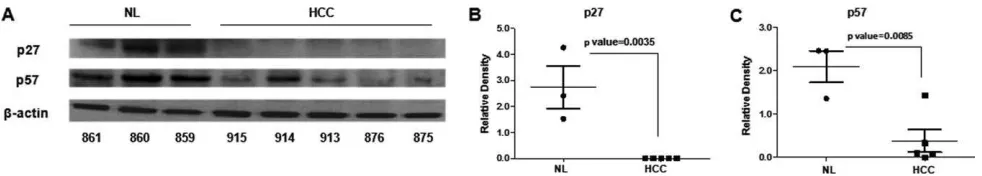
At the molecular level, tumors were characterized by
a further increase in miR-221 expression (Fig. 4).
Other miRNAs typically deregulated in human HCC
were analyzed: miR-21 was up-regulated, whereas
miR-122 and miR-199 were down-regulated, which
are results that mimic the human HCC condition.
The further increase in miR-221 expression was likely
responsible for the strong inhibition detected on its
targets, Cdkn1b/p27, Cdkn1c/p57, and Bmf (Fig. 5
and Supporting Fig. 11).
Anti-miR-221 Can Control
In Vivo
Tumorigenicity.
Previous studies in mice and primates had shown that
AMOs were able to silence miRNAs
in vivo
.
21,22To
sup-port the idea that the up-regulation of miR-221 was
im-portant for promoting and maintaining liver tumors as
well as investigating the potential antitumor activity of
anti-221, we sought to inhibit the endogenous
miR-221 through
in vivo
delivery of anti-miR-221 AMOs.
To assess the effects on miR-221 levels, first, a
group of 3 TG mice were IV injected through the tail
vein with a single dose of an antisense 2’-
O
-methyl
Fig. 2. miR-221
gene
targets
exhibit a reduced expression in
liv-ers
of
TG
mice.
(A)
miR-221
‘‘seed’’
matches
with
3’-UTR
sequences
present
in
mouse
Cdkn1b/p27,
Cdkn1c/p57,
and
Bmf genes. (B) Western blotting
analyses of p27, p57, and Bmf
target
proteins
and
b
-actin
in
healthy livers of TG (TG NL) and WT
(WT NL) mice. (C) Protein
expres-sion was quantified and normalized
versus levels of
b
-actin.
P
values
were calculated using a two-tailed
Student’s
t
test. UTR, untranslated
region.
oligoribonucleotide targeting miR-221 (10 mg/kg).
Forty-eight hours after injection, molecular analysis
revealed a significant down-regulation of miR-221
lev-els, both in liver and plasma of anti-miR-treated mice,
in comparison to untreated controls, thus revealing a
functional antisense inhibition of miR-221
in vivo
(Fig. 6A; Supporting Table 3). These effects were also
accompanied and supported by a concurrent increase in
Cdkn1b/p27 protein expression in the liver (Fig. 6B,C).
Then, to assess the effect of anti-miR-221
oligonu-cleotides on liver tumorigenicity in this TG mouse
model and establish whether miR-221 could represent
an antitumor therapeutic target, a group of 5 mice were
treated with anti-miR-221 AMOs (10 mg/kg at 60, 75,
and 90 days) after IP injection with DENA (at 10
days). Three mice were sacrificed at 120 days and 2 at
150 days of age. Significantly, a reduction in number
and size of tumors was observed in anti-miR-221-treated
mice, in comparison with same-age (4 or 5 months)
mice treated with DENA only (Fig. 7 and Supporting
Fig. 12). These antitumor effects were accompanied by
a persistent, significant decrease of miR-221 expression
in tumors arising in the group of AMO-treated mice.
Discussion
miR-221 is one of the most commonly up-regulated
miRNAs in human cancer, including HCC, and is
considered an ‘‘oncogenic’’ miRNA, as reviewed
recently.
1To date, the only model aimed at proving its
oncogenic role
in vivo
was based on the use of
c-myc-immortalized P53
/liver progenitor cells implanted
into irradiated nude mice. The introduction of
miR-221 into this model promoted tumor progression
in
vivo
and shortened animal survival.
13Because the
reproduction of this model is technically challenging
and difficult to compare with human HCC, we
addressed the issue of proving the
in vivo
tumor-pro-moting activity of miR-221 by the generation of a TG
mouse model that presents a stable increase of
miR-221 in the liver. By using this model, we were able to
provide a formal demonstration of miR-221
in vivo
tumor-promoting capability.
miR-221 TG animals exhibited a strong
predisposi-tion to the development of liver tumors. They
sponta-neously developed visible neoplastic lesions starting at
9 months of age, which were undetectable in WT
mice. If treated with DENA, TGs developed a
signifi-cantly higher number and larger tumor lesions that
became evident much earlier than in WT animals
treated with the same carcinogen.
better defined tumor margin, even if no capsule was
identifiable.
These tumors did not arise on a cirrhotic
back-ground, which is typical of most human HCCs.
However, the livers of TG mice exhibited high levels
of steatosis, a condition that in humans is frequently
observed in the context of metabolic dysfunctions
that predispose to HCC.
23,24Interestingly,
gene-expression profiling of non-neoplastic livers of TG
versus WT mice provided evidence that a different
molecular background driven by the aberrantly
expressed miR-221 existed and was likely responsible
for the differences in liver phenotypes, including the
predisposition to liver cancer. Many of the identified
protein-coding genes were connected to the
modula-tion of IFN-
c
, which was itself expressed at lower
levels in the livers of TG mice. Interestingly, a role of
defective IFN-
c
response was previously shown to be
connected to HCC. Indeed, IFN-
c
, through its
action on hepatocytes or immune cells, could elicit
tumor-suppressive effects by both inhibiting cell-cycle
progression and by initiating apoptosis in models of
HCC.
25-27Similar to human or other mouse models, the
pre-disposition was stronger in males, a result that
indi-cates a protective effect of estrogens and a stimulating
effect of androgen hormones in the development of
HCC, as previously shown.
28At the molecular level, these tumors revealed a
fur-ther increase of miR-221, which was accompanied by
a strong repression of the cell-cycle inhibitors,
Cdkn1b/p27 and Cdkn1c/p57, and the proapoptotic
Bmf proteins. In addition to miR-221, other miRNAs
known to play a key role in human HCC were found
to be dysregulated in the tumors arising in this model.
Among them, the down-regulated 122 and
miR-199 or the up-regulated miR-21 were dysregulated in
the same direction observed in human HCC. Overall,
these findings suggest that this TG mouse
overexpress-ing miR-221 represents a useful
in vivo
liver cancer
model for better understanding HCC and testing new
anticancer approaches.
To confirm the role of the tumor-driving force of
miR-221, we sought to inhibit its activity using an
AMO. It was previously established that silencing
miRNA activity
in vivo
using synthetic
oligoribonu-cleotides is feasible. Indeed, miR-122 inhibition by
AMO administration in mice and primates was shown
as a promising approach to reduce miRNA activity in
the
adult
liver.
21,22In
addition,
evidences
for
anti-miR-221 as a potential anticancer molecule were
provided through the use of intratumor injections of
AMOs targeting miR-221 in PC-3-derived tumors and
in melanoma cell xenotransplants.
29,30Here, we proved
that the use of AMO anti-miR-221 could be effectively
delivered to the liver, block miR-221, and induce a
Fig. 4. miRNA expression in liver nodules versus healthy liver of TG mice. (A-D) MicroRNA expression analysis in liver cancer versus healthy livers
revealed a statistically significant difference of expression levels: Similarly to human HCC, miR-221 and miR-21 exhibited an increased expression,
whereas miR-122 and miR-199 exhibited a down-regulation.
Fig. 5. Cdkn1b/p27 and Cdkn1c/p57 proteins are repressed in liver nodules versus healthy liver of TG mice. (A) Protein levels in tumor and
healthy tissues of TG mice were evaluated by western blotting analysis, revealing a strong reduction of miR-221 target genes in tumor tissues.
(B and C) Protein expression data were normalized versus levels of
b-actin.
significant inhibition of tumor growth. Indeed, the IV
injection of synthetic 2’-
O
-methyl modified
oligonu-cleotides targeting miR-221 in TG mice proved the
ability of these molecules to specifically silence miRNA
expression in the liver, as well as in the circulatory
system. Furthermore, in DENA-treated TG mice,
systemic administration of AMOs led to a significant
containment of liver tumor growth, in comparison to
Fig. 6. Expression of miR-221 and Cdkn1b/p27 in livers after
in vivo
delivery of anti-miR-221 oligonucleotides. A group of TG mice received
one IV injection of an anti-miR-221 synthetic oligonucleotide into their tail vein. (A) Forty-eight hours after injection, liver and blood of mice
were harvested to measure miR-221 expression levels. Quantitative PCR analysis revealed a significant decrease in miR-221 amounts in livers of
treated mice, in comparison to untreated ones. Supporting Table 3 indicates a similar reduction in serum. (B and C) Reduced levels of miR-221
correlated with an increase in Cdkn1b/p27 target protein levels. Protein expression data were normalized versus levels of
b
-tubulin.
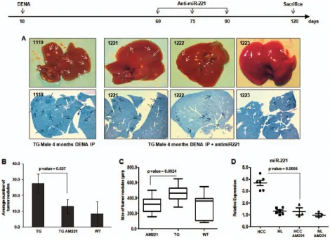
Fig. 7.
In vivo
delivery of AMOs limits tumor growth. A group of TG mice were IP injected with DENA at 10 days of age, and after 2 months,
control animals. This finding has two important
corol-laries: First, it confirms that miR-221 is indeed a
tumor driver for liver cancer, and, second, it
demon-strates that miR-221 can be effectively targeted to
reduce tumor growth. Significantly, this effect was
achieved without appreciable toxicity. For HCC, this
quality appears to be particularly important. In fact,
HCC conveys a very poor prognosis not only because
a small fraction of tumors can be curatively treated,
but also because systemic chemotherapy in advanced
HCC proved to be only marginally effective or too toxic.
In addition to AMOs, the use of miRNA-replacement
approaches was also reported to be effective as an
anti-cancer approach in animal models: miR-26a transduced
by an adeno-associated virus induced a significant
reduc-tion of tumors in a
myc
mouse model of HCC
31;
miR-101 was shown to inhibit tumor cells growth in a nude
mouse xenograft model
32; and miR-31 action could alter
the invasivity of disseminated tumor cells in an
ortho-topical cancer metastatic model.
33Hence, these studies
indicate that the use of miRNAs or anti-miRNAs are
promising approaches in cancer therapy and, possibly,
other noncancer diseases.
The present miR-221 TG animal model represents
an important tool not only for investigating liver
can-cer pathogenesis, but also for testing new miRNA or
anti-miRNA therapeutic approaches.
Acknowledgment:
The authors thank the Transgenic
and Gene Targeting Facility of the Kimmel Cancer
Center (Thomas Jefferson University, Philadelphia,
PA) for their expert production of several lines of
transgenic founders.
References
1. Negrini M, Gramantieri L, Sabbioni S, Croce CM. microRNA involve-ment in hepatocellular carcinoma. Anticancer Agents Med Chem 2011; 11:500-521.
2. Fornari F, Gramantieri L, Ferracin M, Veronese A, Sabbioni S, Calin GA, et al. MiR-221 controls CDKN1C/p57 and CDKN1B/p27 expression in human hepatocellular carcinoma. Oncogene 2008;27:5651-5661. 3. Ciafre SA, Galardi S, Mangiola A, Ferracin M, Liu CG, Sabatino G,
et al. Extensive modulation of a set of microRNAs in primary glioblas-toma. Biochem Biophys Res Commun 2005;334:1351-1358. 4. He H, Jazdzewski K, Li W, Liyanarachchi S, Nagy R, Volinia S, et al.
The role of microRNA genes in papillary thyroid carcinoma. Proc Natl Acad Sci U S A 2005;102:19075-19080.
5. Lee EJ, Gusev Y, Jiang J, Nuovo GJ, Lerner MR, Frankel WL, et al. Expression profiling identifies microRNA signature in pancreatic cancer. Int J Cancer 2007;120:1046-1054.
6. Pallante P, Visone R, Ferracin M, Ferraro A, Berlingieri MT, Troncone G, et al. MicroRNA deregulation in human thyroid papillary carcino-mas. Endocr Relat Cancer 2006;13:497-508.
7. Galardi S, Mercatelli N, Giorda E, Massalini S, Frajese GV, Ciafre SA, Farace MG. miR-221 and miR-222 expression affects the proliferation potential of human prostate carcinoma cell lines by targeting p27Kip1. J Biol Chem 2007;282:23716-23724.
8. Gottardo F, Liu CG, Ferracin M, Calin GA, Fassan M, Bassi P, et al. Micro-RNA profiling in kidney and bladder cancers. Urol Oncol 2007; 25:387-392.
9. Gramantieri L, Ferracin M, Fornari F, Veronese A, Sabbioni S, Liu CG, et al. Cyclin G1 is a target of miR-122a, a microRNA frequently down-regulated in human hepatocellular carcinoma. Cancer Res 2007;67: 6092-6099.
10. Medina R, Zaidi SK, Liu CG, Stein JL, van Wijnen AJ, Croce CM, Stein GS. MicroRNAs 221 and 222 bypass quiescence and compromise cell survival. Cancer Res 2008;68:2773-2780.
11. le Sage C, Nagel R, Egan DA, Schrier M, Mesman E, Mangiola A, et al. Regulation of the p27(Kip1) tumor suppressor by 221 and miR-222 promotes cancer cell proliferation. EMBO J 2007;26:3699-3708. 12. Garofalo M, Di Leva G, Romano G, Nuovo G, Suh SS, Ngankeu A,
et al. miR-221&222 regulate TRAIL resistance and enhance tumorige-nicity through PTEN and TIMP3 downregulation. Cancer Cell 2009; 16:498-509.
13. Pineau P, Volinia S, McJunkin K, Marchio A, Battiston C, Terris B, et al. miR-221 overexpression contributes to liver tumorigenesis. Proc Natl Acad Sci U S A 2010;107:264-269.
14. Gramantieri L, Fornari F, Ferracin M, Veronese A, Sabbioni S, Calin GA, et al. MicroRNA-221 targets Bmf in hepatocellular carcinoma and corre-lates with tumor multifocality. Clin Cancer Res 2009;15:5073-5081. 15. Fu X, Wang Q, Chen J, Huang X, Chen X, Cao L, et al. Clinical
sig-nificance of miR-221 and its inverse correlation with p27(Kip1) in he-patocellular carcinoma. Mol Biol Rep 2011;38:3029-3035.
16. Altavilla G, Trabanelli C, Merlin M, Caputo A, Lanfredi M, Barbanti-Brodano G, Corallini A. Morphological, histochemical, immunohisto-chemical, and ultrastructural characterization of tumors and dysplastic and non-neoplastic lesions arising in BK virus/tat transgenic mice. Am J Pathol 1999;154:1231-1244.
17. Koen H, Pugh TD, Goldfarb S. Centrilobular distribution of diethylni-trosamine-induced hepatocellular foci in the mouse. Lab Invest 1983; 49:78-81.
18. Koen H, Pugh TD, Goldfarb S. Hepatocarcinogenesis in the mouse. Com-bined morphologic-stereologic studies. Am J Pathol 1983;112:89-100. 19. Solt DB, Medline A, Farber E. Rapid emergence of carcinogen-induced
hyperplastic lesions in a new model for the sequential analysis of liver carcinogenesis. Am J Pathol 1977;88:595-618.
20. Kramer MG, Barajas M, Razquin N, Berraondo P, Rodrigo M, Wu C, et al.In vitroand in vivocomparative study of chimeric liver-specific promoters. Mol Ther 2003;7:375-385.
21. Elmen J, Lindow M, Schutz S, Lawrence M, Petri A, Obad S, et al. LNA-mediated microRNA silencing in non-human primates. Nature 2008;452:896-899.
22. Krutzfeldt J, Rajewsky N, Braich R, Rajeev KG, Tuschl T, Manoharan M, Stoffel M. Silencing of microRNAsin vivowith ’antagomirs’. Na-ture 2005;438:685-689.
23. Yatsuji S, Hashimoto E, Tobari M, Taniai M, Tokushige K, Shiratori K. Clinical features and outcomes of cirrhosis due to non-alcoholic steatohepatitis compared with cirrhosis caused by chronic hepatitis C. J Gastroenterol Hepatol 2009;24:248-254.
24. Ariz U, Mato JM, Lu SC, Martinez Chantar ML. Nonalcoholic steato-hepatitis, animal models, and biomarkers: what is new? Methods Mol Biol 2010;593:109-136.
25. Komita H, Homma S, Saotome H, Zeniya M, Ohno T, Toda G. Inter-feron-gamma produced by interleukin-12-activated tumor infiltrating CD8þT cells directly induces apoptosis of mouse hepatocellular carci-noma. J Hepatol 2006;45:662-672.
26. Detjen KM, Murphy D, Welzel M, Farwig K, Wiedenmann B, Rose-wicz S. Downregulation of p21(waf/cip-1) mediates apoptosis of human hepatocellular carcinoma cells in response to interferon-gamma. Exp Cell Res 2003;282:78-89.
27. Matsui M, Machida S, Itani-Yohda T, Akatsuka T. Downregulation of the proteasome subunits, transporter, and antigen presentation in hepa-tocellular carcinoma, and their restoration by interferon-gamma. J Gas-troenterol Hepatol 2002;17:897-907.
28. Nakatani T, Roy G, Fujimoto N, Asahara T, Ito A. Sex hormone de-pendency of diethylnitrosamine-induced liver tumors in mice and che-moprevention by leuprorelin. Jpn J Cancer Res 2001;92:249-256. 29. Mercatelli N, Coppola V, Bonci D, Miele F, Costantini A, Guadagnoli
M, et al. The inhibition of the highly expressed miR-221 and miR-222 impairs the growth of prostate carcinoma xenografts in mice. PLoS One 2008;3:e4029.
30. Felicetti F, Errico MC, Bottero L, Segnalini P, Stoppacciaro A, Biffoni M, et al. The promyelocytic leukemia zinc finger-microRNA-221/-222 pathway controls melanoma progression through multiple oncogenic mechanisms. Cancer Res 2008;68:2745-2754.
31. Kota J, Chivukula RR, O’Donnell KA, Wentzel EA, Montgomery CL, Hwang HW, et al. Therapeutic microRNA delivery suppresses tumorigenesis in a murine liver cancer model. Cell 2009;137: 1005-1017.
32. Su H, Yang JR, Xu T, Huang J, Xu L, Yuan Y, Zhuang SM. Micro-RNA-101, down-regulated in hepatocellular carcinoma, promotes apoptosis and suppresses tumorigenicity. Cancer Res 2009;69: 1135-1142.