Microphthalmia-associated transcription factor
mutations are
associated with white-spotted coat color in swamp buffalo
Y. Yusnizar*
†‡1, M. Wilbe
†§1, A. O. Herlino
†, C. Sumantri
¶, R. Rachman Noor
¶, A. Boediono**,
L. Andersson
†††and G. Andersson
†*Graduate School, Bogor Agricultural University (IPB), 16680 Bogor, Indonesia.†
Department of Animal Breeding and Genetics, Swedish University of Agricultural Sciences (SLU), SE-750 07 Uppsala, Sweden.‡
Research Center for Biotechnology, Indonesian Institute for Sciences (LIPI), 16912 Cibinong, Indonesia.§
Department of Immunology, Genetics and Pathology, Uppsala University, SE-751 08 Uppsala, Sweden. ¶
Department of Animal Productions and Technology, Faculty of Animal Sciences, Bogor Agricultural University (IPB), 16680 Bogor, Indonesia. **Department of Anatomy, Physiology and Pharmacology, Faculty of Veterinary Medicine, Bogor Agricultural University (IPB), 16680 Bogor, Indonesia.††
Department of Medical Biochemistry and Microbiology, Uppsala University, Uppsala, Sweden.
Summary A candidate gene analysis of themicrophthalmia-associated transcription factor(MITF) gene was used in an attempt to identify the genetic basis for a white-spotted coat color phenotype in the Asian swamp buffalo (Bubalus bubalis carabanensis). Ninety-three buffaloes—32 solid,
38 spotted and 23 white individuals—were Sanger-sequenced for allMITFexons as well as
highly conserved intronic and flanking regions.MITFcDNA representing skin and iris tissue from six spotted, nine solid and one white buffaloes was also Sanger-sequenced to confirm detected mutations. Two independent loss-of-function mutations, a premature stop codon (c.328C>T, p.Arg110*) and a donor splice-site mutation (c.840+2T>A, p.Glu281_ Leu282Ins8), both of which cause white-spotted coat color in swamp buffaloes, were identified. The nonsense mutation leads to a premature stop codon in exon 3, and likely removal of the resulting mRNA via nonsense-mediated decay pathway, whereas the donor splice-site mutation leads to aberrant splicing of exon 8 that encodes part of a highly conserved region ofMITF. The resulting insertion of eight amino acid residues is expected to perturb the leucine zipper part in the basic helix-loop-helix leucine zipper (bHLH-Zip) domain and will most likely influence dimerization and DNA binding capacity. Elec-trophoretic mobility shift assay was performed using mutant and wild-type MITF proteins and showed that the mutant MITF protein resulting from the splice-site mutation decreased
in vitroDNA binding capacity compared to wild-type MITF. White-spotted buffalo bulls are sacrificed in funeral ceremonies in Tana Toraja, Indonesia, because they are considered holy, and our results show that genetic variation causes a tie to the cultural use of these buffaloes.
Keywords Bubalus bubalis, donor splice-site mutation, MITF, nonsense mutation
Introduction
The domestic Asian water buffalo (Bubalus bubalis) is divided into two subspecies: river buffalo (Bubalus bubalis bubalis) and swamp buffalo or the Carabao (Bubalus bubalis caraba-nensis) (Castillo 2004). The swamp buffalo is an
econom-ically important domestic animal used for meat production and as a draft animal in Indonesia as well as in other parts of South-East Asia.
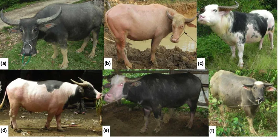
The swamp buffalo exhibits several coat color phenotypes and have either solid, white or spotted coats. The solid phenotype is characterized by dark gray coat color, whereas the white phenotype appears like a dilution phenotype rather than an extreme form of white spotting. White-spotted buffalo are found exclusively in a subpopulation that exists in the Tana Toraja region, South Sulawesi, Indonesia. Buffaloes with the spotted phenotype are classi-fied according to Toraja cultural traditions into four different subclasses based on the spotted phenotype and iris color (Fig. 1).
Address for correspondence
G. Andersson, Department of Animal Breeding and Genetics, Swedish University of Agricultural Sciences (SLU), SE-750 07 Uppsala, Sweden. E-mail: [email protected]
1These authors contributed equally to this work.
Accepted for publication 09 June 2015
676 ©2015 The Authors.Animal Geneticspublished by John Wiley & Sons Ltd
There is no clear information about when and why people started to keep white-spotted buffaloes, but reference is made to a cultural tradition calledAluk To Dolo(a rule of life that was brought to Tana Toraja by immigrants from Indochina in the years 7000 to 5000 before present). This tradition plays a key role in the funeral ceremonies, called ‘Rambu Solo’, and during these funeral ceremonies, male white-spotted buffaloes are sacrificed, according to Torajan tradition, thereby providing a ‘ride to heaven’ for the deceased. This tradition has continued even after 1920 when many Toraja people converted to Christianity.
The spotted phenotype implicates a defect in the migra-tion and survival of melanocytes given that pigmentamigra-tion is essentially normal in the pigmented areas. The spotted swamp buffaloes show altered pigmentation in both coat and iris (eye) color. This suggested a number of well-defined coat color genes,that is,MITF, SOX10, EDN3,EDNRBand
PAX3, affecting both coat and eye pigmentation as candi-date genes (Hugheset al.1994; Baldwinet al.1995; Edery
et al. 1996; Read & Newton 1997; Smith et al. 2000; Bondurandet al. 2007; Pingaultet al.2010). We hypoth-esized that, among these genes, the prime candidate gene was the gene encoding the microphthalmia-associated transcription factor (MITF) that has been shown to be associated with coat color, eye color and hearing disorder phenotypes, known as Waardenburg syndrome and Tietz syndrome in human (Tassabehji et al. 1994; Smith et al.
2000; reviewed in Pingaultet al.2010) and in several other mammals, for example, mice (Steingrimsson et al. 2003,
2004), dogs (Karlsson et al. 2007; Baranowska K€orberg
et al. 2014), horse (Hauswirth et al. 2012) and cattle (Philippet al.2011).
Materials and methods
DNA and RNA samples
Blood samples were collected from a total of 93 buffaloes, 32 solid, 38 spotted (four Lotong Boko, eight Toddi’, nine Saleko, 17 Bonga) and 23 white individuals. Phenotypic data also were collected at the same time. Genomic DNA from blood samples was prepared according to standard protocols.
Skin and iris tissue samples were collected from six spotted, nine solid and one white buffaloes. Samples were stored in RNAlater (Qiagen) at 4°C for 24 h and then kept at 80°C until RNA extraction was performed. Tissue samples were homogenized in b-mercaptoethanol-buffer RLT (containing guanidine thiocyanate) solution. Total RNA was extracted using a standard extraction protocol (RNeasy Mini Kit; Qiagen). Total RNA was stored in RNase-free water. The quality of total RNA was evaluated using the Agilent RNA 6000 Nano Kit (Agilent Technologies).
PCR amplification and DNA sequencing
DNA quality was measured using a NanodropTM
1000 spectrophotometer. A total of nine exons, highly conserved
(a) (b) (c)
(d) (e) (f)
intronic regions and flanking regions based on sequence conservation as defined by comparison with 29 mammals (Lindblad-Tohet al.2011) were amplified using PCR primer pairs (Table S1) designed based on theBos taurusgenome reference, NM_001001150.2 (UCSC Genome Browser assembly ID: October 2011, Baylor Btau_4.6.1/bosTau7). PCR amplifications were prepared in 25-ll reactions, which included 19 PCR buffer II, 2.5 mM of MgCl2, 0.2 mM of
dNTP, 0.06 U of AmpliTaqâ
Gold (Applied Biosystems), 0.5lM of forward and reverse primers and 10 ng of
genomic DNA. The PCR profile included an initial denatu-ration step at 94°C for 10 min, followed by 10 touchdown cycles (denaturation at 94°C for 30 s, annealing at 65°C for 30 s, followed by extension at 72°C for 30 s), 30 cycles of amplification (denaturation at 94°C for 30 s, annealing at 55°C for 30 s and extension at 72°C for 30 s) and an additional extension step of 7 min at 72°C. PCR products were purified with ExoCiAP mix containing alkaline phos-phatase, Exo1 buffer and Exo1 at 37°C for 1 h and 85°C for 15 min. The purified PCR products were sequenced using Sanger sequencing. Nucleotide sequences were aligned usingCODON CODE ALIGNER(Codon Code Corporation),
which was also used for SNP calling. Sequencing data for both genomic DNA and cDNA (see below) were deposited in the European Nucleotide Archive (ENA) (Study accession: HG917419–917433), available at www.ebi.ac.uk/ena and Table S2.
cDNA sequencing
Reverse-transcription PCR was performed to produce cDNA from total RNA samples. The RT-PCR protocol included 5 mMof MgCl2, 1 mMof dNTP, 19PCR Buffer II, 0.8 U of
RNase inhibitor, 1lMof RNA oligo (dT), 5 U of MiLV-RT
enzyme, RNase-free water and 2lg of RNA samples. The PCR profile included initiation at 25°C for 10 min, ampli-fication at 45°C for 80 min and extension at 15°C for 5 min. cDNA sequences were generated using Sanger sequencing, andCODON CODE ALIGNER was used for sequence
analysis. Primer pairs were designed based on the cattle (Bos taurus) genome assembly NM_001001150.2 (UCSC Genome Browser assembly ID: October 2011, Baylor Btau_4.6.1/bosTau7).
Transcription/translation system for PCR-generated DNA
PCR-generated DNA was translated into protein using the TnTâ
T7 Quick Kit (Promega). The TnTâ
quick master mix was thawed and other components were thawed at room temperature and thereafter stored on ice. The final volume of each reaction was 50ll and included 40ll of TnTâ
quick master mix, 1ll of 1 mMmethionine, 0.5lg of
PCR-generated DNA template, 1ll of T7 TnTâ
PCR enhancer
and nuclease-free water. The reaction was incubated at 30°C for 90 min and stored at80°C.
Electrophoretic mobility shift assay
Electrophoretic mobility shift assays (EMSAs) were performed using a LightShiftTM
Chemiluminescent EMSA Kit (Thermo Scientific). Probe and competitor DNAs used in EMSA included wild-type MITF (50-CCAACATGTGCACTC
CAC-30) and mutantMITF(50-CCAAACTGGTCACTCCAC-30).
The wild-type MITF probe was labeled with biotin using a Biotin 30End DNA Labeling Kit (Thermo Scientific). EMSAs
were performed by incubating wild-type and mutant MITF proteins in binding buffer (10 mM of Tris-HCl, pH 7.5, 50 mM of NaCl, 1 mM of dithiothreitol, 50 ng/ll of poly (dIdC), 1 mMof EDTA, 5% glycerol, 5 mMof MgCl2, 50 mM
of KCl and 4 pmol of unlabeled MITF DNA) on ice for 20 min. Thereafter, 20 fmol Biotin End-Labeled MITF DNA was added to each reaction with a total volume of 20ll, and the reactions were incubated at room temperature for 20 min. Then, 5ll of 59loading buffer was added to each 20-ll binding reaction. MITF proteins were separated from DNA by electrophoresis through 5% Criterion TBE gel from Bio-Rad at 120 V for 70 min and then transferred to a nylon membrane at 100 V for 60 min. Transferred DNA was cross-linked for 15 min with the membrane face down on a trans-illuminator equipped with 312-nm bulbs. The membrane was exposed using a CCD camera.
Statistical analysis
Statistical analyses were performed using the statistical program VASSARSTATS (http://faculty.vassar.edu/lowry/
VassarStats.html). A 292 table of allele frequencies was used for Fisher’s exact test.
Results
position chr22: 32 297 683 (NM_001001150.2) in intron 8 (Fig. 3).
Twenty-three of 38 spotted individuals (60.5%) were heterozygous for the nonsense variant, whereas all solid and all white individuals were homozygous for the wild-type allele. The nonsense variant showed a highly significant association with white spotting (P=1.39107; Table 1).
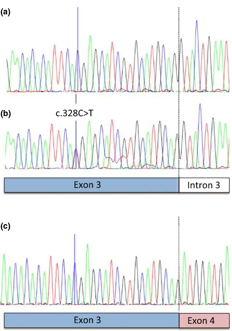
To confirm that MITF mRNAs were expressed from the mutant alleles detected in genomic DNA, MITF cDNAs derived from mRNA that was isolated from skin and irises of six different spotted individuals were analyzed (Table 1). RT-PCR analysis was performed on cDNA made from these mRNA preparations. In mRNA prepared from skin obtained from Saleko type spotted buffaloes carrying the nonsense variant (Fig. 1c), mRNA molecules containing the nonsense variant were undetectable, suggesting that this mutant transcript is degraded by the nonsense-mediated decay pathway (Maquat 2004).
Seven of 38 spotted individuals (18.4%) and one of the 32 solid buffaloes (3.1%) were heterozygous for the splice-site variant (P=0.05, Table 1). The presence of mRNA expressed in spotted individuals carrying the splice-site variant allele was confirmed by RT-PCR analysis performed on mRNA prepared from skin obtained from Bonga type individuals (Fig. 1e). The capacity of the mutant MITF protein produced by aberrant splicing to bindin vitroto the well-established MITF target site (50-CACGTG-30) was
evaluated by EMSAs using in vitrotranslated mutant and wild-type MITF protein. The mutant protein showed a lower efficiency in binding to the MITF binding site in vitro
(Fig. 4). Schematic views of the wild-type and mutant MITF proteins are shown in Fig. S1.
Discussion
Here we have identified two MITF mutations associated with the white-spotted coat color phenotype in swamp buffaloes. As expected for loss-of-function mutations in an essential gene, either one of the two mutations was found only in the heterozygous form.MITF mRNA carrying the nonsense mutation was undetectable in our mRNA prepa-rations from iris and skin, most likely because such mRNAs are degraded by the nonsense-mediated mRNA decay pathway. The mRNA containing the splice-site variant was detected at similar levels as the wild-typeMITFmRNA. There was considerable phenotypic variability in spotted coat color among the 23 animals that were heterozygous for the nonsense variant. Some individuals showed massive spotted color, whereas others had only a very small white spot on the forehead. This phenotypic variation is likely caused by genetic variation at loci interacting withMITF.
There are many examples of MITFmutations associated with hypopigmentation and white-spotted phenotypes in different mammalian species. In German Fleckvieh cattle, a dominant and recessive lethal p. Arg210Ile missense muta-tion has been identified as responsible for hypopigmentamuta-tion, heterochromia iridis, colobomatous eyes and bilateral hear-ing loss (Philipp et al. 2011). Three variant mutations, a proximal melanocyte-specific M promoter replacing a thy-mine with 11 nucleotides, a small deletion in exon 5 and ade novo missense mutation in exon 6, were detected causing splashed white and macchiato in horses (Hauswirth et al.
2012). In white-spotted dogs, genome-wide association studies identified a region overlapping the melanocyte-specificMITF-M promoter to be associated with white coat color (Karlssonet al. 2007). Subsequent functional studies revealed that regulatory mutations located in the MITF-M promoter influenced transcription where a simple repeat polymorphism was shown to be a key factor underlying white spotting (Baranowska Korberg€ et al.2014).
The absence of MITF mutations in the white buffalo confirms our hypothesis that these do not represent an extreme form of white spotting. Importantly, among the
(a)
(b)
(c)
spotted individuals, none of them were homozygous for the mutations. According to Mendelian segregation, 25% of calves produced from matings between two spotted buffaloes will be either homozygous recessive or composite
heterozygotes that may lead to embryonic lethality, at least for animals that are homozygous for the nonsense mutation.
The mutations detected using genomic DNA was con-firmed by sequencing cDNA derived from mRNA that was isolated from skin and iris of six different spotted individuals. The splice-site mutation leads to aberrant splicing of exon 8, and the resulting protein translated from such aberrantly spliced mRNA is expected to disturb the leucine zipper part in the basic helix-loop-helix zipper (bHLH-Zip) domain of MITF and will most likely influence dimerization and DNA binding capacity. MITF belongs to the bHLH-Zip transcrip-tion factor family and the bHLH-Zip domain is highly conserved among both vertebrates and invertebrates (Steingrimsson et al. 2004). There was a clear trend that individuals that carry the splice-site variant show a less pronounced white-spotting phenotype compared with most of the animals carrying the nonsense variant, suggesting that this is a partial loss-of-function allele,
(a)
(b)
(c)
(d)
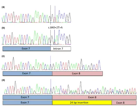
Figure 3 Sanger sequencing of gDNA and cDNA in solid and spotted buffaloes revealed a heterozygous donor splice-site mutation in
intron 7 inMITFresulting in 24-bp insertion (a) Wild-type gDNA sequence in solid buffaloes. (b) A donor splice-site mutation (NM_001001150.2) c.840+2T>A, p.Glu281_Leu282Ins8 at position chr22: 32 297 683 in intron 7 was identified in spotted buffaloes. Exon–intron borders in exon 7 are shown. (c) Wild-type cDNA sequence including exon borders of exons 7 and 8. (d) The heterozygous splice-site mutation resulted in one wild-type cDNA allele and one cDNA allele containing an insertion of 24 extra bp (eight amino acids).
Table 1 Genotype frequencies of theMITFnonsense variant c.328C>T, p.Arg110*(chr22: 32 322 2421) and the splice-site variant
c.840+2T>A, p.Glu281_Leu282Ins8 (chr22: 32 297 6831) in swamp buffaloes with different coat color phenotypes.
Phenotype
Nonsense variant Splice-site variant
CC CT TT P2 TT AT AA P2
Solid 32 0 0 31 1 0
Spotted 15 23 0 1.39107 31 7 0 0.05
White 23 0 0 23 0 0
1Nucleotide positions are given according to the cattle reference
genome assembly NM_001001150.2 (UCSC Genome Browser assem-bly ID: October 2011, Baylor Btau_4.6.1/bosTau7).
2Fisher’s exact test (one-tailed) comparing the allele frequencies in solid
whereas the nonsense mutation results in a null allele. The observation of a solid colored buffalo carrying the splice-site variant suggests either that this is due to a phenotype misclassification or that this mutation is not fully penetrant.
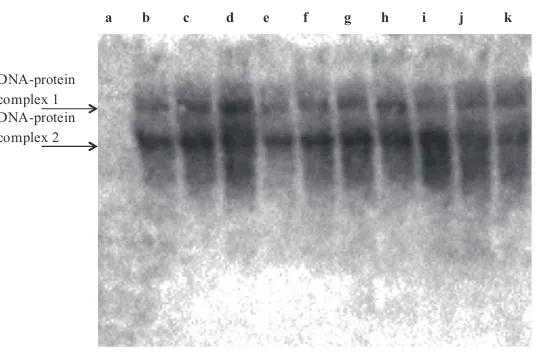
To assess thein vitroDNA–protein interaction effect of the mutant MITF protein, we performed EMSAs using MITF-specific consensus oligonucleotides and in vitro translated mutant and wild-type MITF protein. Two specific complexes were detected, most likely representing dimers and multi-mers. The mutant protein was shown to bind with lower efficiency compared to wild-type protein (Fig. 4, lanes b vs. e; c vs. g). Thus, the mutant MITF protein maintained its
in vitrobinding profile but with decreased binding efficiency. Increasing amounts of in vitro translated mutant MITF protein was added toin vitrotranslated wild-type MITF to evaluate whether mutant MITF could interfere with the binding activity of wild-type MITF, but we could not ascertain that mutant MITF had any negative effect on wild-type MITF target interaction. Most likely, the insertion of the eight amino acid residues negatively influences dimerization capacity because the insertion is located immediately before the third leucine in the leucine zipper domain (Fig. S1). Dimerization of MITF is required for its ability to bind DNA (Steingrimssonet al.2004). In mice, the DNA binding domain of Mitf (Mitfmi, Mitfmi-wh, Mitfmi-b, Mitfmi-H, Mitfmi-enu5, Mitfmi-bc2and Mitfmi-ew) was affected by semi-dominant mutations (Steingrimssonet al.2004).
Moreover, eight of 38 spotted individuals did not carry any of the twoMITFmutations that were identified in our study (Table 1) and none were composite heterozygous for the two mutations as expected for loss-of-function mutations. Thus, there must be at least one additional mutation affecting white spotting in this population of swamp buffaloes.
In conclusion, we have identified two independentMITF
mutations responsible for white-spotting phenotypes in swamp buffaloes. The fact that at least two mutations are causing white spotting in swamp buffaloes is consistent with the huge cultural importance these animals have had.
Strong positive selection over a considerable time period has led to the accumulation of multiple mutations causing a white-spotting phenotype. This is an example of the cultural importance of white color in domesticated animals. Another famous example is white horses caused by the Greying-with-age inSyntaxin 17, which have had a huge impact on human culture worldwide (Rosengren Pielberg
et al.2008).
Ethics statement
The Swedish Board of Agriculture has given the permission to import (Dnr 6.2.18-2166/13) and use biological samples from Bubalus bubalis and to use the animal byproducts for research purposes (Dnr 38-10019/12; Dnr 38-9492/12). All animal work has been conducted according to the national and international guidelines for animal welfare. Veterinary division in North Toraja, South Sulawesi, Indonesia, has approved this study (Permit number 553/ 10/P3/01-10).
Acknowledgements
We wish to acknowledge the head and staff of Animal Division of North Toraja Government for supporting this study and collecting samples. Finally, we wish to thank all of the farmers who provided the buffalo samples for this study.
Competing interests
A patent application has been filed and is pending.
References
Baldwin C.T., Hoth C.F., Macina R.A. & Milunsky A. (1995) Mutations inPAX3 that cause Waardenburg syndrome type I: ten new mutations and review of the literature.American Journal of Medical Genetics58, 115–22.
DNA-protein complex 1 DNA-protein complex 2
a b c d e f g h i j k
Baranowska K€orberg I., Sundstr€om E., Meadows J.R.S.et al.(2014) A simple repeat polymorphism in theMITF-M promoter is a key regulator of white spotting in dogs.PLoS One9, e104363. Bondurand N., Dastot-Le Moal F., Stanchina L. et al. (2007)
Deletions at theSOX10gene locus gene Waardenburg syndrome types 2 and 4.American Journal of Human Genetics81, 1169–85. Castillo L. (2004) New scientific name of the domesticated swamp buffalo, the carabao–Bubalus bubalis carabanensis. In:Proceedings of the 7th World Buffalo Congress, Makati City, Philippines. Edery P., Attie T., Amiel J., Pelet A., Eng C., Hofstra R.M.W.,
Martelli H., Bidaud C., Munnich A. & Lyonnet S. (1996) Mutation of theendothelin-3gene in the Waardenburg-Hirsch-sprung disease (Shah-Waardenburg syndrome).Nature Genetics
12, 442–4.
Hauswirth R., Haase B., Blatter M.et al.(2012) Mutations inMITF
and PAX3 cause “splashed white” and other white spotting phenotypes in horses.PLoS Genetics8, e1002653.
Hughes A.E., Newton V.E., Liu X.Z. & Read A.P. (1994) A gene for Waardenburg syndrome type 2 maps close to the human homologue of the microphthalmia gene at chromosome 3p12-p14.1.Nature Genetics7, 509–12.
Karlsson E.K., Baranowska I., Wade C.M. et al. (2007) Efficient mapping of Mendelian traits in dogs through genome-wide association.Nature Genetics39, 1321–8.
Lindblad-Toh K., Garber M., Zuk O.et al., (2011) A high-resolution map of human evolutionary constraint using 29 mammals.
Nature,478, 476–82.
Maquat L.E. (2004) Nonsense-mediated mRNA decay: splicing, translation and mRNP dynamics.Nature Reviews Molecular Cell Biology5, 89–99.
Philipp U., Lupp B., Momke S., Stein V., Tipold A., Eule J.C., Rehage J. & Distl O. (2011) AMITFmutation associated with a dominant white phenotype and bilateral deafness in German Fleckvieh cattle.PLoS One6, e28857.
Pingault V., Ente D., Dastot-Le Moal F., Goossens M., Marlin S. & Bondurand N. (2010) Review and update of mutations causing Waardenburg syndrome.Human Mutation31, 1–16.
Read A.P. & Newton V.E. (1997) Waardenburg syndrome.Journal of Medical Genetics34, 656–65.
Rosengren Pielberg G., Golovko A., Sundstr€om E.et al.(2008) A cis-acting regulatory mutation causes premature hair graying and susceptibility to melanoma in the horse.Nature Genetics40, 1004–9.
Smith S.D., Kelley P.M., Kenyon J.B. & Hoover D. (2000) Tietz syndrome (hypopigmentation/deafness) caused by mutation of MITF.Journal of Medical Genetics37, 446–8.
Steingrimsson E., Arnheiter H., Hallsson J.H., Lamoreux M.L., Copeland N.G. & Jenkins N.A. (2003) Interallelic complementa-tion at the mouseMitflocus.Genetics163, 267–76.
Steingrimsson E., Copeland N.G. & Jenkins N.A. (2004) Melanocytes and the microphthalmia transcription factor net-work.Annual Review of Genetics38, 365–411.
Tassabehji M., Newton V.E. & Read A.P. (1994) Waardenburg syndrome type 2 caused by mutations in the human microph-thalmia (MITF) gene.Nature Genetics8, 251–5.
Supporting information
Additional supporting information may be found in the online version of this article.
Figure S1 Schematic representation of MITF exons. The estimated positions of the encoded helix-loop-helix DNA binding domain (HLH DBD) and the leucine zipper part in the basic helix-loop-helix zipper (bHLH-Zip) domain in the wild-type individual. The splice-site mutation is expected to disturb the leucine zipper part in the bHLH-Zip domain because of the eight additional amino acid residues in exon 8.
Table S1Primers used to amplify gDNA.