Plasma iron, zinc, and copper levels in heart
disease patients in Dr. Sardjito Hospital
Yogyakarta
Pramudji Hastuti1*, Lukman Endro2, HAH Asdie3, Tri Hartati3, ASM Sofro4
1Department of Biochemistry, 2Medical student, 3Department of Internal Medicine, Faculty of
Medicine, Gadjah Mada University, Yogyakarta,4Faculty of Medicine, YARSI University, Jakarta,
Indonesia
ABSTRACT
Heart disease is a major cause of death in both men and women in industrialized countries. In fact, many individuals, who develop heart disease, have normal cholesterol and blood pressure levels. It suggests that other risk factors may also play a role. Iron (Fe), zinc (Zn) and copper (Cu) may promote CHD by increasing lipid peroxidation and causing oxidant-induced damage in various organs. These minerals are essential as cofactors for functional enzymes in the body. The hypothesis that Fe and Cu depletion protects against ischemic heart disease has generated significant debate, but this hypothesis cannot be rejected until stronger evidence that high Fe stores increase the incidence of CHD or death from myocardial infarction is proven. This research was aimed to find out the levels of Fe, Cu, and Zn plasma as risk factors for heart disease patients, compared with those in hyperlipidemic patients and controls. In this study, we examined the Fe, Zn and Cu levels in plasma of CHD patients, hyperlipidemic patients, and healthy controls by atomic absorption spectrophotometer. Thirty five CHD patients, 31 hyperlipidemic patients, and 26 healthy controls. Age, body weight, body height, BMI, systolic and diastolic blood pressure of each group were not significantly different (p > 0.05). There were higher cholesterol and lower HDL-C levels in CHD patients compared with those in hyperlipidemic patients and controls. There were significantly higher triglyceridemia level in hyperlipidemic patients compared to those in CHD patients and controls. Iron level in CHD patients were not significantly different than those in hyperlipidemic patients and controls. Zinc level in CHD patients were not significantly different compared with those in controls, but in hyperlipidemic patients, they were significantly lower than those in CHD patients and controls. Copper level in CHD and hyperlipidemic patients were significantly lower than those in controls. In this research, it could be concluded that the decrease in Cu level might be one of the risk factors of CHD.
Keywords: iron-zinc-copper-heart disease-risk factor
INTRODUCTION
Until now, studies on iron (Fe) and copper (Cu) as risk factors of CHD is still controversial.1-3
Hypothesis suggested in 1981 and known as “iron hypothesis” that the decrease in Fe level might prevent ischemic heart disease had generated debates. Studies were continued to prove the hypothesis. It was reported that there was a relationship between serum ferritin level and the risk of heart attack.4Abnormal Fe stores might caused
damage in various organs induced by oxidants. Iron stores in the heart were involved in the development of heart fibrosis. In CHD, there was an increase in Fe level in intimal lesion, compared with healthy control.3,4
Zinc and Cu are essential minerals in the human body. More than 100 types of enzymes need Zn as cofactors for metabolic pathways important for growth and development. In addition, there was also evidence that Zn and Cu were also involved in the pathogenesis of CHD. Low Zn level was assumed
to cause atherosclerosis.5,6 Patients with dilated
cardiomyopathy had higher level of serum Cu and lower of serum Zn than healthy controls.7
Copper is the third most essential mineral after Fe and Zn in human body.5 Free Cu can not be
found in considerable amount in the body, and almost all are bound by transport proteins, storage proteins, and Cu-containing enzymes. Copper is important for various functions of enzymes, including cytochrome oxidase, superoxide dysmutase (SOD) functioning as antioxidants, tyrosinase which active in pigmentation, dopamine hydroxylase producing cathecolamines, lysyl-oxidase that forms collagen and elastin, coagulation factor V, and ceruloplasmin functioning as oxidant, and participate in Fe metabolism and Cu transport. Outside the body, Cu is known as pro-oxidants and is often used to oxidize LDL. Ceruloplasmin protein which contained Cu had been found to stimulate oxidized-LDL in test tube, causing several experts assumed that the increase in Cu level would increase the risk of atherosclerosis by increasing the oxidation of LDL, but the available evidences were limited. In addition, SOD and ceruloplasmin were known to have antioxidant activities, causing several experts assumed that Cu deficiency would increase the risk of CHD, instead of the Cu excess.8,9 In the body,
Cu was changed into cupro- and cupri- forms. The ability of Cu to give electrons explained its important role in redox reaction and in catching free radicals.5,10
Lack of Cu intake caused abnormality in the formation of blood vessel wall and caused pathological changes, causing CHD. It was also shown that Cu/Zn ratio had an inverse correlation with blood pressure.11
The aim of the study was to find out the plasma levels of Fe, Cu, and Zn plasma of CHD patients, compared with those in hyperlipidemic patients and healthy controls.
MATERIALS AND METHODS
Subjects
Subjects of the study were CHD patients from Dr. Sardjito Hospital Yogjakarta who were diagnosed by internists, and samples were taken by consecutive random sampling. Controls were hyperlipidemic patients who visited Dr. Sardjito
Hospital Yogjakarta and did not suffer from CHD. Hyperlipidemic patients were those who had blood lipid levels outside the normal range. Normal controls were elderlies who joined elderly sport group in Banteng village, Sinduadi, Sleman, Yogjakarta, who had blood lipid levels in the normal range. A detailed explanation of the procedures was given to all subjects and written informed consents were obtained from all of them. The protocol of this study has been approved by the Health Research Ethics Committee of Faculty of Medicine, Gadjah Mada University, Yogyakarta.
Chemical analysis
Ten mL blood sample was taken from each subject after fasting, and put into mineral-free test tubes containing anticoagulant heparin. Iron, Cu, and Zn plasma levels were measured with atomic absoprtion spectrophotometer (AAS). Body weight, body height, systolic and diastolic blood pressure were measured. Total cholesterol, triglyceride, LDL-C, and HDL-C levels were measured with enzymatic method (CHOD-PAP) with Dyasis kit. Patient was grouped as dyslipidemic if his/her HDL-C level was less than 40 mg%, LDL-C level was more than 160 mg%, tryglyceride level was more than 160 mg%, and total cholesterol level was more than 200 mg%, and control was subject who had normal lipid levels.
Data analysis
The results from were analyzed with analysis of variance and t-test. Result was considered significant if p < 0.05.
RESULTS
TABLE 1. Average age, body weight, body height, BMI, blood pressure and lipid profiles of CHD patients, hyperlipidemic patients, and controls
CHD patients
Body weight (kg) 63.00 ±9.00 62.42 ± 11.07 62.56 ± 9.30 >0,05
Body height (m) 162.3 ± 5.0 157.2 ± 37.6 161.4± 5.3 >0,05
BMI 23.8 ± 3.0 25.2 ± 3.7 24.0 ± 3.35 >0,05
Systolic BP (mmHg) 140.61 ± 17.13 138.06 ± 19.95 137.31 ± 19.56 >0,05
Diastolic BP (mmHg) 83.94 ± 10.29 87.10 ± 9.57 89.04 ± 12.96 >0,05
HDL-C (mg/dL) 45.142 ± 14.684 48.816 ± 10.014 50.833 ± 7.429 >0,05
LDL-C (mg/dL) 126.98 ± 38.272 143.39 ± 35.025 97.430 ± 25.010 >0,05
Cholesterol (mg/dL) 199.740 ± 32.974 154.490 ± 66.470 154.710 ± 37.580 <0,05 Triglyceride (mg/dL) 132.900 ± 71.069 239.010 ± 48.816 117.560 ± 51.476 <0,05
FIGURE 1. Age, body weight, body height, BMI, and blood pressure of CHD patients, hyperlipidemic patients, and
controls
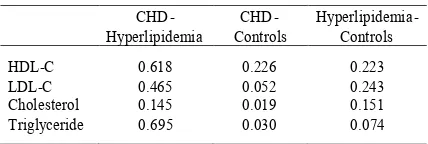
TABLE 2. Significance of lipid profiles between CHD patients, hyperlipidemic patients, and controls
CHD
-HDL-C 0.618 0.226 0.223
LDL-C 0.465 0.052 0.243
Cholesterol 0.145 0.019 0.151
Triglyceride 0.695 0.030 0.074
Data in TABLE 1 shown that age, body weight, body height, BMI, and blood pressure of CHD patients, hyperlipidemic patients, and controls were not significantly different (p > 0.05). The lowest average of HDL-C level was found in CHD patients and was not significantly different with hyperlipidemic patient and controls (p > 0.05). The highest average of LDL-C level was found in
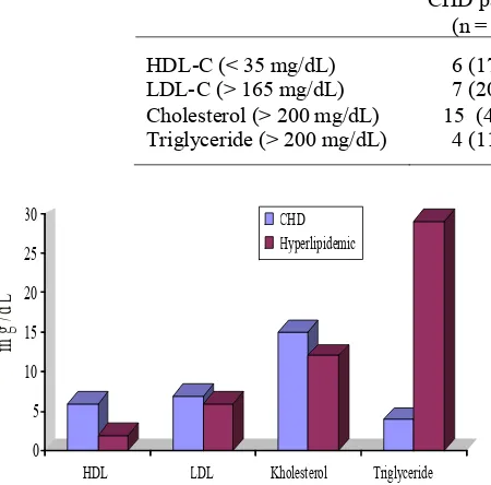
hyper-FIGURE 2. HDL-C, LDL-C, cholesterol, and triglyceride levels of CHD patients, hyperlipidemic patients, and controls
lipidemic patients but not significantly different with CHD patients and controls (p>0.05). Cholesterol levels of CHD patients were significantly higher than those in controls (p < 0.05), but there were not significantly different between CHD patients and hyperlipidemic patients also between hyperlipidemic patients and controls (p > 0.05). Triglyceride levels in hyperlipidemic patients were not significantly different with those in CHD patients and controls (p> 0.05), but there were significantly different between CHD patients and controls (p< 0.05).
The total number of CHD and hyperlipidemic patients who had dyslipidemia are shown in FIGURE 3, while the significance and Odd Ratio of blood lipoprotein abnormality in CHD patients and hyperlipidemic patients are shown in TABLE 3.
0
TABLE 3. Significance and Odd Ratio of blood lipoprotein abnormality in CHD patients and hyperlipidemic patients
CHD patients (n = 35)
Hyperlipidemic
patients (n= 31) p OR
HDL-C (< 35 mg/dL) 6 (17%) 2 (6%) 0.172 3.00
LDL-C (> 165 mg/dL) 7 (20%) 6 (19%) 0.948 1.04
Cholesterol (> 200 mg/dL) 15 (43%) 12 (39%) 0.734 1.19
Triglyceride (> 200 mg/dL) 4 (11%) 29 (94%) 0.000 0.01
TABLE 3 showed that there were 43% CHD patients had hypercholesterolemia, while in hyperlipidemic patients, there were 94% had high level of triglyceride in blood. Odd ratio showed that patients with low HDL-C level would have 3 times increased risk of CHD compared with patients with normal HDL-C level. High concentrations of LDL-C and cholesterol in LDL-CHD patients were not different and not risk factors for CHD. Triglyceride level in FIGURE 3 : The total number of CHD and hyperlipidemic
patients who had dyslipidemia
hyperlipidemic patients was high and significantly different with CHD and this high level was not risk factor for CHD.
The Fe, Zn, and Cu levels of CHD patients, hyperlipidemic patients, and controls are shown in FIGURE 4 and TABLE 4, while the significance of Fe, Zn, and Cu levels in CHD patients, hyper-lipidemic patients, and controls are shown in TABLE 5.
FIGURE 4. Iron, Zn, and Cu levels of CHD patients, hyperlipidemic patients, and controls
TABLE 4. Iron, Zn, and Cu levels of CHD patients, hyperlipidemic patients, and controls
CHD patients Hyperlipidemic patients Controls
Iron (Fe)μg/dL 106.65 ± 70.86 266.61 ± 228.42 141.25 ±132.87
Zinc (Zn)μg/dL 153.66 ±51.91 110.49 ± 43.34 156.70 ±34.17
Copper (Cu)μg/dL 134.90 ± 55.19 136.14 ± 84.84 209.73 ± 41.76
TABLE 5. Significance of Fe, Zn, and Cu levels in CHD patients, hyperlipidemic patients, and controls
CHD
-Hyperlipidemia CHD-Control
Hyperlipidemia-Controls
Iron (Fe)μg/dL 0.615 0.536 0.438
Zinc (Zn)μg/dL 0.049 0.944 0.048
Copper (Cu)μg/dL 0.613 0.046 0.042
0 5 10 15 20 25 30
HDL LDL Kholesterol Triglyceride CHD
Hyperlipidemic
m
g
/d
L
0 50 100 150 200 250
Iron Zinc Copper
CHD Hyperlipidemic Control
g
/d
TABLE 4 and 5 showed that Fe level in CHD was not significantly different (p > 0.05) with those in hyperlipidemic patients and controls. It showed that plasma Fe level was not a risk factor of CHD. Zinc level was lower in hyperlipidemic patients and significantly different (p < 0.05) with those in CHD patients and controls, but Zn level between CHD patients and controls was not significantly different (p > 0.05). It could be concluded that Zn was not a risk factor for CHD, but risk factor for high blood lipid levels. Meanwhile, Cu level in CHD and hyperlipidemic patients was significantly lower than those in controls (p < 0.05), while Cu level between CHD and hyperlipidemic patients was not significantly different (p > 0.05). It could be concluded that Cu might be one of the risk factors of CHD and high blood lipid level.
DISCUSSION
Studies on animals and human have reported that Cu was related to lipid metabolism and that Cu deficiency significantly decreased plasma cholesterol level. Those were consistent with the results of this study which reported that low Cu level caused an increase in cholesterol level in CHD and hyperlipidemic patients. Besides, the increase in cholesterol level would increase LDL-C level and decrease HDL-C level, and increase the risk of CHD. Study on Cu-deficient rats showed that their life expectancy were decreased to 75%. The heart of patients who died of ischemic heart diseases showed hypertrophy, fibrosis, and edema. These pathological changes were also found in Cu-deficient animals.5
Copper deficiency can contribute to some cardiovascular risks. Aortic aneurysms may be genetic condition that related to a defect in the ability to store or absorb Cu. Copper deficiency, due to its effect on ceruloplasmin, may cause an Fe-deficiency anemia which can only be corrected with Cu supplementation as it impairs Fe absorption, reduces heme synthesis and increases Fe accumulation in storage tissues.12
From anatomical, chemical and physiological points of view, there were similarities between Cu-deficient animals and ischemic heart disease patients. There were an increase in cholesterol level, uric acid level, and blood pressure, and a decrease in
glucose tolerance in Cu deficiency.5,8,13,14Copper
deficiency increased the level of lipid peroxide in liver mitochondria. Besides, SOD activity was decreased, and there were also a decrease in catalase and glutathione peroxidase activities, other antioxidants that did not need Cu for their activities. In addition, Cu deficiency decreased the activity of SOD and prostacyclin synthesis in aorta, and increased lipoprotein sensitivity and heart muscle against peroxidation, which gave strong evidence of important role of Cu in the prevention of free-radicals-induced damage on cardiovascular system.5
Salonen was the first who reported that there was a significant relationship between serum ferritin level and the risk of heart attack. The study showed that men with serum ferritin
200
g/L had twice increased risk to have heart attack.4A study by Brandschet al.15in rats which were
given Fe-high diet genetically did not affect their antioxidant status, but it increased the formation of cholesterol oxidation products, particularly if they were given diet high in double-bond unsaturated fatty acid. Sempos2and Bozziniet al.1showed that there
were no strong evidence supporting this. A study by Ishizaka et al.16 on rats which were given
angiotensin II infusion for 7 days showed Fe deposition in the heart, which probably involved in the development of heart fibrosis induced by angiotensin II. In addition, the high level of Fe might increase the formation of neointima in the condition of high angiotensin II level in circulation, but it was not occurred in cathecolamine administration. Study by Stadleret al.3who observed patients with arterial
diseases showed the increase in Fe and Cu levels in injured intima compared with healthy subjects. The study showed that there was Fe accumulation in lesion, that may support the development of the disease. Study on post-menopausal women who drank alcohol and given by Fe and Zn diet showed that high heme Fe intake might be dangerous, but Zn intake was beneficial on the mortality of cardiovascular system with the availability of compounds that interfered with Fe hemostatis such as alcohol.17It was estimated that the increase in
CONCLUSION
This study concluded that:
1. In CHD patients, there were an increase in cho-lesterol level and decrease in HDL-C level, while in hyperlipidemic patients, there were an in-crease in triglyceride level.
2. The decrease in Zn level was risk factor of hyperlipidemia.
3. The decrease in Cu level was a risk factor of CHD and hyperlipidemia.
ACKNOWLEDGMENT
The authors would like to thank the Dean of Faculty of Medicine, Gadjah Mada University and the Director and Staff of Internal Medicine, Dr. Sardjito Hospital for their permission and assistence to perform this research.
REFERENCES
1. Bozzini C, Girelli D, Tinazzi E, Oliveri O, Stranieri C, Bassi A, Trabetti E, Faccini G, Pignatti PF, Corrocher R. Biochemical and genetic markers of iron status and the risk of coronary artery disease : an angiography-based study. Clin Chem 2002; 48: 622-8.
2. Sempos CT. Do body iron stores increase the risk of developing coronary heart disease. Am J Clin Nutr 2002; 76(3): 501 – 3.
3. Stadler N, Lindner RA, Davies MJ. Direct detection and quantification of transition metal ions in human atherosclerotic plaques : evidence for the presence of elevated levels of iron and copper. Arterioscler Thromb Vasc Biol 2004; 24: 949-50.
4. Salonen JT, Nyyssonen K, Korpela H, Tuomilehto J, Seppanen R, Salonen R. High stores iron levels are associated with excess risk of myocardial infarction in Eastern Finnish men, Circulation 1992; 86: 803 – 11.
5. Gissen AS. Copper: the maligned minerlal. Vibrant life 2006; in http://www. oralchelation.com/technical/ copper1.htm
6. Kelishadi R, Alikhassy H, Amiri M. Zinc and copper status in children with high family risk of premature cardiovascular disease. Ann Saudi Med 2002; 22: 5-6. 7. Wilson L. Copper toxicity syndrome. Center for
Develompent 2009; 1-24.
8. Kok FJ, Van Duijn CM, Hofman A, Van der Voet GB, De Wolff FA, Paays CH, Valkenburg HA. Serum copper and zinc and the risk of death from cancer and cardiovascular disease. Am J Epid 1988; 128(2): 352-9.
9. Roland A, Patterson RA, Leake DS. Measurement of copper-binding sites on low density lipoprotein.Arterioscler Thromb Vasc Biol 2001; 21: 594-602.
10. Turnlund JR, Jacob RA, Keen CL, Strain JJ, Kelley DS, Domek JM, Keyes WR, Ensunsa JL, Lykkesfeldt J, Coulter J. Long-term high copper intake: effects on indexes of copper status, antioxidant status, and immune function in young men. Am J Clin Nutr 2004; 79(6):1037-44.
11. Sullivan JL. Irin and the genetics of cardiovascular disease. Circulation 1999; 100: 1260 – 3.
12. Watts DL. The nutritional relationships of copper. J Orthomol Med 1989; 4(2): 99-108.
13. Clevay LM. Lack of a recommended dietary allowance for copper may be hazardous to your health. J Am College Nutr 1998; 17(4): 322-6.
14. Klevay LM. Cardiovascular disease from copper deficiency -a history. J Nutr 2000; 130(2S Suppl): 489S-92S.
15. Brandsch C, Ringseis R, Eder K. High dietary iron concentrations enhance the formation of cholesterol oxidation products in the liver of adult rats fed salmon oil with minimal effects on antioxidants status. J Nutr 2002; 132(8): 2263 – 9.
16. Ishizaka N, Saito K, Mitani H, Yamazaki I, Sata M, Usui S, Mori I, Ohno M, Nagai R. Iron overload augments angiotensin II-induced cardiac fibrosis and promotes neointima formation. Circulation 2002; 106(14): 1840-6. 17. Lee DH, Folsom AR, Jacobs DR. Iron, zinc, and alcohol