Ingestion of Host Immunoglobulin by
Sarcoptes scabiei
SIMSON TARIGAN
Balai Penelitian Veteriner, PO Box 151, Bogor 16114
(Diterima dewan redaksi 6 Desember 2004)
ABSTRAK
TARIGAN, S. 2005. Internalisasi imunoglobulin induk semang oleh Sarcoptes scabiei. JITV 10(1): 35-40.
Skabies adalah salah satu penyakit yang sangat penting pada manusia dan hewan. Cara pengendalian yang bertumpu pada pemakaian akarisida tidak memuaskan karena tidak sustainable, mahal dan tidak ramah lingkungan. Vaksinasi, yang diperkirakan merupakan cara penanggulangan alternatif yang paling baik, adalah cara yang sustainable, berpotensi lebih murah dan ramah lingkungan. Perkembangan teknologi biokimia protein dan rekayasa genetika telah memungkinkan pengembangan vaksin anti parasit, suatu hal yang sebelumnya tidak mungkin dilaksanakan. Penelitian ini bertujuan membuktikan apakah
Sarcoptes scabiei yang hanya hidup pada lapisan tanduk kulit dan tidak menghisap darah memakan imunoglobulin induk semangnya. Hal ini penting dilakukan karena kalau tungau tersebut tidak memakan imunoglobulin induk semangnya pengembangan vaksin tentu tidak memungkinkan. Potongan mikrotom tungau dan jaringan kulit dari seekor kambing penderita kudis yang diproses secara rutin direaksikan dengan anti IgG kambing berlabel peroksidase kemudian hasil reaksi divisualisasi dengan larutan diaminobenzidine. Untuk menentukan apakah IgG yang dimakan sudah terfrakmentasi oleh enzim proteolitik, dilakukan analisis imunobloting terhadap protein tungau yang diekstraksi dari tungau dan difraksinasi dengan SDS-PAGE. Untuk mengkuantifikasi banyaknya IgG yang dimakan oleh tungau digunakan Elisa menggunakan IgG yang dipurifikasi dari serum kambing sebagai kontrol. Penelitian ini menunjukkan bahwa IgG ditemukan pada usus tungau tetapi tidak semua tungau yang diamati mengandung IgG. Imunoglobulin yang dimakan seperti yang ditunjukkan analisis imunobloting sebagian besar masih utuh. Hasil penelitian ini memberikan indikasi bahwa vaksin skabies memungkinkan untuk dikembangkan dari protein membran saluran pencernaan S. scabiei.
Kata Kunci: Sarcoptes scabiei, Imunoglobulin
ABSTRACT
TARIGAN, S. 2005. Ingestion of host immunoglobulin by Sarcoptes scabiei. JITV 10(1):35-40.
Scabies is one of the most important diseases in human and veterinary medicine. The available control measures that rely on acaricides are unsustainable, costly and environmentally unfriendly. Vaccination which is supposedly the most attractive alternative control, is sustainable, potentially cheap and environmentally friendly. Recent development in protein biochemistry and recombinant technology have facilitated the development of anti-parasite vaccine which in the past was impossible. One prerequisite for the anti-parasite-vaccine development is that the parasite has to ingest its host immunoglobulin. This study, therefore, was designed to determine whether Sarcoptes scabiei, a non blood-feeding parasite that resides on the avascular cornified layer of the skin, ingest its host immunoglobulin. Sections of routinely processed mites and skin from a mangy goat were probed with peroxidase-conjugated-anti-goat IgG and the immune complex was visualised with diaminobenzidine solution. To determine whether the ingested IgG was still intact or had been fragmented by the proteolytic enzymes, immunoblotting analysis of SDS-PAGE- fractionated proteins extracted from washed mites was performed. Quantification of IgG was done by an Elisa using purified goat IgG as control. This study showed that IgG in the mites confined to the mite’s gut only, and only a fraction of mite population ingested the IgG. The ingested IgG, as shown by immunoblot analysis, was mostly still intact. This study indicates that development of anti-scabies vaccines is reasonable.
Key Words:Sarcoptes scabiei, Immunoglobulin
INTRODUCTION
Scabies or sarcoptic mange is apparently one of the most neglected diseases worldwide. It is estimated that over 300 million of people every year are infected globally (ARLIAN, 1989). In animals, the mites have been known to infest more than 40 species from 17 families and seven orders of mammals (ZAHLER et al., 1999). The prevalence of sarcoptic mange in many animals species are very high
was reported that sarcoptic mange is the single most important disease in goats, and the second most common disease in all farm animals after Newcastle disease in poultry (ANONIMOUS, 1994). The prevalence of sarcoptic mange in the goat population in Indonesia fluctuates considerably, from less than 5% to nearly 100%. The mortality rate of the disease is reported to be surprisingly high, 67-100% in young and around 11% in mature goats (BROTOWIJOYO, 1987; MANURUNG et al., 1987). The economic losses due sarcoptic mange in goats has been estimated at USD 5 million annually (PARSON and VERE, 1984). A high prevalence of sarcoptic mange in goats is also reported in Malaysia (DORNY et al., 1994), India (PARIJA et al., 1995) and Libya (GABAJ et al., 1992).
In spite of substantial losses inflicted by this mite, control measures that have been developed against this ectoparasites is limited. The means of controlling disease in animals and man rely on the treatment of affected individuals with acaricides. This practices, however, have many drawbacks which are getting more and more serious. Prolonged and improper use of any acariside may promote resistance of parasites against the pesticide. The consumer of food products increasingly demand that the food supply should be free of all chemical residues, whether or not they are known to be harmful (DONALD, 1994). Some insecticides may be harmful to the environment or to other non-target insecticides. Animals injected with Ivermectin have been reported to contain the chemical in their excretes that affect a variety of dung-colonising insects (WALL, 1992).
Because of the serious downside of acarisides, alternative methods of control are seriously sought. Vaccination are considered to be the most attractive alternative because it fulfils the criteria of sustainable control measure, specific against its target parasite and its effects are limited to the vaccinated individuals (DONALD, 1994). Development of vaccine against a metazoan parasites has greatly facilitated by the advancement in the protein biochemistry and recombinant technology. A high resolution protein separation technology and sensitive detection system are required in the isolation of the protective antigen and a sophisticated recombinant technology is essential to produce cheaply the functional recombinant protective antigen as the main component of a vaccine.
Willadsen and his co-workers has successfully developed the first commercial vaccine against ectoparasite (Boophilus microplus) (WILLADSEN, 2001). The active component of the vaccine is a recombinant protein of a glycoprotein lining the midgut lumen of the tick. The glycoprotein (Bm86) does not recognised by host’s immune system and therefore is called novel or ‘concealed’ antigen’. However, when animals vaccinated with the glycoprotein, the conferred
antibody is capable of protecting the vaccinated animals against reinfestation. Similar approach may also be feasible with S. scabiei if the mites ingest its host immunoglobulin. Whether the parasites ingest the immunoglobulin is still unknown because the mites are not blood-feeding parasites and they reside not deeper than the stratum corneum of the skin which is devoid of vascularisation. This study, therefore, was design to address this question.
MATERIALS AND METHODS
Source of Sarcoptes scabiei
Goat was experimentally infested with S. scabiei by a previously described procedure (TARIGAN, 1998). After being severely infested, the goats were euthanised, the coat was clipped, and skin showing encrustation was scraped. Having kept overnight (18 hours) at 4°C, the skin scrapings were placed around the centre of a Petri disc which in turn was put under a stereo microscope. A ray of light was directed towards the centre of the disc and mites migrating towards the centre of the dish, attracting by the light, were suck up using a specially designed apparatus (TARIGAN, 1998).
Immunohistology
hydrochloride (Sigma) in PBS containing 1 µl/ml H2O2 for 5 minutes followed by washing in running tap water for 5 minutes. The sections were counterstained with haematoxylin for 4 minutes, dehydrated with ethanol, cleared in xylol and mounted in DePex (BDH Chemicals).
Enzyme linked immunosorbent Assay (ELISA)
One thousand of adult mites were washed once with 1% solution of SDS in PBS to remove host immunoglobulin that might attached to the surface of mites, and twice with PBS to remove any residual detergent. After the washings, the mites were homogenised in 1 ml PBS, clarified by centrifugation, and the supernatant was collected and concentrated to a 100-µl volume by a Vivaspin-0.5-ml concentrator (Vivascience, Germany). Protein concentration of the supernatant was determined by Bradford’s methods using bovine serum albumin as control, then the supernatant was kept at -20°C until used (BRADFORD, 1976). The concentrated supernatant was diluted serially (two fold) from 1:50 to 1: 512,000 in 0.1 M carbonate buffer pH 9.6. A 100-µl volume from each dilution was added to the well of a 96-well-microtitration plate (Maxisorp, Nunc™, Denmark) and left overnight at room temperature (± 25°C). As a control or standard, goat immunoglobulin G (IgG) (4 mg/ml) purified from goat serum using a protein-G sepharose column (Amersham Biosciences) was diluted serially (two fold) from 1:1000 to 1,024,000 in carbonate buffer. The coated wells, after washing twice with PBST (PBS containing 0.05% Tween 20), were blocked with 5% solution of non-fat-skim milk for 2 hours. Having washed 5 times with PBST, proxidase-anti-goat IgG (Jeckson ImmunoReasearch Laboratories, West Grove, USA) diluted in PBS at 1: 20,000 was added to each well and incubated at room temperature for 2 hour. Washed plates were developed in the dark for 30 minutes with a chromogenic substrate mixture of ABTS/H2O2 and the resulting green colour was quantitated at 410 nm using a microplate reader.
Immunoblotting
About 100 mg mites, after being washed with 1% SDS solution and PBS, were homogenised in 5 ml PBS, clarified by centrifugation and the supernatant was collected. The supernatant (10 µl), after being mixed with 10 µl sample buffer and heated for 10 minutes at 95°C, was electrophoresed in a SDS-PAGE Mini Preotean II (Bio-Rad Laboratories) using stacking and separating gels containing 4 and 12 % monomer of acrylamide, respectively. Purified goat IgG (10 µl) was also loaded to the well next to the mite proteins, and electrophoresis was carried out through the gels at 100
volts until the leading dye reached about 1.5 cm above the bottom of the gel. After electrophoresis, proteins from the acrylamide gel were transferred onto a nitrocellulose membrane using a transfer buffer consisting of 25 mM Tris, 192 mM glycine, 20% methanol in an electrophoretic transfer cell (MiniTransblot, Bio-Rad Laboratories) at 100 volts, 350 mA for 2 hour . After the transfer, the membrane was soaked for 1 hours in 0.1% solution of non-fat-skim milk to blocked unoccupied sites in the membrane, incubated in anti goat IgG diluted in PBS at 1: 10,000, and immune complexes were visualised by staining with Diaminobenzidine tetra hydrochloride (DAB).
RESULTS
Probing section of skins prepared from a mangy goats with peroxidase-labelled-anti-goat IgG localised IgG to the extravascular space and to the invading mites. In the skin, the immunoglobulin was found mainly in the superficial dermis and only scanty in the deep dermis, epidermis and in the crust above the epidermis. In the mites, the IgG was concentrated in a sac-like structure which was presumably the gut of the mite (Figure 1A). Immunohystochemistry performed on the section of mite pellets revealed that IgG was contained only in some, approximately halfe of the mite population (Figure 1B). It was difficult to determine what stage of the mites internalised the IgG but based on the size, it seemed that the majority of the IgG-containing mites were adult stage. Close examination on the location of the IgG revealed the immunoglobulin confined only to the gut of the mites. No IgG was found on the cuticle or in other organs of the mites (Figure 1C).
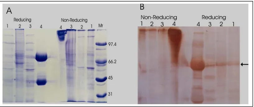
prominent but, as in control IgG, several faint bands were also seen (Figure 2 B).
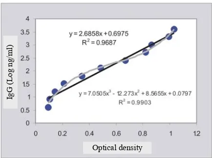
Quantification of the IgG by Elisa generated a linear correlation between the concentration of the control IgG and Elisa OD’s with correlation coefficient (R2) of 0.97. An even higher correlation coefficient (R2= 0.99) was obtained when the concentration was cubically correlated with the OD (Figure 3). When the regression equations were used to estimate the amount of IgG ingested by the mites, it gave unexpected results. Although a positive correlation between the number of mites and the amount of IgG was observed, halving the number of the mite did not followed by halving the amount of IgG (Table 1). The determination of IgG content of a mite by interpolation of mite-samples’ OD into the regression equation indicated that the IgG content reversed the mite concentration (number of mite per millilitre extraction buffer). The highest IgG content of 1.83 ng/mite was obtained in the extract containing 6 mite/ml.
Table 1. Immunoglobulin-G content of Sarcoptes scabiei
Mite/ml OD Total IgG (ng) IgG (ng)/mite
200 0.188 20.09* 0.10
100 0.180 18.51 0.19
50 0.176 17.60 0.35
25 0.192 20.87 0.83
12.5 0.174 17.30 1.33
6.25 0.137 10.99 1.83
3.12 0.086 5.33 1.78
Mean ± SD IgG (ng) per mite 0.92 ± 0.68
Note* derived from interpolation of OD into the regression equation (Figure 2)
Figure 1. Localization of immunoglobulin G by immunohistochemistry. Photograph in (A) is to show the distribution of IgG in skin and mites. (B) is to demonstrate that only a fraction of mites ingested the IgG. (C) revealed that the IgG confine to the mite’s gut only
Figure 2. Correlation between purified goat IgG and Elisa’s optical density (OD)
DISCUSSION
This study demonstrated that S. scabiei undoubtedly ingest the immunoglobulin of its host. The fact that IgG was confined only to the gut’s lumen indicates that the IgG was actively ingested by the mites, not passively diffused into the body through the cuticle. The fact that no IgG was seen attached to the cuticle of the S. scabiei was in contrast to that seen in Psoroptes ovis (PETTIT et al., 2000). As a matter of fact, the total amount of IgG in the washings was reported to be higher than that in the psoroptic-mite homogenate. It would have been expected that S. scabiei had higher amount of IgG attached to their cuticle because they burrow the skin compared to the non-burrowing Psoroptes ovis. The lack of IgG on the mite cuticle could be associated with the fact that S. scabiei burrowed the skin at a distance to the exudates (TARIGAN, 2003).
Since the mites acquired the IgG from the exudates, it was expected that the immunoglobulin would have been profoundly degraded by the proteolytic enzymes produced by inflammatory cells, skin microflora and the mites. A recent study by NOVIANA et al. (2004) indicated that skin of scabies-infested animals contained substantial activity of proteases derived from the mast cells. However, the immunoblotting analysis in the present study indicated the reverse, the IgG extracted from the mite’s gut was practically still intact. Only small amount of the IgG was observed to be degraded. Protease activities of S. scabiei are apparently lower than those ofpsoroptic mites. In the psoroptic mites, the mite proteases have been demonstrated to degrade intensely various substrates including host IgG and haemoglobulin (NISBET and BILLINGSLEY, 1999; PETTIT et al., 2000).
The fact that S. scabiei ingests its host IgG and that the immunoglobulin is slowly digested in its gut as demonstrated in this study supports the feasibility of
vaccine development. If a membrane protein lining the mite’s gut is isolated then used to immunise animals, protective immunity will be conferred provided that the membrane protein has a vital function to the survival of the mites and that its function is inactivated by the ensuing antibodies. It should be realised, however, that identification and purification of such membrane protein, especially for the microscopic, burrowing S. scabiei, will be laborious, costly and technically demanding (ROSS et al., 1992).
Having determined that S. scabiei ingests intact IgG from its host and that the IgG is not rapidly digested by the mites, a further question regarding the adequacy of ingested IgG may evolve. Will the amount of IgG antibody ingested by the mites from animals that have been vaccinated with appropriate membrane proteins be sufficient to cause death or significant damage to the mite?. Although this is an important question, this study was not intended to address to the question because it can only be answered by vaccination and challenge studies. Nevertheless, some findings obtained from this study addresses some of the question. The finding that only a portion of the mites at a point of time ingest IgG may suggest that vaccination will not provide protection that rapidly eliminate invading mites. The intoxication of mites occur only after they ingest sufficient quantity of immunoglobulin from their vaccinated host. This condition is exaggerated by the fact that the mites acquire IgG from the exudates not directly from the blood vessels which means elimination of invading mites occur only after the development of inflammatory reactions. In addition, the actual diet of the mites is still unknown, therefore, it is unknown whether ingestion of its host blood is vital for the survival or reproduction of the mites. If the blood is not vital for the mites they may avoid ingesting the immunoglobulin which in turn resulted in ineffective vaccination.
Optical density
IgG
(Log
Attempt to quantify the amount of IgG ingested by the mites was made in this study but the results were unsatisfactory. The concentrations of IgG in the mite homogenate did not parallel the total number of mites. This problem could not be attributed to the ELISA that used to quantify the IgG because such problem did not observed with the control IgG. With the control IgG, a very close correlation between the IgG concentration and the Elisa’s OD was observed indicating that the Elisa could be used to quantify IgG precisely. The illogical correlation between the number of mite and its IgG content is supposedly associated with the inconsistent solubility of the IgG in the extraction buffer or inconsistent binding of the extracted IgG to the microtitration plate. Quantifying the amount of ingested IgG, however, was only a supplementary objective of this study. The finding that intact immunoglobulin was ingested by the mites is much more important because it implies that developing anti-scabies vaccine is feasible.
CONCLUSION
Although Sarcoptes scabiei is not a blood-sucking ectoparasite and resides on the avascular cornified layer of the skin, it ingest its host immunoglobulin. The ingested immunoglobulin does not rapidly digest by the mites. These suggest that developing an anti-scabies vaccine from a relevant protein lining the mite’s gut is feasible.
ACKNOWLEDGMENTS
This research was supported by Balitvet Research Projects (Anggaran APBN 2000/2001). The author express appreciation for the excellent technical assistance of Mr.Yudi Mulyadi and Mr. Ismath.
REFERENCES
ANONIMOUS. 1994. In: Proceedings Rapat Koordinasi Bidang Kesehatan Hewan se-Jawa ke X tahun 1994. Yogyakarta, 3-5 Juli 1994.
ARLIAN, L. G. 1989. Biology, host relations and epidemiology of Sarcoptes scabiei. Annu. Rev. Entomol. 34: 139-161. BRADFORD, M. M. 1976. A rapid and sensitive method for
quantitation of microgram quantities of protein utilizing the principle of protein-dye binding. Anal. Biochem. 72: 248-254.
BROTOWIJOYO, M. D. 1987. Scabies pada hewan dan permasalahannya. Bulletin FKH UGM 7: 1-5.
DONALD, A. 1994. Parasites, animal production and sustainable development. Vet. Parasitol. 54: 27-47.
DORNY, P., T. VAN WYNGAARDEN, J. VERCRUYSSE, C. SYMMEONS and A. JALIA. 1994. Survey on the importance of mange in the aetiology of skin lesions in peninsular malaysia. Trop. Anim. Health Prod. 26: 81-86.
FORCHHAMMER, M. C. and T. ASFERG. 2000. Invading parasites cause a structural shift in red fox dynamics. Proc. R. Soc. Lond B. Biol. Sci. 267: 779-786.
GABAJ, M. M., W. N. BEESLEY and M. A. AWAN. 1992. A survey on farm animals in Libya. Ann. Trop. Med. Parasitol. 86: 537-542.
MANURUNG, J., BERIAJAYA and M. KNOX. 1987. Pengamatan pendahuluan penyakit kudis pada kambing di Kabupaten Pandeglang, Jawa Barat. Penyakit Hewan
19: 78-81.
NISBET, A. J. and P. F. BILLINGSLEY. 1999. Immunological control of scab mites: Digestive enzymes as candidate compounds. Vet. Parasitol. 83: 231-239.
NOVIANA, D., W. H. D, Y. OTSUKA and Y. HORII. 2004. Proliferation of protease-enriched mast cells in sarcoptic skin lesions of raccoon dogs. J. Comp. Pathol. 131: 28-37.
PARIJA, B., S. MISRA and P. SAHOO. 1995. Changing pattern in the epidemiology of caprine mange in bhubaneswar.
Indian Vet. J. 72: 536-538.
PARSON, S. A. and D. T. VERE. 1984. Benefit-cost analysis of the bakitwan project, Australian Development Assistance Bureau, Canberra, Australia.
PETTIT, D., W. D. SMITH, J. RICHARDSON and E. A. MUNN. 2000. Localization and characterization of ovine immunoglobulin within the sheep scab mite, psoroptes ovis. Vet. Parasitol. 89: 231-239.
ROSS, L., D. SMITH, D. H. KEMP and P. WILLADSEN. 1992. Vaccination against ticks. In: Animal Parasite Control Utilizing Biotechnology. W. K. YONG (Ed.). CRC Press, London. pp. 303-331.
TARIGAN, S. 1998. Metode pengembangbiakan dan pemanenan tungau Sarcoptes scabiei. Pros. Seminar Nasional Teknologi Peternakan dan Veteriner, Bogor, 1-2 Desember 1998. Puslitbang Peternakan, Bogor. hlm. 1009-1017.
TARIGAN, S. 2003. Histopathological changes in naive and sensitised goats caused by Sarcoptes scabiei. JITV 8: 114-121.
WALL, R. 1992. The environment impact of avermectin use in livestock. Parasitol. Today 8: 255-256.
WILLADSEN, P. 2001. The molecular revolution in the development of vaccines against ectoparasites. Vet. Parasitol. 101: 353-367.
ZAHLER, M., A. ESSIG, R. GOTHE and H. RINDER. 1999. Molecular analyses suggest monospecificity of the genus sarcoptes (acari: Sarcoptidae). Int. J. Parasitol.