*corresponding author: [email protected]
Comparison of Bcl-xL protein expression
in placental trophoblast cells between
pregnancy complicated by severe
preeclampsia and normotensive pregnancy
Diah Rumekti Hadiati1*, Arsi Palupi1, Mohammad Hakimi1, Sofia Mubarika Haryana2 1Department of Obstetrics and Gynecology, 2Department of Histology and Cell Biology,
Faculty of Medicine, Universitas Gadjah Mada, Yogyakarta, Indonesia. DOI: http://dx.doi.org/10.19106/JMedSci005001201804
ABSTRACT
Preeclampsia is one of the main causes of maternal and perinatal mortality and morbidity. The pathogenesis of preeclampsia remains unclear until now. It is believed that regulation of apoptosis in trophoblast cells plays an important role in the pathophysiology of preeclampsia. Failure of spiral arteries remodeling will eventually lead to placental hypoxia lead to excessive trophoblast apoptosis. The molecular mechanism of apoptosis is very complicated involving many signaling molecules included 2 proteins. The Bcl-2 protein group consists of proapoptosis proteins (Bax) and apoptosis inhibitor proteins (Bcl-2 and Bcl-xL). The aimed of this stuty was to compare the expression of Bcl-xL protein in placental trophoblast cells of pregnancy complicated by severe preeclampsia with that normotensive pregnancy. This study was an observational study with cross sectional design involving 43 pregnancy patients with severe preeclampsia and 38 normotensive pregnancy who treated in Dr. Sardjito General Hospital, Yogyakarta from October 2011 until March 2012. Placenta samples were obtained from all subjects for Bcl-xL protein expression analysis using immunohistochemistry technique. Data were analyzed using independent t-test, chi-square test, and logistic regression. A p value <0.05 was considered significant. Significant difference in Bcl-xL protein expression in trophoblast cells of pregnancy complicated by severe preeclampsia (1.29 ± 0.12) compared to that normotensive pregnancy (1.71 ± 0.14) was reported (p = 0.00). In addition, logistic regression test showed that diagnosis of severe preeclampsia had a statistically significant role in Bcl-xL protein expression (p= 0.000). In conclusion, the expression of Bcl-xL protein is lower in pregnancy complicated by severe preeclampsia compared to normotensive pregnancy.
ABSTRAK
INTRODUCTION
Preeclampsia is one of the main causes of maternal and perinatal mortality and morbidity. The pathogenesis of preeclampsia remains unclear until now. However, it is believed that the failure of spiral arteries remodeling will eventually leads to placental hypoxia. This theory may not be the main cause of preeclampsia, but at least it is involved in the pathogenesis of this disease.1, 2
Apoptosis has an important role not only in the development of placenta but also in the pathophysiology of pregnancy complicated by preeclampsia. Apoptosis of trophoblast cells is increasing with gestational age and this increase has been studied as a complication of pregnancy with preeclampsia and intrauterine growth restriction (IUGR). Although this hypothesis is still under study, it is believed that regulation of apoptosis in trophoblast cells plays a key role in the pathophysiology of preeclampsia.3, 4
The molecular mechanism of apoptosis in human is very complicated involving many signaling molecules included Bcl-2 proteins. The Bcl-2 protein family consists
and Bcl-xL. During pregnancy, Bcl-2 is found
in placenta since irst trimester of pregnancy
until third trimester and the concentration is decreasing with gestational age. In pregnancy complicated by preeclampsia, regulators of placental apoptosis are expressed
diferently. Several recent studies have
found that expression of Bcl-2 and Bcl-xL as antiapoptotic molecules in patients with severe preeclampsia and IUGR are lower than patients with normal pregnancy.3, 4
The purpose of this study was to compare the expression of Bcl-xL protein in placental trophoblast cells of pregnancy complicated by severe preeclampsia with normotensive pregnancy. This study also aimed to evaluate
the efect of maternal age, gestational age, and
maternal mean arterial pressure (MAP) in the expression of Bcl-xL protein.
MATERIALS AND METHODS
Subjects
This was an observational study with cross sectional design. The population were patients with severe preeclampsia and normotensive
patients who treated in Dr. Sardjito General
observasi dengan rancangan potong lintang ini melibatkan 43 pasien wanita hamil dengan preeklamsia berat dan 38 wanita hamil normotensi yang dirawat di RSUP Dr. Sardjito, Yogyakarta antara Oktober 2011 dan Maret 2011. Sampel plasenta diambil dari semua subjek untuk pemeriksaan ekspresi protein Bcl-xL menggunakan teknik imunohistokimia. Data yang diperoleh dianalisis denga uji t independen, uji chi square dan uji regresi logistic. Nilai p<0.05 digunakan sebagai dasar menyatakan perbedaan nyata. Dijumpai perbedaan nyata ekspresi protein Bcl-xL pada sel trofoblas kehamilan dengan preeklamsia berat (1,29 ± 0,12) dibandingkan dengan kehamilan normotensi (1,71 ± 0,14) (p=0,00). Selain itu, uji regresi logistik menunjukkan diagnosis preeklamsia berat mempengaruhi secara nyata terhadap ekspresi protein Bcl-xL (p=0,000). Dapat disimpulkan bahwa ekspresi protein Bcl-xL lebih rendah pada kehamilan dengan preeklamsia berat dibandingkan dengan kehamilan normotensi.
were patients with severe preeclampsia in 28-40 weeks of gestational age and agreed to be included in the study. The exclusion criteria were presence of comorbid diseases such as chorioamnionitis, chronic hypertension, diabetes, systemic lupus erythematosus, sickle cell disease, thyroid diseases, heart diseases, bronchial asthma, seizure which was caused by other etiologies beside preeclampsia, HIV,
and fetus with major congenital disorder.
Written inform consent were obtained from
each patient after suicient information was
given.
Protocol of study
Samples were taken from the placenta immediately after the baby was born. Samples
were then sent to Histology Laboratory,
Faculty of Medicine, Universitas Gadjah Mada, Yogyakarta, Indonesia. Samples
of placental tissue were stained using immunohistochemistry technique to measure the expression of Bcl-xL protein. The expression of Bcl-xL protein was reported to be positive if brown color was found in cytoplasm or cell membrane. The expression of Bcl-xL protein was measured using semiquantitative immunohistochemical scoring system
(HSCORE). The formula was HSCORE =
∑ Pi (i+1), where Pi was percentage of cells which are stained positively with Bcl-xL immunostaining and i was intensity of staining
with diferent grades (0=negative; 1=weakly positive; 2=moderately positive; 3=strongly
positive). The HSCORE measurement was conducted by two observers with concealment of sample’s identity. Inter observer agreement was tested using kappa test and the result of kappa value was 0.88 which showed that there was a strong agreement between two
observers. Expression of Bcl-xL was observed
with microscop using high magniication (400x) in 5 ields of view from each samples.
The protocol of the study was approved by
the Medical and Health Research Ethics Committee, Faculty of Medicine, Universitas Gadjah Mada, Yogyakarta.
Statistical analysis
All numerical data were presented as mean
± standard deviation (SD). Statistical analysis
was conducted using independent t test to
evaluate the diference in mean between two
groups. Bivariate analysis using chi-square test was used to evaluate correlation between two categorical variables. Multivariate analysis using logistic regression was applied to evaluate the relationship between independent variable (preeclamptic vs normotensive group), confounding variables (patient’s age, gestational age, and mean arterial pressure), and dependent variable (Bcl-xL protein expression). A p value less than 0.05 was
considered statistically signiicant.
RESULTS
Placenta samples were obtained from 43 patients with pregnancy complicated by severe preeclampsia and 38 patients with normotensive pregnancy. All samples were stained using immunohistochemistry technique to evaluate the expression of Bcl-xL protein
Characteristics of subjects of normotensive and severe preeclamptic groups are shown
in TABLE 1. No signiicantly diferent of
patient’s age of severe preeclamptic compared to normotensive group was observed (p>0.05).
Meanwhile, there were signiicant diferences
TABLE 1. Characteristics of subjects (mean ± SD) of normotensive and severe preeclamptic groups
Variable Normotensive
Group (n=38)
Severe Preeclamptic Group (n=43)
Mean Diference
(95% CI) p
Patient’s age (years) Gestational age (weeks) Mean arterial pressure (mmHg)
28.42 ± 6.77 35.67 ± 3.01 126.29 ± 17.00
28.37 ± 7.08 38.39 ± 1.85 88.95 ± 6.44
0.5 (-3.0136 – 3.116) -2.72(-3.82 - -1.63) 37.34 (31.74-42.94)
0.974 0.000 0.000
In this study, the expression of Bcl-xL protein was observed in decidual trophoblast. In accordance with early onset preeclampsia theory which stated that the failure of spiral artery remodelling occured in decidual
FIGURE 1. Intensity of color in Bcl-xL protein staining
layer. The change in intensity of brown color in trophoblast cells of decidual layer corresponded to expression of Bcl-xL as
shown in FIGURE 1.
Intensity score 0 Intensity score 1
Intensity score 2 Intensity score 4
TABLE 2. The Bcl-xL protein expression (mean ± SD) in placenta of severe preeclamptic and normotensive groups
Variable
Severe Preeclamptic Group (n=43)
Normotensive Group (n=38)
Mean Diference
(95% CI) p
Bcl-xL Expression 1.29 ± 0.12 1.71 ± 0.14 - 0.42 (-0.47 – -0.36) 0.00
Receiver operating characteristic (ROC)
analysis was conducted to determine the
cutof point of Bcl-xL protein expression.
The analysis found that the area under curve
(AUC) was 0.93 or 93% with cutof point of 1.495 as shown in FIGURE 2.
FIURE 2. ROC curve of Bcl-xL protein expression
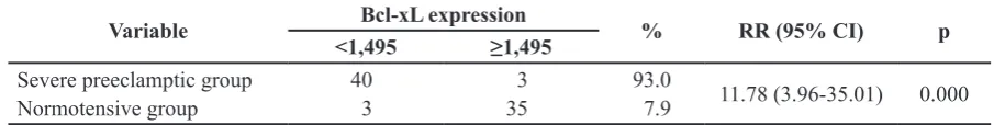
TABLE 3 shows bivariate analysis
between independent and dependent variable. The pregnant patients with severe preeclampsia had more chances to have expression of
Bcl-xL protein valued less than 1.495 compared to that normotensive patients, with relative risk
of 11.78 (3.96-35.01) and p=0.000.
TABLE 3. Bivariate analysis between independent and dependent variable
Variable Bcl-xL expression % RR (95% CI) p
<1,495 ≥1,495
Severe preeclamptic group
Normotensive group 40 3
3 35
93.0
7.9 11.78 (3.96-35.01) 0.000
Bivariate analyses between confounding variables and the expression of Bcl-xL protein
Bcl-TABLE 4. Bivariate analyses between confounding variables and expression of Bcl-xL protein
Variables Bcl-xL expression % RR (95% CI) p
<1,495 ≥1,495
Patient’s Age
<20 and >40 years old 20-40 years old
Mean Arterial Pressure (MAP) >123 mmHg
In contrary, gestational age and MAP afected the expression of Bcl-Xl protein (RR =0.38
(0.25-0.58; p<0.001) and [RR= 2.67 (1.83-3.91); p<0.001], respectively.
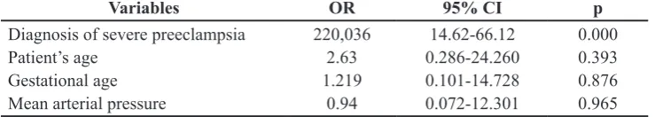
Multivariate analysis using logistic regression between independent variable, dependent variable, and confounding variable
is shown in TABLE 5. Diagnosis of severe
preeclampsia consistently afected the expression of Bcl-xL protein (p=0.000), while gestational age and MAP did not afect the
expression of Bcl-xL protein (p>0.05).
TABLE 5. Multivariate analysis using logistic regression between independent variable, dependent variable, and confounding variable
Variables OR 95% CI p
Diagnosis of severe preeclampsia Patient’s age
No signiicantly diferent in patient’s age between severe preeclamptic group and normotensive group was found in this study. Previous studies reported that women with severe preeclampsia were older than normotensive pregnancy although it was
not statistically signiicant.5-7 Meanhwile, other studies found that preeclampsia were more common in women with advanced
younger women. Advanced maternal age is an independent risk factor for adverse outcomes
in irst-time mothers with preeclampsia.8, 9 Mean gestational age in severe
preeclamptic group was signiicantly lower
than normotensive group indicating an inhomogenous distribution in research samples. This result was in accordance with a study by Zhang et al.7 which found that mean gestasional age in severe preeclamptic group
al.10 found no diference in gestasional age between preeclamptic group and control group.
This study also found that MAP in severe preeclamptic group was higher than in normotensive group. This could be understood clearly because in preeclampsia there will be an increase in blood pressure.
This study found that expression of Bcl-xL protein as antiapoptotic molecule in patients with preeclampsia was lower than normotensive patients. This was in
accordance with a study by Shu et al.11 which found that Bcl-xL expression was down-regulated in preterm preeclampsia, but not in term preeclampsia and controls. Allaire et al.10 concluded that the presence of apoptotic marker could be used as a sign of intrauterine hypoxia. Hung et al.12 found that hypoxia and prolonged hypoxia-reoxigenation seemed to cause more reduction in the levels of Bcl-xL,
although the diference was not statistically signiicant. Meanwhile, a study by Zhang
et al.7 found that Bcl-xL mRNA expression levels was unchanged in severe preeclamptic placentas when compared to control. In contrast, Whitehead et al.13 found signiicantly
increased placental RNA expression of Bcl-xL
in early onset FGR (fetal growth restriction),
PE complicated by FGR, and PE without FGR
compared with preterm controls. This perhaps
relects a disordered regulation of apoptosis in
placental dysfunction that is as yet not clearly understood.
No correlation between maternal age and
expression of Bcl-xL protein as antiapoptotic molecule was observed in this stuudy. In contrast, Kavathia et al.14 reported that there was a positive linear correlation between apoptosis and person’s age. The discrepancy could be caused by group arrangement in this study which was based on risk factor of preeclampsia, where patient with age<20
years old and >40 years old were considered in high risk, and patient between 20-40 years old were in low risk.
This study also found that there was a correlation between gestational age and expression of Bcl-xL protein. In normal condition, apoptotic activity is increasing with gestational age. Previous study by
Smith et al.15 which compared apoptosis in
normotensive pregnant women from irst
trimester and third trimester found that there was an increase in apoptosis index in third trimester. In normal condition, antiapoptotic expression is decreasing with gestational age as shown in a study by Kim et al.16 which found that that there was a decrease in expression of Bcl-2 protein, an antiapoptic molecule, in third trimester.
Abnormality in apoptosis stimulation in patients with essential hypertension showed that antiapoptotic factors concentration in those patients were decreased and it could be caused by ischemia.17 This was in accordance with this study where bivariate analysis between MAP and expression of Bcl-xL found
that patients with MAP≥123 mmHg had 2.67
increases in probability to have expression of Bcl-xL <1.50 compared to patients with MAP<123 mmHg.
CONCLUSIONS
In conclusion, this study found that expression of Bcl-xL protein is lower in pregnancy complicated by severe preeclampsia compared to normotensive pregnancy. In addition, diagnosis of severe preeclampsia
consistently afect in the expression of Bcl-xL
protein.
ACKNOWLEDGEMENTS
We would like to thank all subjects who
REFERENCES
1. Steegers EA, von Dadelszen P, Duvekot JJ, Pijnenborg R. Pre-eclampsia. Lancet 2010;
376(9741):631-44.
h t t p : / / d x . d o i . o r g 1 0 . 1 0 1 6 / S 0 1 4 0
-6736(10)60279-6
2. Levy R. The role of apoptosis in preeclampsia.
Isr Med Assoc J 2005; 7(3):178-81.
3. Straszewski-Chavez SL, Abrahams VM, Mor
G. The role of apoptosis in the regulation of
trophoblast survival and diferentiation during
pregnancy. Endocr Rev 2005; 26(7):877-97.
http://dx.doi.org/10.1210/er.2005-0003
4. Heazell AE, Buttle HR, Baker PN, Crocker
IP. Altered expression of regulators of caspase activity within trophoblast of normal pregnancies and pregnancies complicated by
preeclampsia. Reprod Sci 2008;
15(10):1034-43.
h t t p : / / d x . d o i . o r g / 1 0 . 1 1 7 7 / 1933719108322438
5. Jasovic-Siveska E, Jasovic V, Stoilova S. Previous pregnancy history, parity,
maternal age and risk of pregnancy induced
hypertension, Bratisl Lek Listy 2011;
112(4):188-191.
6. Sharp AN, Heazell AEP, Baczyk D, Dunk CE, Lacey HA, Jones CJP, et al. Preeclampsia is associated with alterations in the p53 pathway
in villous trophoblast. PLoS ONE 2014;
9(1):e87621.
h t t p : / / d x . d o i . o rg / 1 0 . 1 3 7 1 / j o u r n a l .
pone.0087621
7. Zhang Z, Yang X, Zhang L, Duan Z, Jia L,
Wang P, et al. Decreased expression and
activation of stat3 in severe preeclampsia. J Mol His 2014; 46(2):205-219.
http://dx.doi.org/10.1007/s10735-015-9613-8
8. Macdonald-Wallis C, Tilling K, Fraser A, Nelson SM, Lawlor DA. Established
preeclampsia risk factors are ralted to patterns
Study of Parents and Children, J Hypertens 2011; 29(9):1703-11.
h t t p s : / / d o i . o r g / 1 0 . 1 0 9 7 /
HJH.0b013e328349eec6
9. Lamminpaa R, Vehviläinen-Julkunen K, Gissler M, Heinonen S. Preeclampsia
complicated by advance maternal age: a registry-based study on primiparous women
in Finland 1997-2008, BMC Pregnancy Childbirth 2012; 12:47.
https://doi.org/10.1186/1471-2393-12-47
10. Allaire AD, Ballenger KA, Wells SR, McMahon MJ, Lessey BA. Placental
apoptosis in preeclampsia. Obstet Gynecol
2000; 96(2):271-6.
h t t p : / / d x . d o i . o rg / 1 0 . 1 0 9 7 / 0 0 0 0 6 2 5 0 -200008000-00022
11. Shu C, Liu Z, Cui L, Wei C, Wang S, Tang JJ, et al. Protein proiling of preeclampsia placental
tissues. PLoS ONE 2014; 9(11):e112890.
h t t p : / / d x . d o i . o rg / 1 0 . 1 3 7 1 / j o u r n a l .
pone.0112890
12. Hung TH, Chen SF, Liou JD, Hsu JJ, Li MJ,
Yeh YL, et al. Bax, bak and mitochondrial
oxidants are involved in hypoxia-reoxygenation-induced apoptosis in human
placenta. Placenta 2008; 29(7):565-83.
h t t p : / / d x . d o i . o r g / 1 0 . 1 0 1 6 / j .
placenta.2008.03.005
13. Whitehead CL, Walker SP, Lappas M, Tong S. Circulating RNA coding genes regulating
apoptosis in maternal blood in severe early onset fetal growth restriction and
pre-eclampsia. J Perinatol 2013; 33:600-4. http://dx.doi.org/10.1038/jp.2013.16
14. Kavathia N, Jain A, Walston J, Beamer BA, Fedarko NS. Serum markers of apoptosis
decrease with age and cancer stage. Aging
2009; l(7):652-63.
http://dx.doi: 10.18632/aging.100069
h t t p : / / d x . d o i . o r g / 1 0 . 1 0 1 6 / S 0 0 0 2
-9378(97)70438-1
16. Kim PKM, Zamora R, Petrosko P, Billiar T. The regulatory role of nitric oxide in apoptosis.
Int Immunopharmacol 2001; 1(8):1421-41. h t t p : / / d x . d o i . o r g / 1 0 . 1 0 1 6 / S 1 5 6 7
-5769(01)00088-1
17. Kaufmann P, Black S, Huppertz B. Endovascular trophoblast invasion:
implications for the pathogenesis of intrauterine growth retardation and
preeclampsia. Biol Reprod 2003; 69(1):1-7.