International Journal of Science and Research (IJSR)
ISSN (Online): 2319-7064Index Copernicus Value (2013): 6.14 | Impact Factor (2013): 4.438
Volume 4 Issue 6, June 2015
www.ijsr.net
Licensed Under Creative Commons Attribution CC BY
Hyperglicemia IncreasedThe Expression of TLR2
mRNA and Coronin-1A Level in Lung Mice which
were infected by Mycobacterium Tuberculosis
Wande IN
1, Sutirta Yasa IWP
2, Linawati NM
31, 2Department of Clinical Pathology, Medical FacultyUdayana UniversityDenpasar-Bali. Indonesia (0361) 222510
3
Department of Histology Medical FacultyUdayana UniversityDenpasar-Bali. Indonesia (0361) 222510
Abstract: The present study was conducted to evaluate the expression ofTLR2mRNAandCoronin-1A level in micewhich wereinfected withMycobacterium tuberculosis (M.tb) and hyperglycemia.A total of24mice weredivided into 2 groups and all were infectedwith M.tb,then control group were injectedof 5 μl a sodium citrate buffer intraperitoneallyandtreatment groupwere injected150mg/kgbwstreptozotocin (STZ)dissolved in 5 μlsodiumcitratebuffer intraperitoneally to make hyperglycemia.Lung tissue harvestingconductedforRT-PCR examinationfor expression of TLR2 mRNA and Coronin-1A level at 3 and 5 week. In this study found the expression of TLR2 mRNA and Coronin-1A level in mice infectedwith M.tband hyperglycemia which wereexaminedin the fifth week were signifikan higher than others.
Keywords: TLR2 mRNA, Coronin-1A, Mycobacterium tuberculosis
1.
Introduction
TLR2 and TLR6 found to play a role in stimulating production of IL-1β. TLR2 also contribute for IL-12 secretion by macrophages.1 In TLR2-deficient mice showed granuloma formation is not perfect, when it infected with a high dose of Mycobacterium tuberculosis (M.tb) there is suseptibility increase compared to normal mice. TLR2-deficient mice also showed impaired in control of M.tb chronic infection.2 In Type 1 diabetes showed increase expression of TLR2 and TLR4 mRNA on peripheral blood monocytescompared to healthy. Final target TLR such as nuclear factor B, myeloid differentiation factor 88 (MYD 88), Trif, and phosphorylated IL-1 receptor-associated kinase was significantly increased in type 1 diabetes. TLR2 and TLR4 expression is significantly associated with glycosylated hemoglobin.3In type 2 diabetes with tuberculosis, type 1 (IL-12, IL-2 and IFN-) and natural
(IL-1β, IL-6, TNF, IL-8, and granulocyte monocyte-CSF) cytokines are consistently increasedin tuberculosis with chronic hyperglycemia compared with tuberculosis patients without diabetes.4
TLR is important in killing process of mycobacterium by macrophages through induction of intracellular antimicrobial peptide cathelicidin. TLR polymorphism was significantly associated with tuberculosis in humans.In protein analysis, composition of phagosome contain M.tb indicate the presence of very strong proprietary protein plays a role in M.tb survival, named TACO (tryptophan-aspartatecontaining coat protein) which is now referred as Coronin-1A. In both patients with active and latent tuberculosis showed increased levels of Coronin-1A compared to healthy.5
2.
Material and Methods
A total of 24 Balb/c mice, male, 20-25 gr, 8 weeks old were randomly placed in four cages and adapted for 2 weeks. Group I and II consisted of each 12 mice which were placed in cages 1,2,3 and 4. In the first day after adaptation, both of groups were infected by 60 µl (with 105 CFU/ml of concentration) M.tb H37Rv intranasally. In grup of treatment also injected with 150 mg/ kgbw STZ dissolved in
5 μM sodium citrate buffer (pH 4.5) after overnight fasting, while in group of control only received intraperitoneal injection of sodium citrate buffer. Two weeks after intervention, blood sugar level were measured, state diabetes mellitus if the fasting glucose level> 200 mg / dl and unchangeduntil5th week. On the third and fifth week after initial treatment, there were 12 mice of each group terminated. The pulmonary organs were prepared divided in two part. First part were submerged in RNA later (Qiagen) for examining TLR2 mRNA and other part were submerged in PBS for examining Coronin-1A with ELISA methods (due to BT-lab kit procedure).
3.
Results
a. The expression of TLR2 mRNA
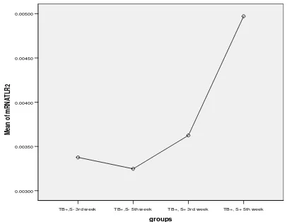
Mean relative of TLR2 mRNA expression in K1: groups of tuberculosis (+) without hyperglycemia (STZ-) in third week is 0.003376. In K2: groups of tuberculosis (+) without hyperglycemia (STZ -) infifth week is 0.003247. In P1: groups of tuberculosis (+) with hyperglycemia (STZ +) in third week is 0.003625. In P2: groups of tuberculosis (+) with hyperglycemia (STZ +) in fifth week 0.004972. Based on One Way ANOVA found there were significant differences in TLR2 mRNA expressionbetween groups (p = 0.013). In post hoc test with LSD found there were significant difference(p <0.05)in TLR2 mRNAexpression between groups P2 vs P1; P2 vs K2; and P2 vs K1. While there were no significant difference in TLR2 mRNA expression between grups K1 vs K2 (p = 0.807); between
International Journal of Science and Research (IJSR)
ISSN (Online): 2319-7064Index Copernicus Value (2013): 6.14 | Impact Factor (2013): 4.438
Volume 4 Issue 6, June 2015
www.ijsr.net
Licensed Under Creative Commons Attribution CC BY
K1 vs P1 (p = 0.639) and also between P1 vs K2 (p = 0.478). The curve of mean relative TLR2 mRNA expression in group of study were showed in Fig.1
Figure 1: Mean relative curve ofTLR2 mRNA expression in groups of study
b. Coronin-1A level
Mean of Coronin-1A level in groups P1 (210,2) pg/ml; P2 (350,4) pg/ml; K1 (174,2) pg/ml and K2 (175,6) pg/ml. Normality test with Shapiro-Wilk result in normal distribution (p> 0.05). Homogeneity test with Levene's test showed homogen data (p = 0.391). Based on One Way ANOVA test found there were significant differences between groups in Coronin-1A levels (p = 0.00). Post hoc test with LSD also found there were significant difference (p = 0.00) in Coronin-1A level between P2 vs K2; P2 vs K1; and P2 vs P1). While there were no significant difference in Coronin-1A level between grups K1 vs K2 (p = 0.693); between K1 vs P1 (p = 0.246) and also between P1 vs K2 (p = 0.265). Mean of Coronin-1A level (pg/ml) in all group of treatment showed in Fig.2
Figure 2: Coronin-1A levels(pg/ml) curve in all groups of treatment
4.
Discussion
a. TLR2 mRNA expression
In vivo study with MyD88 deficient mice compared to TLR deficient mice showed MyD88 deficient mice were highly susceptible to respiratory infections by M.tb than TLR-deficient mice. Susceptibility of TLR2-TLR-deficient mice to M.tb varies from several different studies, whereas mice with TLR4 deficiency did not show high susceptibility to M.tb infection. Report mentions that TLR9 deficient mice
were susceptible to M.tb infection and more vulnerable if its deficient in both TLR2 and TLR9. These findings indicatedthat TLRs was important in first binding to mycobacteria. The latest report said that mice deficient TLR2 / TLR4 / TLR9 showed phenotype abnormalities milder than MyD88-deficient mice. Further study was needed to know if any other molecules besides TLRs participate in MyD88 signalling. TLR or MyD88 is an independent component of natural immunity involved in induction of adaptive immune response during M.tb infection.6In experimental studies with ratwas given STZ to make diabetic and infected M.tb intravenously, showed decrease in IL-12 and IFN-level in lung, liver and lymph compared to control mice that were not diabetic. There was also a decrease in expression of inducible nitric oxide monocytes surface compared to healthy individuals. The final target TLR such as nuclear factor B, myeloid differentiation factor 88 (MYD 88), Trif, and phosphorylated IL-1 receptor-associated kinase was significantly increased in diabetes type 1. There was increasing TNF- and IL-1βlevel in these patients. The expression of TLR2 and TLR4 was significantly associated with glycosylated hemoglobin.3
In this study demonstrated the expression of TLR2 mRNA was higher in group of treatment with tuberculosis and hyperglycemia which was examined in fifth week after treatment compared to a group with tuberculosis without hyperglycemia.
b. Coronin -1A level
Coronin-1A is regulated by Phox protein through deployment of 'C' protein kinase to phagosome then subsequently occur phosphorylation P47phox and Coronin complex. Coronin phosphorylated dissolved and disposal of Coronin layer cause fusion of phagosomewith endosome. Cholesterol, epigallocatechin-3-Galates (EGCG, the main component in green tea polyphenol), vitamin D3 + retinoic acid and chenodeoxycholic acid acid + retinoic acid regulate Coronin-1A gene transcription. Through receptor-Ck-dependent signaling, cholesterol is known to regulate SREBP and PPAR- transcriptional factor. EGCG was known to work through SP-1 transcription factors, in which vitamin D + retinoic acid and chenodeoxycholic acid + retinoic acid is known to work through VDR / RXR and FXR / RXR heterodimers. This stage showed epigenomik Coronin-1A gene control plays a role in the evolution of micobacterial phagosome.5
In this study, an increase of Coronin 1A level in group with tuberculosis and hyperglycemia were examined in the fifth week compared with the other treatment groups, showed the length of tuberculosis suffer and accompanied by hyperglycemia causes Coronin 1A level increased. This situation blocked fusion of phagosome and lysosome so phagocytosis against micobacterium not occur, which
International Journal of Science and Research (IJSR)
ISSN (Online): 2319-7064Index Copernicus Value (2013): 6.14 | Impact Factor (2013): 4.438
Volume 4 Issue 6, June 2015
www.ijsr.net
Licensed Under Creative Commons Attribution CC BY
increased the number of bacterial colonie and also increased tuberculosis severity.
Studies with type 1 diabetic mice induced by alloxan showed that neutrophils and alveolar macrophages impaired in cytokine secretion and phagocytosis, but with insulin administration can recover some of the circumstances. The main finding in chronic diabetes was on immune response to mycobacteria by slowing natural immune response that mediate production of interferon-. This effect is caused by disorders of rapid inflammatory response by infected alveolar macrophages, while dendritic cells migration from lungs to local lymph nodes.8
5.
Conclusion
It could be concluded there were increasing of TLR2 mRNA expression and Coronin-1A level in lung mice which were infected with M.tb and hyperglycemia examined in the fifth week compared with other groups of treatment.This may be due to many inflammatory factors that act as ligands of TLR that can spur an increase in the expression of TLR especially TLR2.
References
[1]Kleinnijenhuis J., M. Oosting, LAB Joosten, Netea MG, and Crevel RV 2011. Innate immune recognition of Mycobacterium tuberculosis. Clinical and Developmental Immunology. Vol. 2011, article ID 405 310, p. 1-12.
[2]Drennan MB, Nicolle D., Quesniaux VJE, Jacobs M, Allie N., J. Mpagi, Fremond C., H. Wagner, Kirschning C., and Ryffel B. 2004. Toll-like receptor 2-deficient mice succumb to tuberculosis tuberculosis infection. American Journal of Pathology, vol. 164, no. 1, p. 49-57. [3]Devaraj S., Dasu MR, J. Rockwood, Winter W., Griffen
SC, Jialal I. 2008. Increased Toll-like receptor (TLR) 2 and TLR4 expression in monocytes from Patients with type 1 diabetes: further evidence of a proinflammatory state , Journal of Clinical Endocrine Metabolic, vol. 93 (2), p. 578-583.
[4]Restrepo BI, Fisher-Hoch SP, Pino PA, A. Salinas, MH Rahbar, F. Mora, Cortes-Penfield N., and McCormick JB 2008. Tuberculosis in poorly controlled type 2 diabetes: Altered cytokine expression in peripheral white blood cells. Clinical Infectious Diseases, vol. 47, p. 634-641. Available from: http://cid.oxfordjournals.org. Accessed: December 19th, 2011.
[5]Constantoulakis P., Filiou E., N. Rovina, Chras G., Hamhougia A., Karabela S., A. Sotiriou, C. Roussos, and Poulakis N. 2010. In vivo expression of innate immunity markers in Patients with mycobacterium tuberculosis infection. BMC Infectious Diseases, vol., 10, no. 243, p. 1-10.
[6]Saiga H., Y. Shimada, and Takeda K. 2010. Review article: innate immune effectors in mycobacterial infection. Clinical and Developmental Immunology. Vol. 2011, no. 347 594, p. 1-8.
[7]Yamashiro S., K. Kawakami, K. Uezu, Kinjo T., Miyagi K., Nakamura K., Saito A. 2005. Lower expression of Th1-related cytokines and inducible nitric oxide synthase in mice with streptozotocin-induced diabetes mellitus
infected with Mycobacterium tuberculosis. Clinical and Experimental Immunology, doi: 10.1111 / j.1365-2249.2005.02677.x, no. 139, p. 57-64.
[8]Knapp S. 2012. Diabetes and infection: Is there a link? A mini-review. Gerontology, DOI: 10.1159 / 000345107.p. 1-6. Available from: www.karger.com/ger. accessed: Pebruary 20th, 2013.