Halkidou K, Gnanapragasam VJ, Mehta PB,
Logan IR, Brady ME, Cook S, Leung HY, Neal DE,
Robson CNExpression of...
Article in Oncogene · May 2003
DOI: 10.1038/sj.onc.1206342 · Source: PubMed
CITATIONS
136
READS
18
9 authors, including:
Some of the authors of this publication are also working on these related projects:
CamCaPView project Vincent J Gnanapragasam University of Cambridge
164PUBLICATIONS 1,948CITATIONS
SEE PROFILE
Ian R Logan
Newcastle University
17PUBLICATIONS 1,043CITATIONS
SEE PROFILE
David Edgar Neal University of Oxford
1,043PUBLICATIONS 22,906CITATIONS
SEE PROFILE
Craig N Robson Newcastle University
202PUBLICATIONS 7,117CITATIONS
SEE PROFILE
Expression of Tip60, an androgen receptor coactivator, and its role in
prostate cancer development
Kalipso Halkidou
1, Vincent J Gnanapragasam
1, Piyush B Mehta
1, Ian R Logan
1, Mark EBrady
1,
Susan Cook
1, Hing Y Leung
1, David ENeal
1,2and Craig N Robson*
,11Prostate Research Group, School of Surgical and Reproductive Sciences, University of Newcastle, Framlington Place, Newcastle upon Tyne NE2 4HH, UK;2Oncology Centre, Addenbrooke Hospital, University of Cambridge, Cambridge CB2 2QQ, UK
Prostate cancer (CaP) is initially androgen sensitive and responsive to hormone ablation therapy. However, cancer growth recurs despite androgen deprivation in the majority of cases of advanced disease. The molecular basis of this progression still remains unknown. The significance of androgen receptor (AR) coactivator proteins in this androgen-dependent malignancy is only beginning to emerge. In the present study, we examined the role of Tat interactive protein, 60 kDa (Tip60), an AR coacti-vator, in CaP progression. In hormone refractory CaP biopsies, we observed a nuclear accumulation of Tip60 expression in contrast to a more diffuse distribution pattern observed in benign prostate hyperplasia and primary CaP. Furthermore, in both the prostate xenograft model CWR22 and the LNCaP CaP cell line, we observed that androgen withdrawal promoted upregulation of Tip60 as well as nuclear accumulation. In contrast, androgen exposure resulted in decreased Tip60 expression that was more closely linked to a cytoplasmic presence. Chromatin immunoprecipitation analysis revealed Tip60’s recruit-ment to the PSA gene promoter in both androgen-dependent and -inandrogen-dependent cell lines. Thus, in vitroand in vivo data support a possible role for Tip60 in the molecular pathway leading to the development of andro-gen-independent CaP following long-term androgen de-privation therapy.
Oncogene(2003)22,2466–2477. doi:10.1038/sj.onc.1206342
Keywords: Tip60; co-activator; CWR22 xenograft;
hor-mone refractory; prostate cancer
Introduction
Prostate cancer (CaP) is the second most prevalent malignancy in UK men, accounting for more than, 9000 deaths each year. Both CaP incidence and death rates increased significantly over the last three decades, in England and Wales (Majeedet al., 2000). One in every four men diagnosed will eventually die from this disease.
CaP is initially androgen sensitive and responsive to hormone ablation therapy, the standard treatment for metastatic disease (Catalona, 1994). However, despite the high (80%) rate of response to hormone treatment, the median duration of response is less than 3 years (Crawford, 1992). Consequently, nearly all dependent CaP eventually relapse as fatal hormone-independent disease (hormone refractory, HR), while the molecular basis of this progression still remains unknown.
Androgens regulate prostate growth through the androgen receptor (AR), a member of the nuclear hormone receptor superfamily. Transcriptional inter-mediary factors (coactivators and corepressors) have been identified, which enhance androgen responsiveness through interaction with the AR ligand-binding domain (Tsai and O’Malley, 1994). Inspite of the increasing number of AR coactivators expressed in the prostate, the physiological significance of these cofactors and their role in CaP progression has not been clarified. For example, the AR coactivator ARA70 (Yeh and Chang, 1996) may be involved in prostate carcinogenesis and consists of a key mediator of androgen–oestrogen synergism (Tekur et al., 2001). RAP250, a recently identified nuclear receptor co-activator, is highly ex-pressed in reproductive organs including the prostate and ovary (Cairaet al., 2000); however, its role remains unclear. FHL2 is an AR-specific co-activator expressed in prostatic epithelium (Muller et al., 2000). We previously identified Tat interactive protein, 60 kDa (Tip60) as an AR coactivator and showed that it potentiates AR transcriptional activity both in vitro
and in vivo (Brady et al., 1999). We further
demon-strated that Tip60 is a coactivator specific for Class I nuclear hormone receptors, including oestrogen and progesterone receptor (Gaughanet al., 2001). Tip60 is a histone acetyltransferase belonging to the MYST protein family (Yamamoto and Horikoshi, 1997). Its intracellular localization has not yet been clarified; however, reports to date place it in all cellular compartments. A C-terminal truncation of Tip60 has been reported to be expressed in the nucleus (Yamamoto and Horikoshi, 1997). Subsequently, full-length Tip60 has been found in association with membrane-bound receptors in a study that verified both nuclear Received 18 April 2002; revised 11 December 2002; accepted 20
December 2002
*Correspondence: CN Robson; E-mail: [email protected]
and cytoplasmic expression (Sliva et al., 1999). Recent studies suggest that Tip60 shuttles between the cytoplasm and the nucleus in response to stimuli such as endothelins (Lee et al., 2001) and serum starvation (Sheridanet al., 2001).
AR coactivator molecules, like Tip60, may modulate the growth response of CaP to androgens or antiandro-gens. One could envisage that changes in the expression levels and/or the localization of Tip60 might play an essential role in the development and progression of CaP. Therefore, Tip60 could constitute a crucial therapeutic target for CaP.
Our aim, in the present study, was to investigate the involvement of Tip60 in CaP progression towards the androgen-independent state. We systematically screened the subcellular localization and the expression of Tip60 at the protein level in benign, malignant and HR CaP samples, obtained by transurethral resection of the prostate (TURP). We also studied Tip60 mRNA and protein expression in the androgen-responsive CaP cell line LNCaP, and protein expression in the CaP xenograft model, CWR22. ChiP assays were performed to investigate the mechanism responsible for Tip60’s involvement in transcription of AR-targeted genes, in both androgen-dependent and -independent CaP cell lines. Our results demonstrate an upregulation of Tip60 mRNA and protein expression combined with a shift in Tip60’s cellular distribution from predominantly cyto-plasmic to nuclear localization as the disease progresses towards hormone resistance, a status at which, accord-ing to our findaccord-ings, Tip60 remains actively involved in transcription of target genes.
Results
Generation of an anti-Tip60 rabbit polyclonal antibody
The specificity of the antibody was confirmed following expression of FLAG-tagged Tip60 in COS-7 cells. A single band was apparent in the transfected cells, which was absent in untransfected cells. Subsequent reprobing of the blot with a FLAG antibody confirmed the band to be Tip60 (data not shown).
Expression of Tip60 in benign and malignant prostate tissue
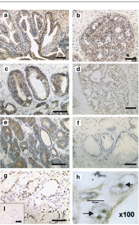
Immunohistochemistry was performed to determine the expression levels and distribution of Tip60 protein in 10 cases of BPH, 43 clinical cases of untreated CaP of different grade and stage and 15 cases of HR cancer. Histological evidence of BPH is found in more than 50% of men above the age of 45 years. Positive glandular staining for Tip60 was observed in all 10 cases of BPH examined, while the distribution of the signal was either cytoplasmic (Figure 1a) (5/10) or both cytoplasmic and nuclear (5/10) (Table 1).
We also examined Tip60 expression in normal bronchus, ileum, kidney and breast tissue. The staining profile, especially in breast (Figure 1b), was similar to
that observed for BPH, with high levels of expression particularly in the cytoplasmic compartment.
The staining pattern observed for the 43 cases of CaP ranged from high expression in both cellular compart-ments (12%) to a complete lack of expression (28%). Intermediate cases showing solely nuclear (37%) or cytoplasmic (23%) staining were also observed (Figure 1c–f and Table 1).
We found a statistically significant (P=0.008, Fischer’s exact test) correlation between signal localiza-tion and metastatic lesions at the time of diagnosis. Data on metastatic lesions were available for 36 out of the 43 patients in the CaP group. These cases were distributed into two groups based on the presence of nuclear signal. We observed that nuclear Tip60 inversely correlated with metastasis at the time of diagnosis (all 36 patients Figure 1 Expression of Tip60 protein in BPH, normal breast and prostate cancer. (a) Tip60 protein is solely expressed in the cytoplasm in 50% of the BPH cases. (b) Tip60 is expressed in normal breast tissue.(c–f)different staining patterns of Tip60 in prostate cancer cases. Tip60 was observed to be expressed throughout the cell (c), nuclear (d), cytoplasmic (e) or was undetectable (f). (g) Tip60 protein shows exclusively nuclear staining in hormone refractory prostate cancer. (h) image captured by the oil immersion lens (100) showing nucleolar accumulation of Tip60 in HR CaP (scale¼20mm), (i) negative control (—, 80mm)
were newly diagnosed and hence had received no prior hormonal ablation treatment) (Table 2).
Tip60 is predominantly nuclear in HR cancer
Tip60 expression in HR cancer was investigated in 15 cases. A distinct expression pattern was revealed, with Tip60 exclusively expressed in the nucleus in 87% (13/ 15) of cases (Figure 1g, Table 1), while in the remaining 13% (2/15) the signal was present in both cellular compartments. The specificity of the staining was also confirmed using a commercially available C-terminal antibody (Upstate Biotech) that resulted in the same nuclear-specific staining pattern (data not shown). Interestingly, in the majority (10/15) of the HR cases, the nuclear signal accumulated in subnuclear structures (Figure 1h, arrows). The above pattern was focal, with areas of the slide showing expression of Tip60 in a diffuse nucleoplasmic pattern with the presence of nuclear aggregates, whereas some areas exhibited specific, possibly nucleolar, staining (Figure 1h). The negative control failed to show any staining (Figure 1i). AR and prostate-specific antigen (PSA) expression were also examined (data not shown). All tumours expressed high levels of PSA. AR expression was lost in 5/15 cases, consistent with the reported loss of AR (20– 30%) in HR CaP (Kinoshitaet al., 2000).
Nuclear accumulation of Tip60 in the CWR22 model in response to androgen deprivation
The CWR22 prostate xenograft model was used to further investigate the effect of androgen deprivation on Tip60 distribution. CWR22 is a serially transplantable CaP xenograft that causes a marked elevation of the PSA blood levels in nude mice. In response to surgical or chemical castration, the tumour regresses along with an up to 3000-fold fall in PSA blood levels (Wainsteinet al., 1994; Nagabhushan et al., 1996). Transplanted animals were untreated (Figure 2a, b), treated with testosterone supplements (Figure 2c, d) or underwent orchidectomy (Figure 2e, f). Tumour sizes significantly changed in
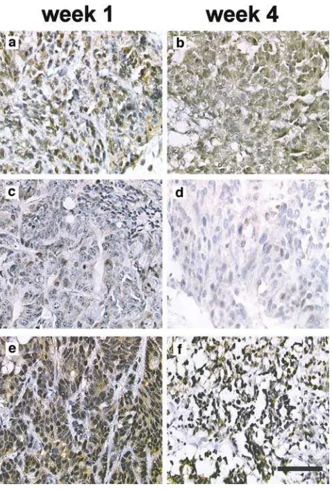
response to treatment over a period of 3 weeks confirming the model was androgen responsive (Gna-napragasamet al., 2002). In addition, PSA levels were induced by testosterone treatment and reduced by castration as previously demonstrated (Wainsteinet al., 1994). Tip60 was diffusely expressed in both cellular compartments in the untreated group at the end of week 1 (Figure 2a). After 3 weeks, there was a slight decrease in Tip60 expression levels with mainly cytoplasmic expression (Figure 2b). The difference in Tip60 expres-sion was particularly striking between samples taken Table 1 Case distribution according to the staining pattern for Tip60, within the BPH, CaP and HR cancer groups
Staining pattern Nuclear Cytoplasmic Nuclear and cytoplasmic Negative Total no. of cases
BPH 0 5/10 (50%) 5/10 (50%) 0 10
CaP 16/43 (37%) 10/43 (23%) 5/43 (12%) 12/43 (28%) 43
HR 13/15 (87%) 0 2/15 (13%) 0 15
Table 2 Nuclear expression of Tip60 is significantly inversely correlated with presence of metastasis at the time of diagnosis
Metastasis status Tip60’s localization
Nuclear +ve Nuclearve
Positive 3 (18.7%) 13 (65%)
Negative 13 (81.3%) 7 (35%)
Total 16 20
P=0.008, Fishcher’s exact test
Figure 2 Tip60 protein is upregulated and localized to the nucleus following castration in the CWR22 xenograft model. The human primary prostate cancer CWR22 xenograft model was used to investigate Tip60 protein expression. Animals were either left untreated (a,b), implanted with testosterone pellets (c,d) or castrated(e,f). Animals were killed at week 1 (a, c, e)or week 4 (b, d, f). At 4 weeks after castration, Tip60 localized almost exclusively in the nucleus(f). Testosterone decreased the staining intensity and resulted in mainly cytoplasmic localization of Tip60 (d) (—, 80mm)
after 1 and 4 weeks of testosterone treatment (Figure 2c, d, respectively). Testosterone treatment attenuated Tip60 levels and led to a more diffuse signal distribution (Figure 2d). Castration (Figure 2e, f) caused predomi-nantly nuclear localization as well as a slight upregulation of Tip60 protein levels. Within the group of animals killed at 4 weeks, the tissue samples obtained from the castrated animals group (Figure 2f) showed the most intense nuclear signal as opposed to a weak diffuse Tip60 signal in the case of the testosterone-supplemented group (Figure 2d). Interestingly, the latter exhibited weaker expression compared to the corresponding control (Figure 2b), which was under the influence of the much lower endogenous mouse androgen levels. Samples obtained at 2 and 3 weeks of treatment failed to show a substantial shift in the staining pattern (data not shown).
Tip60 expression and cytoplasmic/nuclear distribution is regulated by hormone treatment in LNCaP cells and transfected COS7 cells
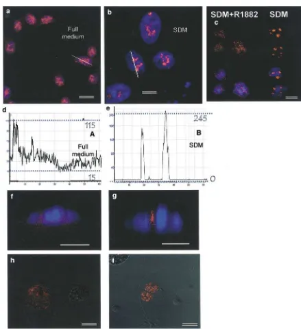
The effect of androgens on Tip60 expression and localization was investigated in the androgen-respon-sive, AR-expressing LNCaP prostate cancer cell. Cells were grown in full medium (FM) containing steroid hormones and growth factors (Figure 3a, f) or in steroid depleted medium (SDM) (see Materials and methods, Figure 3b, g). The results supported the previously observed trend: androgen deprivation caused nuclear localization and upregulation of Tip60. This trend was seen to increase with time (data not shown), with maximum expression and nuclear accumulation ob-served following 3 days of hormone depletion (Figure 3b and histogram b). On the contrary, growth in FM resulted in low and diffuse protein expression through-out both cellular compartments (Figure 3a and histo-gram a). A similar trend was found when SDM grown cells (Figure 3d) were compared to cells grown in SDM supplemented with the synthetic androgen R1881 (10 nm) for a period of 3 days (Figure 3c), therefore
suggesting that androgen presence is sufficient to cause the above redistribution. To further support the above trend, the immunofluorescence signal was quantified using the LCS 2.00.585 software and paired two-sample
t-test performed on 20 randomly selected cells per optical field for each treatment (absence (SDM) or presence of R1881 (SDM+R1881)), in three indepen-dent experiments. A statistically significant upregulation of the Tip60 signal was observed upon androgen withdrawal (P¼0.02) (data not shown). The negative control (no primary antibody) failed to show any fluorescent signal (Figure 3e). Figure 3f and g corre-sponds to A and B, respectively. Z-series were collected to illustrate the intranuclear pattern of Tip60 in the different conditions. The diffuse nucleoplasmic signal in FM (Figure 3f) becomes concentrated in the interchro-matin space in the absence of hormones and growth factors (Figure 3g).
COS7 cells are lacking the AR and were transiently transfected with Tip60-RFP. Signal was visualized within living cells to rule out any artifact caused by
fixation. COS7 cells cultured in FM displayed a diffuse Tip60 signal in both cellular compartments (Figure 3h), whereas cells grown in SDM displayed a distinct nuclear speckled pattern (Figure 3i). RFP images were over-lapped with DIC/Nomarski images to display the subcellular signal distribution and verify cell viability during scanning (see Materials and methods). The above trend was supported by indirect immunofluorescence carried out for endogenously expressed Tip60 in COS7 cells grown in the presence (SDM+R1881) or absence (SDM) of androgens (data not shown).
Subnuclear rearrangement of Tip60 in response to androgen deprivation
We report a speckled distribution of both endogenous and transiently transfected Tip60 in the nucleus of LNCaP and COS7 cells grown in SDM, as opposed to a less prominent speckled pattern along with a more diffuse nucleoplasmic signal, when the cells were maintained in androgen-containing medium. Fluores-cence signal quantification enabled us to study the intranuclear distribution of Tip60 in both the presence and absence of androgens (Figure 3a, d and correspond-ing histograms).
As illustrated by the ‘line’ quantification, SDM causes not only an upregulation of Tip60 protein levels, but also an accumulation of nuclear structures not overlapping with chromatin 4,6-diamidino-2-phenylin-dole (DAPI stain) (histogram 3b – two distinct peaks and no nucleoplasmic signal). Cells grown in FM displayed a weak, diffuse cytoplasmic signal, less prominent nuclear speckled pattern with lower peaks and a uniform nucleoplasmic background expression (histogram 3a). Similar quantification data were ob-tained when the fluorescence signal in SDM versus SDM+R1881 was measured (data not shown). To further support the quantification data, Z-series were collected. Figures 3f and g correspond to a and b, respectively, and illustrate the intranuclear pattern of Tip60 under the different conditions. The diffuse nucleoplasmic signal in FM (Figure 3f) becomes concentrated in the interchromatin space in the absence of hormones and growth factors (Figure 3g).
Androgen depletion upregulates Tip60 protein levels in LNCaP cell lysates
We verified the above observations by performing Westerns on LNCaP cell lysates, in the corresponding cases studied by immunofluorescence. Androgen pre-sence caused a downregulation of Tip60 protein levels, compared to cells grown in SDM (Figure 4).
Study of intracellular protein levels of Tip60 by flow cytometry
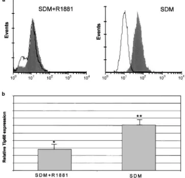
Flow cytometry was used to examine intracellular expression of Tip60 in LNCaP cells (Figure 5). The unique advantage of this technique is that it provides a collection of individual measurements from a large
number of discrete cells, rather than making a bulk estimation. The difference of the means between the negative control and the sample stained for Tip60 was calculated in three individual experiments (Figure 5b). Pairedt-test was used to assess the statistical significance of the difference in each condition (SDM+R1881 and
SDM). In both cases, the shift between the unstained control and the test sample for Tip60 was statistically significant with P-values of 0.04 and 0.002, for SDM+R1881 and SDM, respectively. These results confirm that androgen depletion causes upregulation of Tip60 protein levels.
Figure 3 Tip60 is upregulated and translocates to the nucleus in response to androgen withdrawal. Endogenous Tip60 was detected by immunofluorescence in LNCaP cells cultured in either FM (a) or SDM(b), for three days. Histogramsaandb(corresponding to Figures 3a and b), line scans measuring Tip60 signal intensity in cells cultured in FM or SDM.X-axis represents the line pixel number and Y-axis the relative signal intensity (see text). Figures (c, d) illustrate endogenous Tip60 in LNCaP cells in the presence (SDM+R1881) or absence (SDM) of androgen, respectively. Upper panel: Tip60, lower panel: Tip60/DAPI overlay.(e)Control LNCaP cells showing no background immunofluorescence.(f, g), Z-series corresponding to a and b, respectively, showing nuclear localization throughout the entire thickness of the nucleus in a punctuate pattern in the absence of androgens (g)or diffusely throughout the nucleoplasm in FM(f). COS7 cells were transfected with Tip60-RFP and the fluorescence image was overlapped with the corresponding DIC image to show cellular morphology in living cells. A diffuse pattern throughout both cellular compartments was observed in FM(h)compared to an exclusively nuclear pattern in SDM (I) (¼, 20mm; — , 8mm)
Tip60 mRNA is upregulated in androgen-depleted conditions
To determine whether Tip60 regulation is controlled at the transcriptional level, we assayed mRNA levels in LNCaP cells following different hormone treatments using RT–PCR. Cells were grown in SDM or SDM+10 nmR1881, for 3 days (Figure 6).
Two different starting amounts of cDNA were used to enable semiquantification of our results. The difference
between the treatments followed a logarithmic scale in both cases of the cDNA input used (data not shown). Our data indicate Tip60’s upregulation at the transcript level in the absence of androgens (lane 1). A reduction in Tip60 mRNA levels was apparent following 8 h of exposure to 10 nmR1881, with highest effect noted after a 72 h exposure (data not shown). Similarly, androgen
Figure 4 Tip60 protein is upregulated in androgen-depleted conditions. Western analysis showing the difference in Tip60 protein levels between cells grown either in the absence (SDM – lane 1) or presence (SDM+R1881 – lane 2) of androgens.a-tubulin loading control in lower panel
Figure 5 Upregulation of Tip60 protein levels upon androgen deprivation. LNCaP cells were grown in SDM in either the presence or absence of R1881 for 3 days and then analysed for expression of Tip60 using flow cytometry.(a), Open histogram indicates the negative control (no primary antibody); filled histogram, Tip60. (b), Column indicates mean (7s.d.) of three independent experiments. *Po0.05, **Po0.01 as determined by Student’st-test difference of the means compared with the control group
Figure 6 Androgen withdrawal increased expression of Tip60 mRNA. RT–PCR was performed on RNA extracted from LNCaP cells treated in different conditions, to evaluate the effect of hormone deprivation on Tip60 expression. Lane 1, 72 h SDM; lane 2, 72 h SDM+R1881 (10 nm). Lower panel represents b-actin loading control
(0.001–100 nm) applied for 3 days caused a
dose-dependent reduction in Tip60 mRNA levels (data not shown). In conclusion, the trend observed at the protein level corresponds to a shift in the expression pattern of Tip60 at the mRNA level. However, we cannot rule out an additional regulation at the translational level.
Tip60’s role in transcription of androgen-regulated PSA gene in androgen-dependent and -independent cell systems
To further understand Tip60’s involvement in
transcrip-tion in vivo, chromatin immunoprecipitation (ChIP)
assays were performed in the AR-positive, androgen-dependent CaP cell line LNCaP and in an AI (androgen-independent) LNCaP derivative. The promoter of the PSA gene was chosen as the target (Cleutjens et al., 1996), since androgen-induced PSA synthesis is a well-characterized even in LNCaP cells (Younget al., 1992;
Lee et al., 1995) and PSA has been used as a
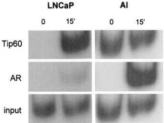
prostate-specific tumour marker (Brawer, 2000). AI is an LNCaP derivative that expresses the AR at higher levels compared to LNCaP cells, but is androgen independent in terms of cell growth and proliferation, and its cell cycle profile remains unaffected by androgen manipula-tion. PSA expression is preserved, and this gene remains androgen regulated (data not shown, manuscript in preparation). Tip60 was recruited to the PSA promoter after androgen stimulation in both cell lines. Specifi-cally, Tip60 showed a high basal recruitment in the absence of androgen in AI cells, while there was no recruitment in the absence of stimulus in LNCaP cells (Figure 7). Following 15 min of androgen stimulation, Tip60 was found bound to the promoter in both cases. AR, on the other hand, was more strongly recruited to the PSA promoter following androgen stimulation in the case of AI compared to LNCaP/PSA levels, monitored by Western analysis, were found to be induced at similar levels after androgen stimulation in both systems (data not shown). Genomic DNA control reactions (input) as well as negative water controls were performed
along-side the immunoprecipitated samples. Together, our results show a distinctively different recruit-ment pattern of Tip60 and the AR to the PSA promoter of androgen-dependent and -independent CaP cell lines. Recruitment of the AR to the PSA promoter is a rapid, ligand-dependent event in both LNCaP and AI cells. In contrast, Tip60’s recruitment profile is altered in the absence of androgen in AI, unlike LNCaP cells. We can therefore argue that Tip60’s recruitment is not ligand dependent in this androgen-independent AR- and PSA-expressing cell model that closely simulates HR CaP.
Discussion
We previously demonstrated that Tip60 interacts directly with the AR and enhances its androgen-induced transcriptional activity (Brady et al., 1999). The distribution pattern and the expression levels of this coactivator in prostate tissue have not yet been addressed. Indeed, very little is known about the expression of coregulators in CaP. To gain a better understanding of Tip60’s role in the biology of the disease, we generated an antibody to study Tip60’s expression profile in both benign and malignant prostate tissue. We investigated endogenous expression of Tip60 in BPH, malignant tissue of different grades, and in HR CaP. The mouse xenograft model CWR22 and an androgen-responsive cell line, LNCaP, were also exam-ined. Lastly, ChIP assays were carried out to further elucidate the molecular mechanism underlying Tip60’s involvement in transcription of AR target genes in both androgen-dependent and -independent systems.
The contribution of transcriptional coactivators in cancer development remains unclear. However, there have been reports where upregulation and/or dysfunc-tional subcellular trafficking of coactivator molecules correlates with a more malignant phenotype. For example, the expression levels of several coactivators (TIF2, AIB1, CBP and PCAF) are upregulated during breast cancer development, and correlate with the aggressive phenotype of advanced disease (Kurebayashi
et al., 2000). In contrast, Chenet al. (2001) reported that the rhabdomyosarcoma phenotype associates with a dysfunctional subcellular localization of p160 family members. In the present study, we observed a change in the expression and localization of Tip60 in advanced CaP. Furthermore, our results agree with a recent report (Gregoryet al., 2001), where overexpression and nuclear localization of both SRC1 and TIF2 coincided with the onset of CaP recurrence. Immunoperoxidase staining was exclusively nuclear in 87% of HR cancer, in over one-third of androgen-responsive CaP and in none of the BPH cases (Table 1). In the mouse CWR22 xenograft model, Tip60 localized specifically in the nucleus of cancer cells in castrated animals. The exclusively nuclear localization of Tip60 in the majority of cases of HR CaP may be, in part, responsible for the increased AR sensitivity to extremely low levels of Figure 7 ChIP assay demonstrating Tip60’s involvement in
circulating androgens and its transcriptionally active state even after castration. Furthermore, prolonged androgen deprivation may lead to increased Tip60 nuclear recruitment, as shown by HR cases and the CWR22 model, and thus activate alternative, androgen-independent signalling pathways and account, at least in part, for the aggressive HR phenotype of advanced CaP. Data concerning the metastatic state of the newly diagnosed CaP patients were available for 36 out of a total of 43 cases. There was a statistically significant (P¼0.008) correlation between the localization of Tip60 and metastasis at the time of diagnosis. Nuclear expression of Tip60 correlated with a less aggressive phenotype, while cases where Tip60 was excluded from the nuclear compartment had a significantly higher incidence of metastasis. Interestingly, nuclear expression of BAG-1, a multifunctional protein promoting cell survival, proliferation and metastasis, was found to be significantly inversely correlated with tumour grade and directly associated with improved overall survival (Cutresset al., 2001).
The above results do not contradict our observation of Tip60’s predominantly nuclear expression in HR CaP. This group of patients received long-term hormone ablation treatment, and biopsies were obtained when the tumour ceased to respond to androgen deprivation. In contrast, the group of the 36 CaP patients consisted of newly diagnosed cases and metastasis data corresponded to the time point of diagnosis, prior to any hormonal treatment (see Materials and methods). Therefore, these two groups cannot be directly compared. It would be of interest to examine Tip60’s expression pattern in CaP cases before and after the development of androgen unresponsiveness, and thus clarify the effect of androgen deprivation on Tip60’s localization and expression levels. However, our CWR22 data strongly suggest that the hormonal environment affects both these parameters.
Our immunofluorescence data support the previously discussed tissue observations and indicate that reintro-duction of androgens is sufficient to cause Tip60’s cellular redistribution. Interestingly, androgen stimula-tion results in similar changes in terms of protein levels and distribution pattern in two relatively different cellular systems: LNCaP and COS7 cells. The latter does not express the AR; however, androgen treatment may cause a response via an alternative, AR-indepen-dent pathway, possibly involving a different nuclear receptor. This is further supported by our ChIP data, where Tip60 is involved in androgen-dependent tran-scription in an androgen-independent, in terms of growth and proliferation, model system (see below). Our hypothesis is that Tip60 can respond to androgen stimulation even in androgen-independent or AR-negative systems through alternative signalling pathways, which makes it important in advanced, hormone-independent CaP. COS7 cells express Tip60 at similar levels to LNCaP cells (data not shown). Endogenous Tip60 responds to androgen stimuli to a lesser degree (only half of the cells are responsive) but in a similar manner to transiently transfected cells (data not shown); therefore, our results are not caused by
artificially overexpressed Tip60 protein. Furthermore, these results support our hypothesis of a functional, AR-independent but androgen-responsive pathway in these cells.
Our observations, however, focus not only on the subcellular localization of Tip60, but also on the subnuclear distribution of the protein as a response to the presence or absence of hormonal stimuli.
The importance of the subnuclear organization of proteins is revealed by the effects of mislocalization of nuclear proteins (discussed in Kokenet al., 1997). Tip60 has been reported to exhibit a nuclear punctuate pattern (Yamamota and Horikoshi, 1997; Ran and Pereira-Smith, 2000), when transiently overexpressed in cell lines. In our study, upon androgen deprivation, Tip60 exhibited a distinct speckled nuclear distribution similar to that observed for nuclear splicing and transcription factors (Spector, 1993; Zenget al., 1997). Our observa-tions confirm the previously reported speckled nuclear distribution of Tip60, both for endogenous and trans-fected Tip60 protein, supporting Tip60’s potential to be incorporated in distinct nuclear complexesin vivo(Ikura
et al., 2000). Furthermore, our results suggest a link
between Tip60’s subnuclear localization and hormonal stimuli.
Regulation of nuclear receptor expression is a common mechanism for modulating cell responses to hormones and growth factors, and it is well established that nuclear receptor gene expression is hormonally regulated. Our results suggest that Tip60’s expression could be driven by hormonal signals. Similar observa-tions have been made for other coregulatory molecules. For example, SRC-1 is downregulated by dexametha-sone (Kuriharaet al., 2000) and oestrogen (Misitiet al., 1998). RIP140 corepressor expression is increased 2–3 fold by 17b-oestradiol in MCF7 breast cancer cells, while antioestrogens had the opposite effect (Thenot
et al., 1999). The recently isolated human corepressor
SHARP is induced by oestrogen (Shiet al., 2001). These observations suggest that a regulatory mechanism exists, acting via a negative feedback loop, to protect cells against excessive hormone effects. Thus, hormone-induced upregulation of repressor factors and/or down-regulation of coactivator molecules can serve such a regulatory mechanism. A loss of this mechanism in malignant tissue could account for the uncontrolled, abnormal growth. Supporting those observations, our semiquantitative study of Tip60 transcript levels showed an increased attenuation of Tip60’s signal over longer exposure times and increasing androgen concentration. Inspite of the fact that we cannot rule out an additional regulation at the post-transcriptional level there seems to be a regulation of Tip60 mRNA levels by androgens. At the protein level, quantitative immunofluoresence, flow cytometry and Western blot data indicate a similar trend.
In order to describe in a more mechanistic manner, Tip60’s involvement in transcription and its potential implications in the development of HR disease, we looked for putative model systems closely related to the HR state in CaP patients. DU145 and PC3 cells are
well-characterized androgen-independent CaP cell lines. Tip60’s overexpression in a tetracycline-inducible ex-pression system of a PC3 subline led to enhanced transcription of ostrogen receptor pathways (unpub-lished data). However, PC3 cells, like DU145, do not express AR or PSA. These features make them poor representatives of HR tumours, usually characterized by elevated PSA expression and mutated or amplified AR (only one in five HR CaP cases lose AR expression despite an androgen-independent phenotype). There-fore, we studied a cell line (AI) that was androgen-independent in terms of proliferation, yet retained AR expression and maintained PSA responsiveness to androgen stimulation (manuscript in preparation). AI cells are LNCaP derivatives grown for extended periods in androgen-depleted medium. Similar cell lines have been developed and characterized (Soto et al., 1995;
Gaoet al., 1999). A ligand-dependent recruitment of the
AR to the PSA promoter was evident in both LNCaP and AI cell lines. More AR protein was recruited to the PSA promoter, however, in the androgen-independent AI cells. Those cells express AR at higher levels relative to the parental LNCaP cell line, possibly in an attempt to overcome long-term androgen ablation and increase its sensitivity to the extremely low levels of ligand present. On the other hand, Tip60 exhibits a totally altered profile as androgen independence develops. R1881 induced a rapid recruitment of Tip60 to the PSA promoter in LNCaP cells. However, in AI cells, Tip60 was found to be constitutively recruited to the promoter even in the absence of stimulus. This may explain the relatively high expression of PSA noted in AI cells, in an AI environment. It is possible therefore that androgen-dependent genes may remain switched on by the continuous loading of coactivator proteins, like Tip60, to their promoter region even in the absence of androgen.
Our results suggest an androgen-dependent regulation of Tip60 expression and localization. However, this effect may not be because of a direct androgen action on the Tip60 promoter via the activated AR. The AR is an essential mediator of differentiation cell growth and proliferation in androgen-responsive tissues. Therefore, the effect observed may be indirect, caused by induction of proliferation. Androgen and antiandrogen treatments affect the cell cycle profile and may regulate Tip60 levels in the different cell cycle phases. Indeed, our flow cytometry analysis of the profile of cycling cells upon different androgen treatments shows a G1/0 accumula-tion when cells are grown in the absence of androgens (SDM) and an increase in S phase population upon androgen reintroduction (our observations; Burtkhart
et al., 1999; Horikawa et al., 2001). However, this does not rule out the possibility of a direct androgen effect upon Tip60 expression.
Recent studies suggest a two-way interaction between histone acetyltransferase (HAT) complexes and cell cycle regulatory molecules. For example, TRRAP, a member of both Tip60 and PCAF complexes, regulates the activity of E2F and c-Myc (Mcmahon et al., 1998). Tip60 may similarly exert an effect on such regulatory
molecules. Alternatively, cyclin D1, a G1 phase reg-ulatory cyclin, was recently shown to bind to the PCAF HAT domain, displacing the AR and inhibiting its transcriptional activity, in a hormone dependent manner (Reutens et al., 2001). These observations provide further support for a model in which an androgen inducible cell cycle regulatory molecule may interface with the activity of histone-modifying complexes.
In summary, our results indicate that in HR CaP, Tip60 is predominantly found in the nucleus as opposed to a more diffuse distribution pattern observed in BPH and CaP. Upregulation at both the mRNA and protein levels was also demonstrated when androgens are withdrawn both in vitro (cell lines) and in vivo
(CWR22 xenograft model). Those observations suggest a role for Tip60, an AR coactivator, in the progression of CaP to the HR status after hormone ablation treatment. The predominant nuclear localization and upregulation of Tip60 in response to androgen depletion and the consequent development of androgen indepen-dence directly link the coactivator Tip60 to the molecular pathway of progression to androgen insensi-tivity. Additionally, our ChIP data implicate Tip60 in the transcription of androgen-targeted genes like PSA and demonstrate for the first time the constitutive, ligand-independent recruitment of a HAT enzyme and AR coactivator to the PSA promoter, in an AI cell model of HR CaP. Further studies are needed to identify the factors that control Tip60’s expression and its mechanism of action, since this coactivator might constitute a therapeutic target in the most lethal phase of prostate malignancy, that of hormone-independent CaP.
Materials and methods
Patient data
Patients with newly diagnosed CaP (n¼43) as well as those with documented hormone relapsed cancer (n¼15) were identified from a pathology database. Newly diagnosed cases were defined as those without prior hormone therapy and diagnosed at the time of surgery. The presence of metastatic lesions was determined at the time of diagnosis. Hormone relapsed cases were defined as patients with documented resurgence of serum PSA following androgen ablation therapy. All patients had presented with bladder outflow obstruction and required TURP. Sections of breast cancer and normal kidney, bronchus and ileum were identified from archival material. A total of 10 cases of BPH were also examined. Following formalin fixation and embedding in paraffin, 4mm sections were mounted on APES-coated slides for further study.
Generation of Tip60 constructs
PCR was used to amplify full-length Tip60 cDNA from POZ viral vector (Gift from Y Nakatani, Dana-Farber Institute). A FLAG-tagged Tip60 was cloned into mamma-lian expression vector pCMV5 (Invitrogen). Tip60 was also cloned into the expression vector pDSREDN-1 (Clontech) to create Tip60-RFP. DNA sequencing confirmed construct integrity.
Transient transfections of COS-7 cells
Cells were cultured for 48 h in RPMI1640 medium (Life Technologies, Inc). supplemented with 10% fetal calf serum in 35 mm wells (Corning Inc, Costar). Transfections were performed with Superfect reagent (Qiagen) following the manufacturer’s guidelines.
Tip60 antibody
A 15 amino-acid peptide within the conserved MYST homology region (283–297 aa) was synthesized and used to immunize rabbits. The polyclonal antiserum was affinity purified using protein A and peptide affinity columns. Western blotting was performed to check the antibody specificity in lysates from different cell lines. A commercially available antibody (Upstate Biotechnology), directed against the C-terminus of Tip60, was also used.
Western blotting
Sample preparation, electrophoretic separation and Western blotting were performed as described (Bradyet al., 1999).
Immunohistochemistry
Immunohistochemical studies were carried out using the avidin–biotin–peroxidase complex method (ABC), based on the manufacturer’s instructions (Vectastain Elite kit, Vector Laboratories, CA, USA). Antigen retrieval was performed as previously described (Nortonet al., 1994; Krenacset al., 1996).
Brightfield microscopy
Images were acquired using the Leica DMR light microscope (20, 40 and 100 oil immersion lenses) and analysed using the Spot Advanced 3.5.2, 1997–2002 software and Adobe Photoshop 6.00.
CWR22 xenograft model
The CWR22 xenograft model was established as previously described (Gnanapragasam et al., 2002), under UK Home Office regulation with an assigned project license number. When the tumour was palpable (4–6 weeks), animals were allocated in three groups: Group 1, subcutaneous testosterone implant; Group 2, untreated; Group 3, surgically castrated. Animals were killed on the day of randomization (day 0) and subsequently on days 7, 14, 21 and 28. All mice had blood PSA and testosterone levels measured.
Cell culture
LNCaP and COS7 cells were cultured in RPMI1640 media (Sigma) referred to as FM, supplemented with 10% fetal calf serum, 1% glutamine, penicillin (100 U/ml), and streptomycin (100mg/ml), as previously described (Swinnen et al., 1994). SDM was made as previously described (Bradyet al., 1999). AI cells were derived from LNCaP cells following continuous passaging in an environment deprived of androgens for a period of 8 months. AR expression has been preserved, but the cells do not depend upon androgen to proliferate (manuscript in preparation).
Immunofluorescence
LNCaP cells were seeded in eight-well chamber slides (Becton-Dickinson) at densities of 5103cells/well. The cells were fixed with 100% methanol for 30 min at201C and incubated with
the primary antibodies at 41C overnight. The following day, the cells were washed (3) and treated with Alexa 633 nm (1 : 200, Molecular Probes) secondary Ab, for 1 h at RT in the dark. All washes were performed in TBS 50 nM, Triton-X 0.4%/blocking serum 0.03%. Vectashield with DAPI, for nuclear counterstain, was used to mount the slides.
A series of 1mm verticle optical sections through the entire thickness of the tissue was used to produce a Z-series. To quantify fluorescent signal intensity, the intensity of pixels along a line drawn through lateral cell borders was recorded and relative signal strength was calculated. The average pixel intensity, normalized for the cell surface, was measured using the ‘polygonal area’ tool of the LCS 2.00.585 software to allow signal quantification in the presence and absence of androgens.
Confocal microscopy
All images of fixed cells were acquired using the 63 oil immersion lens (1.32 NA DIC) of a Leica TCS SP2 UV laser-scanning microscope. DAPI was visualized with the 351/ 364 nm UV/Ar laser line (max power: 20 mW), FITC was visualized with the 488 nm Ar laser line (max power: 20 mW) and Alexa633 nm was visualized using the He/Ne line (max power: 1.2 mW). All images were analyzed using the LCS 2.00.585 software and Adobe Photoshop 6.0.
Live cell imaging
Cells were grown in 35 mm Petri dishes and visualized with an HCX APO L40 water-dipping lens (0.8 NA) of the Leica TCS SP2 UV laser-scanning microscope. The argon laser spectral line at a wavelength of 488 nm was set at an intensity of no greater than 25% of its total power (1.2 mW) for image collection. This laser intensity caused no cell damage over a total scanning time of 5 min (unpublished data). The scanning time in our experiments was less than 10 s. A 50% neutraliza-tion filter was used to prevent bleaching, To assess cell viability during the scanning period, we monitored cells by differential interference contrast (DIC) optics for changes in cellular morphology, before, during and after scanning. No significant changes were detectable. DIC and fluorescence images were overlaid using Adobe Photoshop 6.0.
RT–PCR
Total RNA was isolated from LNCaP cells using TriZol reagent (GIBCO-BRL), according to the manufacturer’s recommendations. Complementary DNA was synthesized from 2mg of total RNA using MMLV reverse transcriptase (Promega, UK). PCR was performed using standard condi-tions, and included 1/5th of the reverse transcriptase reaction. The oligonucleotide primers used to amplify Tip60 (Accession number: U74667) were 50-AGC GTC ATT TGA CCA AGT GT-30and 50-AGT TCA TAG CTG AAC TCG AT-30(RT1 and RT2, respectively). The thermal profile consisted of initial melt at 951C for 3 min, followed by 30 cycles comprising 951C, 30 s; 561C, 20 s; 721C, 30 s. A 5-min extension was used for the final cycle.b-actin, a housekeeping control gene, was used to indicate cDNA loading. Amplification products were resolved by electrophoresis on 1% agarose gel containing ethidium bromide and visualized by UV. Densitometry of the bands was performed using the Bio-Rad Molecular Analyst (version 1.4) software and the relative numbers were normalized to the correspondingb-actin controls.
ChIP assay
ChIP assays were performed as previously descried (Gaughan et al., 2002). For the PCR reactions the following set of primers were used: F: AGG GAT CAG GGA TCA TCA CA and R: GCT AGC ACT TGC TGT TTC TGC which amplify the 406 to 164 PSA promoter containing androgen-responsive element II (Shanget al., 2002).
FACS analysis for intracellular expression of Tip60
For FACS analysis, cells (5105 cells) were trypsinized and washed with 10% fetal calf serum in PBS. Cells were subsequently washed with cold PBS and fixed with 2% paraformaldehyde for 30 min at 41C. Cells were permeabilized with 0.2% Triton-X for 5 min at 41C and stained with the anti-Tip60 antibody (Upstate Biotech; 1 : 50) for 1 h. All incuba-tions were performed at 41C. After each incubation, cells were washed in PBS containing 0.05/NGS. Cells were incubated for 1 h with a fluorescein-conjugated goat anti-rabbit antibody (Sigma; 1:200), in the dark. Samples were then analysed in a FACScan flow cytometer (Becton-Dickinson). Each analysis included 5000 events collected in list mode and analysed using Lysis II software (Becton-Dickinson) and WinMDI 2.8. Controls included cells stained with secondary antibody alone and cells with no antibody addition, to determine the autofluorescence levels of LNCaP cells. All experiments were independently performed at least three times.
Statistical analysis
To study correlation of metastasis with Tip60’s distribution pattern, statistical analysis was performed using Fischer’s exact test. Student’s t-test was used to analyse FACS and fluorescent quantification data. A P-value of o0.05 was considered statistically significant in all cases.
Abbreviations
AR, androgen receptor; CaP, prostate cancer; ChlP, chroma-tin immunoprecipitation; CWR22, Case Western Reserve prostate xenograft strain 22; RT–PCR, reverse transcription polymerase chain reaction; HR, hormone refractory; SDM, steroid-depleted medium; FM, full medium; CaS, bicalutamide (Casodex); TURP, transurethral resection of the prostate; PSA, prostate-specific antigen; Tip60, Tat interactive protein 60 kDa; AI, androgen independent.
Acknowledgements
We are grateful to Yoshihiro Nakatani for generously providing us with the Tip60 cDNA, and Trevor Booth (Electron Microscopy Unit, Newcastle Medical School) for helpful discussions and excellent technical assistance.
References
Brady ME, Ozanne DM, Gaughan L, Waite I, Cook S, Neal DEand Robson CN. (1999). J. Biol. Chem., 274, 17599–17604.
Brawer MK. (2000).Semin. Surg. Oncol.,18,3–9.
Burkhart BA, Alcorta DA, Chiao C, Isaacs JS and Barrett JC. (1999).Exo. Cell. Res.,247,168–175.
Caira F, Antonson P, Pelto-Huikko M, Treuter Eand Gustafsson JA. (2000).J. Biol. Chem.,275,5308–5317. Catalona WJ. (1994).N. Engl. J. Med,331,996–1004. Chen SL, Wang SC, Hosking B and Muscat GE(2001).Mol.
Endocrinol.,15,783–796.
Cleutjens KB, van Eekelen CC, van der Korput HA, Brinkmann AO and Trapman J. (1996). J. Biol. Chem., 271,6379–6388.
Crawford ED. (1992).Br. J. Urol.,70,33–38.
Cutress RI, Townsend PA, Bateman AC, Johnson PW, Ryder K, Barnes DM and Packham G. (2001).J. Clin. Oncol.,19, 3706–3707.
Gao M, Ossowski L and Ferrari AC. (1999).J. Cell Physiol., 179,336–346.
Gaughan L, Brady ME, Cook S, Neal DE and Robson, CN. (2001).J. Biol. Chem.,276,46841–46848.
Gaughan L, Logan IR, Cook S, Neal DEand Robson, CN. (2002).J. Biol. Chem.,277,25904–25913.
Gnanapragasam VJ, Robson CN, Neal DEand Leung HY. (2002).Oncogene,21,5069–5080.
Gregory CW, He B, Johnson RT, Ford OH, Mohler JL, French FS and Wilson EM. (2001). Cancer Res., 61, 4315–4319.
Horikawa I., Parker ES, Solomon GG and Barrett JC. (2001). J. Cell. Biochem.,82,415–421.
Ikura, T., Ogrysko VV, Grigoriev M, Groisman R, Wang J, Horikoshi M, Scully R, Qin J and Nakatani Y. (2000).Cell, 102,463–473.
Kinoshita H, Shi Y, Sandefur C, Meisner LF, Chang C, Choon A, Reznikoff CR, Bova GS, Friedl A and Jarrard DF. (2000).Cancer Res.,60,3623–3630.
Koken MH, Reid A, Quignon F, Chelbi-Alix MK, Davies JM, Kabarowski JH, Zhu J, Dong S, Chen S, Chen Z, Tan CC, Licht J, Waxman S, de The H and Zelent A. (1997)Proc. Natl. Acad. Sci. USA.,94,10255–10260.
Krenacs L, Harris CA, Raffeld M and Jaffe ES (1996)J. Cell. Pathol.,1, 125–136.
Kurebayashi J, Otsuki T, Kunisue H, Tanaka K, Yamamoto S and Sonoo H. (2000).Clin. Cancer. Res.,6,512–518. Kurihara I, Shibata H., Suzuki T., Ando T., Kobayashi S.,
Hayashi M., Saito I. and Saruta T. (2000).Endocr. Res.,26, 1033–1038.
Lee HJ, Chun M and Kandror KV. (2001).J. Biol. Chem.,276, 16597–16600.
Lee C, Sutkowski DM, Sensibar JA, Zelner D, Kim I, Amsel I, Shaw N, Prins GS and Kozlowski, JM. (1995) Endocrinol-ogy,136,796–803.
Majeed A, Babb P, Kones J and Quinn M. (2000).BJU Int., 85,1058–1062.
McMahon SB, Van Buskirk HA, Drugan KA, Copeland TD and Cole MD. (1998)Cell,94,363–374.
Misiti S, Schomburg L, Yen PM and Chin WW. (1998) Endocrinology,139,2493–2500.
Muller JM, Isele U., Metzger E., Rempel A, Moser M, Pscherer A, Breyer T, Holubarsch C, Buettner R and Schule R. (2000).EMBO J.,19,359–369.
Nagabhushan M, Miller CM, Pretlow TP, Giaconia JM, Edgehouse NL, Schwartz S, Kung HJ, de Vere White RW, Gumerlock PH, Resnick MI, Amini SB and Pretlow TG. (1996).Cancer Res.,56,3042–3046.
Norton AJ, Jordan S and Yeomans P. (1994).J Pathol.,173, 371–379.
Ran Q and Pereira-Smith OM. (2000). Gene, 258, 141–146.
Reutens AT, Fu M, Wang C, Albanese C, McPhaul MJ, Sun Z, Balk SP, Janne OA, Palvimo JJ and Pestell RG. (2001). Mol. Endocrinol.,15,797–811.
Shang Y, Myers M and Brown M. (2002). Mol. Cell., 9, 601–610.
Sheridan AM, Force T, Yoon HJ, O’Leary E, Choukroun G, Taheri MR and Bonvertre JV (2001). Mol. Cell. Biol.,21, 4470–4481.
Shi Y, Dones M, Xie W, Kao HY, Ordentlich P, Tsai CC, Hon M and Evans RM. (2001).Genes. Dev.,15,1140–1151. Sliva D., Zhu YX, Tsai S, Kamine J and Yang YC. (1999).
Biochem. Biophys. Res. Commun.,263,149–155.
Soto AM, Lin TM, Sakabe K, Olea N, Damassa DA and Sonnenschein C. (1995).Oncol. Res.,7,545–558.
Spector DL. (1993).Curr. Opin. Cell. Biol.,5,442–447. Swinnen JV, Esquenet M, Heynes W, Rombauts W and
Verhoeven G. (1994).Mol. Cell. Endocrinol.104,153–162.
Tekur S, Lau KM, Long J, Burnstein K and Ho SM. (2001). Mol. Carcinog.,30,1–13.
Thenot S, Charpin M, Bonnet S and Cavailles V. (1999).Mol. Cell. Endocrinol.,156,85–93.
Tsai MJ and O’Malley BW. (1994). Annu. Rev. Biochem.,63, 451–486.
Wainstein MA, He F, Robinson D, Kung HJ, Schwartz S, Giaconia JM and Edgehouse NL, Pretlow TP, Bodner DR and Kursh ED. (1994).Cancer Res.,54, 6049–6052. Yamamoto T and Horikoshi M. (1997).J. Biol. Chem.,272,
30595–30598.
Yeh S and Chang C. (1996).Proc. Natl. Acad. Sci. USA.,93, 5517–5521.
Young CY, Andrews PE, Montgomery BT and Tindall DJ. (1992).Biochemistry,31,818–824.
Zeng C, Kim E, Warren SL and Berget SM. (1997).EMBO J., 16,1401–1412.
2477