Non-cardioembolic Mechanisms in Cryptogenic Stroke:
Clinical and Diffusion-weighted Imaging Features
Oh Young Bang, M.D., Ph.D., Phil Hyu Lee, M.D., Ph.D., Seung Hyeon Yeo, M.D., Ji Won Kim, M.D., In Soo Joo, M.D., Kyoon Huh, M.D.
Department of Neurology, Ajou University School of Medicine, Suwon, Korea
Background: The role of several cardiogenic risk factors, including patent foramen ovale, in patients with cryptogenic stroke has been extensively studied. However, little attention has been paid to the role of non-cardioembolic causes of cryptogenic stroke. We therefore sought to identify the characteristics of cryptogenic stroke.
Methods: We studied 832 patients with acute infarction in the middle cerebral arterial territory. We divided the patients into four subtypes: 402 with large artery atherosclerosis (LAA), 133 with cardioembolism, 182 with small arterial occlusion (SAO), and 115 with cryptogenic stroke. We compared risk factors and lesion patterns observed by diffusion-weighted imaging (DWI) between patients with cryptogenic stroke and those with stroke of other subtypes.
Results: Both risk factors and DWI lesion patterns differed between the cryptogenic and cardioembolic groups (P<0.05). Risk factors for cryptogenic stroke were similar to those for the LAA and SAO groups. Similarly, DWI lesion patterns for cryptogenic stroke were similar to LAA patients. Large cortical infarcts on DWI were more common in the cardioembolic group than in the LAA or cryptogenic groups (P<0.001). In contrast, deep, non-lacunar (OR 5.02; 95% CI 2.68~9.40; P<0.001) and superficial perforator infarcts (OR 2.23; 95% CI 1.08~4.59; P=0.029) were independently associated with the cryptogenic group.
Conclusions: Our results indicate that non-cardioembolic causes, such as macro- and microangiopathy, are important mechanisms in the pathogenesis of cryptogenic stroke.
J Clin Neurol 1(1):50-58, 2005
Key Words : Ischemic stroke, Diffusion-weighted imaging, Magnetic resonance imaging, Risk factors, Stroke classification
Received : January 31, 2005 / Accepted : Marcy 22, 2005 / Address for correspondence : Oh Young Bang, M.D., Ph.D.
Department of Neurology, Ajou University School of Medicine. San 5 Woncheon-dong Paldal-gu, Suwon, Gyeonggi-do, 442-749, Korea Tel: +82-31-219-5175, Fax: +82-31-219-5178, E-mail: [email protected]
INTRODUCTION
Approximately 40% of ischemic strokes have no clearly defined etiology after extensive work-up; these are termed cryptogenic strokes.1 Knowledge of the clinical and radiological features of cryptogenic strokes will help physicians understand the pathogenic mechanisms involved in stroke development. This
information is essential for focused planning and implementation of primary and secondary prevention programs in patients with cryptogenic stroke.
addition, DWI lesion patterns are associated with specific causes of stroke.5-6 However, data on the clinical and DWI features in patients with cryptogenic stroke have not yet been reported.
Recently, the roles of several cardiogenic risk factors, including patent foramen ovale (PFO), and mitral valve prolapse, have been extensively studied. However, little attention has been paid to the role of non-cardioembolic causes in the development of cryptogenic stroke. We hypothesized that non-cardioembolic mechanisms, such as macro- and microangiopathy, as well as the above cardioembolic risk factors may take an important part in the development of cryptogenic stroke.
In this study, we sought to identify the stroke characteristics of patients with cryptogenic stroke. We conducted a comparative study of radiological features and inflammatory factors, as well as conventional stroke risk factors, in patients with cryptogenic stroke and those with stroke due to other documented causes.
MATERIALS AND METHODS
Patient selection
From January 2001 to October 2004, we prospectively studied consecutive patients with acute symptomatic middle cerebral artery (MCA) infarction who were admitted to the department of Neurology at Ajou University Hospital, Korea. Patients were identified as those who had suffered from focal symptoms, had been observed within 7 days of symptom onset, and showed relevant DWI lesions within the area of the MCA distribution. All patients gave their consent with regard to participating in the study. We excluded patients with (a) other determined causes of stroke such as carotid dissection, hypercoagulable state, vasculitis, and complicated migraine (n=42), (b) two or more causes of stroke (n=26), and (c) incomplete work-ups (n=35). Of 1,577 stroke patients who were admitted during the study period, 832 patients were included in the final analysis; 484 (58%) were men and 348 (42%) were women, with a mean age of 62.3±12.7 years (range, 31-95 years).
Work-up
Patients were evaluated according to a protocol that included demographic data, medical history, vascular risk factors, and the National Institute of Health Stroke Scale (NIHSS) score, as in our previous study.7 Both T2-weighted imaging and DWI were performed using a 1.5T clinical magnetic resonance imaging (MRI) system. All patients underwent diagnostic testing, including vascular studies, echocardiography, electrocardiography, and routine blood tests. The degree of stenosis was measured as described in our previous study.7 Hemostatic markers of prothrombotic tendencies, including protein C, protein S, antithrombin III, and antiphospholipid antibodies (lupus anticoagulant and anticardiolipin IgG and IgM) were checked in all patients <50 years of age. Based on their clinical syndromes, infarct sizes on DWI, and results of vascular and cardiological studies, we divided the patients into four groups: (1) patients with non-lacunar syndrome, larger DWI lesions (≥15 mm), and occlusive lesions on vascular studies [large artery atherosclerosis (LAA) group]; (2) patients who presented with clinical and DWI findings similar to the LAA group but who also showed signs of cardio-embolism (CE group); (3) patients with traditional lacunar syndrome and symptomatic, small, deep infarcts on DWI and no arterial occlusion [small arterial occlusion (SAO) group]; and (4) patients with non- lacunar syndrome or larger DWI lesions but no sign of cardioembolism or arterial occlusion (cryptogenic group). Potential sources of cardioembolism were defined as atrial fibrillation, mitral stenosis, prosthetic valves, myocardial infarction within 6 weeks, intracardiac clot, ventricular aneurysm, and bacterial endocarditis; lacunar syndrome as one of the five traditional lacunar syndromes (pure motor hemiplegia, pure sensory stroke, sensory-motor stroke, ataxic hemiparesis, and dysarthria clumsy hand syndrome); and an occlusive lesion as stenosis of greater than 50% or occlusion on the large intracranial vessels and the internal carotid artery. Digital subtraction angiography was performed in 174 patients, magnetic resonance angiogram in 498 patients, and computed tomographic angiogram in 160 patients.
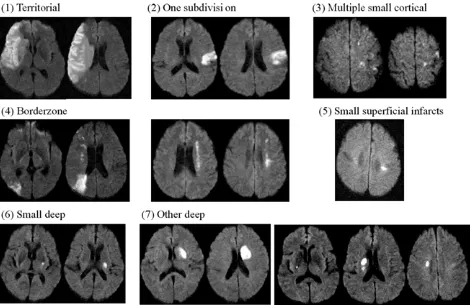
transeso-Figure 1. DWI lesion patterns.
phageal echocardiography (TEE) and/or Holter monitoring to detect paroxysmal atrial fibrillation, if the patient had one of the following characteristics: past vascular events, such as deep venous thrombosis, or pulmonary embolism; palpitations preceding or accompanying stroke onset; or peripheral vascular insufficiency, deep vein thrombosis, or pulmonary embolism.
DWI lesion patterns
Based on observed DWI patterns, patients were grouped as follows: (1) territorial infarcts involving two or more subdivisions; (2) cortical infarcts involving one subdivision; (3) multiple, small cortical infarcts; (4) borderzone infarcts, either cortical or internal; (5) single or multiple superficial infarcts in the centrum ovale; (6) small, deep infarcts; and (7) other deep infarcts, i.e., large striatocapsular lesions or concomitant DWI lesions outside the striatocapsular area (Fig. 1). The MCA territory was divided into three subdivisions, i.e., deep,
superior, and inferior, according to the scheme previously reported.8 Small cortical or centrum ovale infarcts were defined as DWI lesions <1 cm in diameter, and small, deep infarcts were defined as striatocapsular lesions <1.5 cm. Analysis of the DWI data was carried out by two readers (S.H.Y. and P.H.L.) blinded to the clinical data, and inter-observer agreement was 91%. The opinion of a third reader (S.R.Y.) was obtained in cases of disagreement.
Conventional and novel risk factors for stroke
Mechanism of stroke
* differ from cryptogenic stroke, P<0.05.
Table 1. General characteristics of the study population
of antihyperlipidemia medications; cigarette smoking was defined as smoking at the time of the stroke; and previous stroke was identified by patient history, neurological examination, and laboratory tests.
Serum levels of C-reactive protein (CRP) and fibrinogen were evaluated as inflammatory markers, whereas the D-dimer level was checked as a hemostatic marker of ischemic stroke subtypes.9-10 These levels were evaluated in 687 patients (82.6%) who enrolled between August 2002 and October 2004. Peripheral blood samples were drawn from each patient at the time of admission (1.6±2.1 days after symptom onset). Patients in which CRP levels were >10 mg/dl were excluded because of possible coexistent infection (N=29).
Statistical analysis
Differences between the groups were examined using the Chi-square test, Student t-test, or one-way ANOVA with post-hoc analysis. Multiple logistic regression analysis was conducted to determine whether any DWI lesion patterns were independently associated with cryptogenic stroke, over and above the age and sex of
the patient, initial stroke severity, or stroke risk factors. Those that were significant at the 0.2 level were entered into the initial multivariate model. When the most parsimonious model was obtained by backward stepwise elimination of the non-significant factors, each of the excluded variables was again entered separately into the model to test its contribution to the final model. Results are given as OR estimates of relative risk, with 95% CI. Statistical significance was established at the P<0.05 level.
RESULTS
According to the clinical, DWI, angiographic, and cardiological findings, 402 patients were classified in the LAA group, 133 in the CE group, 182 in the SAO group, and 115 in the cryptogenic group.
As shown in Table 1, the clinical features and risk factors of the cryptogenic group were more similar to those of the LAA and SAO groups than to the CE group, especially in terms of age, and the presence of diabetes, hyperlipidemia, or smoking habits.
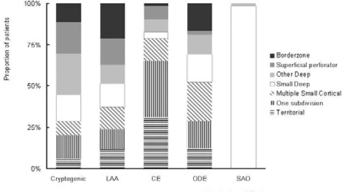
Figure 2. DWI lesion patterns in each stroke subtype.
work-up; TEE or Holter monitoring. Fourteen patients showed abnormalities: PFO with/without atrial septal defect (n=8), aortic arch atheroma (n=1), paroxysmal atrial fibrillation (n=3), and left atrium thrombus (n=2). In contrast, about 40% of patients in the cryptogenic group had stenosis other than significant stenosis of the relevant artery (>50% stenosis of non-relevant vessels or mild stenosis [30~50%] of relevant vessels). The frequency of significant stenosis of non-relevant vessels of the cryptogenic group were intermediate (27 patients; 23.5%); higher than that of the CE group (23 patients; 17.3%) or SAO group (27 patients; 14.9%) but lower than that of the LAA group (143 patients; 35.6%).
The DWI data showed that the lesion pattern in patients with cryptogenic stroke was similar to that of patients in the LAA group (Fig. 2). In the CE group, large cortical infarcts, either territorial (31%) or cortical infarcts involving one subdivision of the MCA territory (35%), were the most frequent type of DWI patterns, whereas border zone infarcts were rarely observed in these patients (2 of 133 patients; 1.5%).
Other deep infarcts on DWI were significantly more prevalent in patients with cryptogenic stroke than in those of the other groups (P<0.001). The frequency of large cortical infarcts in the cryptogenic group was similar to the LAA group (P=0.451) but differed from the CE or SAO group (P<0.01 for both). The frequency of superficial perforator infarcts was similar in the cryptogenic and LAA groups (P=0.389) but was lower
in the CE and SAO groups (P<0.05 for both).
Among the cryptogenic group, the DWI patterns in patients <50 years old were similar to those in patients aged 50 or older (Fig. 3). However, the DWI lesion patterns differed depending on the time of the vascular study (Fig. 4). In cases that the vascular study was delayed, the DWI data showed the characteristics of the LAA group; the frequency of DWI lesion patterns of superficial perforator infarcts was higher and that of large cortical infarcts was lower when the vascular study was conducted after 12 hours of symptom onset than
Figure 4. DWI lesion patterns depending on the time of vascular study in patients with cryptogenic stroke (left) and atherosclerotic stroke (right).
DWI lesion patterns Territorial Cortical involving one subdivision Borderzone
Multiple small cortical Superficial perforator
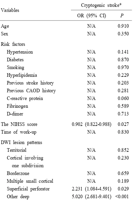
*Compared to those with non-cryptogenic stroke group. N/A; not applicatable
Table 2. Multiple logistic regression test when it was conducted within 12 hours. Such trend was also observed in patients of the LAA group (Fig. 4).
Certain DWI lesion patterns specific to the cryptogenic group were observed; cryptogenic stroke was prevalent in patients who showed other deep infarcts (31%) or superficial perforator infarcts (23%). Such DWI lesion patterns were independently associated with the cryptogenic group after adjustment for other risk factors. Table 2 shows the results of multiple logistic regression analysis and the OR for each factor. Patients with other deep and superficial perforator infarcts were 5 and 2.2 times more likely to have neither significant vascular stenosis nor potential sources of cardioembolism, respectively. The NIHSS score at admission was associated with cryptogenic stroke; the milder the neurological deficit, the more likely physicians failed to document the cause of stroke.
DISCUSSION
Cardioembolic risk factors in cryptogenic stroke
hyperhomo-cysteinemia, and aorta atherosclerosis have been newly implicated. TEE and Holter monitoring have become routine procedures in evaluating stroke patients, especially those with cryptogenic stroke. However, the impact of such routine procedures should be addressed in terms of cost-effectiveness.
Recently, PFO, aortic arch atheroma, and paroxysmal atrial fibrillation have been reported to be risk factors for cardioembolic stroke, especially in patients with cryptogenic stroke.11-14 However, their relationship to stroke occurrence, their prognosis, and the therapeutic implications are not clearly established. First, because of high prevalence of PFO in the normal population, detection of PFO in stroke patients does not necessarily identify it as the cause of stroke.15 Second, the optimal treatment in patients with PFO remains undetermined.16 Third, evidence regarding the therapeutic and prognostic values of Holter monitoring to detect paroxysmal atrial fibrillation is limited and remains inconclusive.17-18
The role of atherosclerosis in the development of cryptogenic stroke
Our results on risk factor profiles show that the cryptogenic group shared characteristics with the LAA and SAO groups, rather than with the CE group. In the present study, patients with cryptogenic stroke were in an atherosclerosis-prone condition. Their risk factor profiles may differ from those in patients with PFO or paroxysmal atrial fibrillation. PFO is associated with cryptogenic stroke in young adults, who are less likely to have traditional risk factors for stroke.11,19
In agreement with previous computed tomography data,6 our DWI data show that different infarct patterns depend on stroke subtypes. The DWI lesion patterns of our patients with cryptogenic stroke were similar to those in patients with LAA, and large cortical infarcts (20%) or concomitant basilar arterial territory infarcts (2.6%) were relatively rare in these patients. It has been reported that stroke in association with PFO occurs commonly in the pial convexity branches, as expected, as well as in the basilar artery distribution area, but rarely in the deep brain structures.11,20-21 In addition, it is possible that spontaneous recanalization occurred
during the subacute phase of ischemic stroke in the cryptogenic group. The DWI lesion pattern was even more similar to the LAA group in patients who underwent vascular studies 12 hours after symptom onset compared to those examined within 12 hours.
We recently reported a high rate of recurrence in patients with cryptogenic stroke, similar to patients with LAA.7 The annual rate of recurrence in cryptogenic stroke patients with cardioembolic risks, such as PFO, is low (1.2~2.4%).22 Atherosclerosis may be responsible for the recurrence of cryptogenic stroke because recurrence is associated with mild stenosis (≤50%) of the relevant artery or >50% stenosis of a non-relevant artery.7
The role of microangiopathy in the development of cryptogenic stroke
In this study, the DWI lesion pattern of other deep infarcts was the most predominant one observed in cryptogenic stroke. Although one-third was associated with mild stenosis (≤50%) of the relevant artery or >50% stenosis of a non-relevant artery, the remainder had normal angiographic results. Microangiopathy may play an important role in the latter group. Although several studies have selected a diameter of 20 mm as the cutoff point of lacunae, most using DWI or conventional MRI have used 15 mm as the size criterion. The conventional 15-mm criterion for lacunae based on DWI may underestimate the diagnosis of SAO as it was suggested.5 Further studies are needed to confirm which diameter-criterion should be accepted for the diagnosis of SAO.
Reasons for differences in the risk factors for cryptogenic stroke among studies
cryptogenic stroke. Most of the studies looking for potential sources of cardioembolism and its association with ischemic stroke have been done in Caucasians or small groups of African-American patients, thus making it difficult to assess and compare the existing risk factor burden for ischemic stroke between other ethnic groups and Asians. However, as with African-Americans, there may be differences between Caucasians and Asians with regard to prevalence of right-to-left cardiac shunt (i.e., PFO), thrombogenic sources (i.e., venous thrombosis), atrial fibrillation, and aortic arch atheroma.23-25
Limitations and conclusions
Our study has several limitations. Although TTE was performed in all patients, more extensive studies should be conducted to document possible embolism in patients with cryptogenic stroke. In this study, about 30% of patients who underwent extensive cardiologic work-up showed abnormalities. Therefore, it should be done if the patient was clinically suspected to have potential sources of cardioembolism. However, our study does not support the use of TEE and Holter monitoring as routine procedures in evaluating cryptogenic stroke patients, because the role of uncommon causes of embolism might have played a small part in our study. Further population-based studies with long-term follow-up and more extensive work-up to document possible causes of embolism are needed.
Our study does not show that cardioembolic stroke from PFO or paroxysmal atrial fibrillation is an important pathogenic mechanism in cryptogenic stroke. It should be stressed, however, that non-cardiogenic mechanisms such as LAA and SAO are equally as important in the pathogenesis of cryptogenic stroke as cardiogenic risk factors such as PFO or paroxysmal atrial fibrillation.
REFERENCES
1. Sacco RL, Ellenberg JH, Mohr JP, Tatemichi TK, Hier DB, Price TR, et al. Infarcts of undetermined cause: the NINCDS Stroke Data Bank. Ann Neurol 1989;25:
382-390.
2. Baird AE, Lovblad KO, Schlaug G, Edelman RR, Warach S. Multiple acute stroke syndrome: marker of embolic disease? Neurology 2000;54:674-678.
3. Gandolfo C, Del Sette M, Finocchi C, Calautti C, Loeb C. Internal borderzone infarction in patients with ischemic stroke. Cerebrovasc Dis 1998;8:255-258.
4. Lee PH, Bang OY, Oh SH, Joo IS, Huh K. Subcortical white matter infarcts: comparison of superficial perforating artery and internal border-zone infarcts using diffusion- weighted magnetic resonance imaging. Stroke 2003;34: 2630-2635.
5. Kang DW, Chalela JA, Ezzeddine MA, Warach S. Association of ischemic lesion patterns on early diffusion-weighted imaging with TOAST stroke subtypes.
Arch Neurol 2003;60:1730-1734.
6. Paciaroni M, Silvestrelli G, Caso V, Corea F, Venti M, Milia P, et al. Neurovascular territory involved in different etiological subtypes of ischemic stroke in the Perugia Stroke Registry. Eur J Neurol 2003;10:361-365.
7. Bang OY, Lee PH, Joo SY, Lee JS, Joo IS, Huh K. Frequency and mechanisms of stroke recurrence after cryptogenic stroke. Ann Neurol 2003;54:227?234.
8. Heinsius T, Bogousslavsky J, Van Melle G. Large infarcts in the middle cerebral artery territory. Etiology and outcome patterns. Neurology 1998;50:341-350.
9. Ageno W, Finazzi S, Steidl L, Biotti MG, Mera V, Melzi D'Eril G, et al. Plasma measurement of D-dimer levels for the early diagnosis of ischemic stroke subtypes. Arch Intern Med 2002;162:2589-2593.
10. Ince B, Bayram C, Harmanci H, Ulutin T. Hemostatic markers in ischemic stroke of undetermined etiology.
Thromb Res 1999;96:169-174.
11. Lamy C, Giannesini C, Zuber M, Arquizan C, Meder JF, Trystram D, et al. Clinical and imaging findings in cryptogenic stroke patients with and without patent foramen ovale: the PFO-ASA Study. Atrial Septal Aneurysm. Stroke 2002;33:706-711.
12. Mas JL, Arquizan C, Lamy C, Zuber M, Cabanes L, Derumeaux G, et al. Patent Foramen Ovale and Atrial Septal Aneurysm Study Group. Recurrent cerebrovascular events associated with patent foramen ovale, atrial septal aneurysm, or both. N Engl J Med 2001;345:1740-1746. 13. Hart RG, Pearce LA, Rothbart RM, McAnulty JH, Asinger
RW, Halperin JL. Stroke with intermittent atrial fibrillation: incidence and predictors during aspirin therapy. Stroke Prevention in Atrial Fibrillation Investi-gators. J Am Coll Cardiol 2000;35:183-187.
14. Amarenco P, Cohen A, Tzourio C, Bertrand B, Hommel M, Besson G, et al. Atherosclerotic disease of the aortic arch and the risk of ischemic stroke. N Engl J Med
15. Konstadt SN, Louie EK, Black S, Rao TL, Scanlon P. Intraoperative detection of patent foramen ovale by transesophageal echocardiography. Anesthesiology 1991;74: 212-216.
16. Homma S, Sacco RL, Di Tullio MR, Sciacca RR, Mohr JP; PFO in Cryptogenic Stroke Study (PICSS) Investigators. Effect of medical treatment in stroke patients with patent foramen ovale: patent foramen ovale in Cryptogenic Stroke Study. Circulation 2002;105:2625- 2631.
17. Rauh R, Fischereder M, Spengel FA. Transesophageal echocardiography in patients with focal cerebral ischemia of unknown cause. Stroke 1996;27:691-694.
18. Schaer BA, Zellweger MJ, Cron TA, Kaiser CA, Osswald S. Value of routine holter monitoring for the detection of paroxysmal atrial fibrillation in patients with cerebral ischemic events. Stroke 2004;35:e68-e70.
19. Cabanes L, Mas JL, Cohen A, Amarenco P, Cabanes PA, Oubary P, et al. Atrial septal aneurysm and patent foramen ovale as risk factors for cryptogenic stroke in patients less than 55 years of age. A study using transesophageal echocardiography. Stroke 1993;24:1865-1873.
20. Steiner MM, Di Tullio MR, Rundek T, Gan R, Chen X, Liguori C, et al. Patent foramen ovale size and embolic
brain imaging findings among patients with ischemic stroke. Stroke 1998;29:944-948.
21. Barinagarrementeria F, Amaya LE, Cantu C. Causes and mechanisms of cerebellar infarction in young patients.
Stroke 1997;28:2400-2404.
22. Nedeltchev K, Arnold M, Wahl A, Sturzenegger M, Vella EE, Windecker S, et al. Outcome of patients with cryptogenic stroke and patent foramen ovale. J Neurol Neurosurg Psychiatry 2002;72:347-350.
23. Rodriguez CJ, Homma S, Sacco RL, Di Tullio MR, Sciacca RR, Mohr JP; PICSS Investigators. Race-ethnic differences in patent foramen ovale, atrial septal aneurysm, and right atrial anatomy among ischemic stroke patients.
Stroke 2003;34:2097-2102.
24. Kizer JR, Silvestry FE, Kimmel SE, Kasner SE, Wiegers SE, Erwin MB, et al. Racial differences in the prevalence of cardiac sources of embolism in subjects with unexplained stroke or transient ischemic attack evaluated by transesophageal echocardiography. Am J Cardiol 2002; 90:395-400.
25. Gupta V, Nanda NC, Yesilbursa D, Huang WY, Gupta V, Li Q, et al. Racial differences in thoracic aorta atherosclerosis among ischemic stroke patients. Stroke