www.elsevier.com / locate / bres
Short communication
Expression of estrogen receptors in the dorsal root ganglia of the
chick embryo
a b ,
*
Sheng Cui , Ronald S. Goldstein
a
China Agricultural University, College of Biology, Beijing 10094, China b
Faculty of Life Sciences, Gonda Research Building, Bar-Ilan University, 52900 Ramat-Gan, Israel
Accepted 15 August 2000
Abstract
Estrogen receptors (ER) are widely distributed in the central nervous system (CNS). Recent studies, to date in rat only, have shown that ER are also expressed in neurons of the dorsal root ganglia (DRG) where they appear to have functional roles. However, no data yet exists about estrogen receptors in the embryonic DRG. In the present study, immunocytochemical staining for ER in the DRG of chick embryos from day 6.5 to 18.5 (Hamburger and Hamilton St. 30–45) of incubation was performed. ER1 cells were first consistently observed at day 8.5 (St. 34), more concentrated in the ventral-lateral portion of the DRG. From day 8.5 to 12.5 (St. 38), the density of ER1cells and the staining intensity increased, with no obvious changes from day (E) 12.5 to 18.5. Although ER is detected mainly in the cytoplasm of embryonic DRG neurones, ER1 cells with nuclear staining are sometimes observed and gradually increase in number during development. ER-immunoreactivity in the DRG at cervical, thoracic and lumbo-sacral levels is similar and no obvious differences in staining were observed between male and female embryos. ER1neurons are also present in the sympathetic ganglia from E8.5 and some primary spinal motoneurons are ER1 beginning at E14.5. The results suggest that estrogen may play a role in the embryonic development of the DRG. 2000 Elsevier Science B.V. All rights reserved.
Theme: Development and regeneration
Topic: Hormones and development
Keywords: DRG; Estrogen receptors; Avian embryo
Estrogen is a hormone that has marked biological affects the CNS of a variety of species, such as rat [3,20], mouse on many kinds of tissues including the nervous system. It [13,25], guinea pig [4], chicken [30], Brazilian opossum is generally thought that estrogen exerts its effects on the [7] and Japanese quail [1] using autoradiography for bound nervous system by acting on intracellular receptors [17,29]. estrogen, immunocytochemistry and detection of ER The effects of estrogen are mediated by specific receptor mRNA. Most research to date on estrogen in the nervous subtypes, including estrogen receptor(ER)-a and ER-b system has focused on the postnatal and adult animals, [14]. Once estrogen is bound to its receptors, it activates although there are reports on the ontogeny of ER in the transcription of target genes [6,17]. Within the central hypothalamus of fetal rat [19,20,29] and chicken embryo nervous system (CNS), estrogen has many functions, [30].
including the regulation of neurotrophic factor expression, There has been much less study of ERs in the peripheral affecting phenotype and controlling synaptic plasticity nervous system compared to the CNS. In recent reports, [2,5,27], although the exact mechanisms of these actions ER immunopositive (ER1) neurons were detected in DRG are not yet clear. Expression of ERs has been described in of the postnatal and adult rat. In these studies, it was demonstrated that estrogen can affect the development and survival of DRG neurons [15,16,21,23,26,28,32]. How-*Corresponding author. Tel.: 1972-3-531-8216; fax: 1
972-3-535-ever, there are not yet any studies of the expression of ER 1824.
E-mail address: [email protected] (R.S. Goldstein). in embryonic DRG. We therefore, examine here the
ontogeny of ER expression in the DRG of the chick neurons appear to have cytoplasmic staining (n58 em-embryo. We found that ER-immunoreactivity is present in bryos for each stage). No additional obvious changes are the DRG of chick embryo from embryonic E7.5 to E18.5 observed through E18.5. The staining of ER is localised (St. 30–45). Expression in DRG at cervical, thoracic and exclusively to the cytoplasm until E12.5. From E12.5 on, lumbo-sacral levels is similar and no obvious differences some small and medium-sized neurons had ER1 nuclei in staining were observed between male and female (Fig. 1G). In a few experiments, sections of more mature embryos. embryos were counterstained with hematoxylin. Examina-Chick eggs were incubated at 37.58C for 6.5, 7.5, 8.5, tion of these sections revealed that all cells that displayed 10.5, 12.5, 14.5, 16.5 and 18.5 days and staged according cytoplasmic staining for ER were neurons, since none of to Hamburger and Hamilton [11]. The embryos were the cells with small, dense nuclei (satellite cells and anaesthetised by cooling in 48C phosphate-buffered saline neuronal precursors) were stained (not shown).
(PBS) and decapitated. The sex of embryos from E8.5 to ER expression in the CNS differs quantitatively by sex E18.5 was determined by examination of the gonads in- [13]. We therefore determined the sex of embryos from situ and the vertebral columns, including surrounding E10.5 on and compared the distribution of ER1 cells tissue, were removed. Fixation overnight in 4% parafor- between DRG in females and males (n54 for each females maldehyde in PBS at 48C ensued, followed by dehydration and males at each stage examined). No obvious differences and embedding in paraffin. Serial sections (6-mm thick) of were observed in number, distribution or quality of stain-whole DRG at the middle of the cervical, thoracic and ing of DRG neurons between the sexes. DRG at different lumbo-sacral levels were cut. axial levels of the animal differ from one another in terms Immunocytochemical localisation of estrogen receptor of size and the proportions of types of sensory neurons was performed using a monoclonal antibody (NCL-ER- [11,18]. Complete sets of serial sections through DRG LH2, Novocastra). The staining pattern for NCL-ER-LH2 from the cervical, thoracic and lumbar levels were stained. is identical to that obtained with the monoclonal antibody No obvious differences in staining of ER between the H222 [24], which has been previously used for the DRG from these different axial levels were found. detection of ER in chick tissues [30]. Antigen retrieval was We observed ER1 cells in the sympathetic ganglia performed by microwaving the sections for 20 min (435 starting at E10.5. By E12.5, virtually all cells in the min) at full power in 0.01 M sodium citrate buffer (pH sympathetic ganglia were ER1, with the staining primarily 6.0), after dewaxing and rehydration. Endogenous per- cytoplasmic (Fig. 1E and F). The cells in the sympathetic oxidase was inhibited by H O in methanol, followed by2 2 ganglia were often more intensely stained than those in the blocking with 10% normal rabbit serum in Tris-buffered DRG. ER1 primary motoneurons in the ventral spinal saline (pH 7.5), and the sections were incubated with cord were consistently observed from E16.5 (Fig. 1H). primary antibody diluted 1:70 for 72 h at 48C. Incubation These appeared to be only a subset of the neurons in the in biotin anti-mouse for 2 h at room temperature followed ventral motor column, but this issue was not investigated and finally avidin–biotin complex was added for 1 h and further.
the peroxidase activity detected using diaminobenzidine. This is the first description of the ontogeny of ER in Controls included the omission of the primary, secondary embryonic DRG, although there have been studies of the and tertiary antibodies, all of which eliminated staining DRG of postnatal and adult rats [21]. ER1 neurons are (not shown). first detected by immunocytochemistry at day 8.5 of In some experiments, alkaline phosphatase–streptavidin incubation. ER expression in DRG then increases and with BCIP/ NBT detection was used. The results obtained reaches its maximum by day 12.5. This time course agrees with both procedures were identical. well with the ontogeny of estrogen immunoreactive and
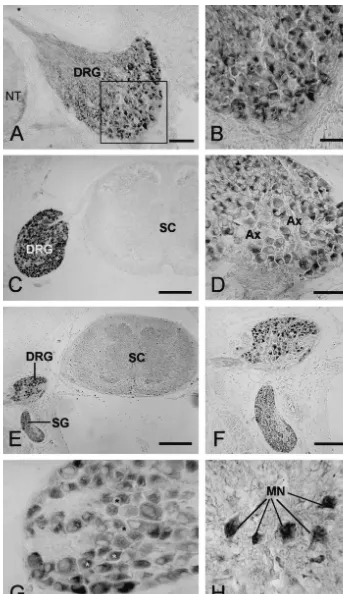
Fig. 1. Micrographs of the transverse sections through chicken embryos stained for estrogen receptor (ER) from 8.5 to 18.5 days of development. (A) Shows a section from an E8.5 embryo. The ventral and part of the medial portions of the DRG contain ER1 cells. Bar550 mm. In (B), higher magnification reveals that the reaction product is localised to the cytoplasm and not the nuclei (asterisks). Bar525mm. (C) At E10.5, staining intensity in the DRG (arrow) is higher than at E8.5, no ER1cells are observed in the neural tube (NT). Bar5200mm. By E12.5 (D), virtually all neurons in the DRG are ER1, reaction remains cytoplasmic. Bar550mm. The areas of the ganglion that are not stained contained axons of the sensory neurons (Ax). E14.5 embryos (E and F) show staining in both the DRG and sympathetic ganglia (SG), but not in the spinal cord (SC). All neurons in both ganglia are ER1at this stage (E). Bar in E525mm, Bar in F5100mm. Some nuclear staining for ER in DRG neurons is observed at E16.5, as shown in G (asterisks). Bar525
binding and estradiol in early development of the zebra finch cytoplasm of CNS neurons in rat [3], Brazilian opossum
telencephalon, J. Neurobiol. 40 (1999) 149–157. [8], guinea pig [4] and Japanese quail [1]. The nuclear
[6] F.E. Murdoch, J. Gorski, The role of ligand in estrogen receptor staining in adult rats [28] may be a function of neuronal regulation of gene expression, Mol. Cell. Endocrinol. 78 (1991) maturity, since the proportion of embryonic ER1 cells C103–C108.
[7] C.A. Fox, L.R. Ross, C.D. Jacobson, Ontogeny of cells containing with nuclear staining to the total ER1 neurons gradually
estrogen receptor-like immunoreactivity in the Brazilian opossum increased throughout in-ovo development.
brain, Dev. Brain Res. 63 (1991) 209–219.
Two types of ER have been described: ERa and ERb. [8] C.A. Fox, L.R. Ross, R.J. Handa, C.D. Jacobson, Localization of ERb was detected in both nuclei and cytoplasm of DRG cells containing estrogen receptor-like immunoreactivity in the
Brazilian opossum brain, Brain Res. 546 (1991) 96–105. neurones and are much more numerous than ERa1
[11] V. Hamburger, H.L. Hamilton, A series of normal stages in the neurons neonatal rat DRG (21). We have not yet used the
development of the chick embryo, J. Morphol. 88 (1951) 49–92. specific antibodies for ER subtypes (ERa and ERb) and [12] J.B. Hutchison, A. Wozniak, C. Byeri, M. Karolczak, R.E. Hutchi-thus distribution of ERa and ERb in developing DRG son, Steroid metabolising enzymes in the determination of brain remains to be elucidated. gender, J. Steroid Biochem. Mol. Biol. 69 (1999) 85–96.
[13] M. Koch, G. Ehret, Immunocytochemical localisation and quantita-During the period studied, there were no obvious sex
tion of estrogen-binding cells in the male and female (virgin, differences in ER expression in embryonic DRG. This is in
pregnant, lactating) mouse brain, Brain Res. 489 (1989) 101–112. agreement with the findings of ER immunocytochemistry [14] G.G. Kuiper, B. Carlsson, K. Grandien, E. Enmark, J. Haggblad, S. in the hypothalamus and adenohypophysis distalis of chick Nilsson, J.-A. Gustafsson, Comparison of the ligand binding spe-cificity and transcript tissue distribution of estrogen receptors alpha embryos [30]. However, the fact that plasma estrogen
and beta, Endocrinology 138 (1997) 863–870. concentration in males is much lower than that in females
[15] F.J. Luzzi, S.A. Scoville, S.M. Bufton, Effect of short-term estrogen [22,31] appears to be inconsistent with the similar ER replacement on trkA mRNA level in axotomized dorsal root expression levels in the DRG of both genders. Aromatase, ganglion neurons, Exp. Neurol. 159 (1999) 433–440.
[16] F.J. Luzzi, S.A. Scoville, S.M. Bufton, Long-term estrogen replace-the enzyme responsible for conversion of androgens into
ment coordinately decrease trkA and beta-PPT mRNA level in estrogen, is widely distributed in the central nervous
dorsal root ganglion neurons, Exp. Neurol. 155 (1999) 260–267. system of embryos. This enzyme is induced by testo- [17] F.E. Murdoch, J. Gorski, The role of ligand in estrogen receptor sterone in the brain [2,12] and there is evidence of higher regulation of gene expression, Mol. Cell Endocrinol. 78 (1991) activity in the CNS of male, compared to female embryos 103–108.
[18] E. Pannese, Electron microscopic study of the development of the [2]. It may be that the equivalent distribution of ER1
satellite sheath in spinal ganglia, J. Comp. Neurol. 135 (1969) neurons in the DRG of embryos of both sexes, is due to the
381–422.
enhanced conversion there of androgens to estrogen in [19] C. Pasqualini, D. Guivarch, Y.V. Boxberg, F. Nothias, J.D. Vincent, P. males, in parallel to the CNS. Vernier, Stage- and region-specific expression of estrogen receptor alpha isoforms during ontogeny of the pituitary gland, Endocrin-ology 140 (1999) 2781–2789.
[20] R.J. Pasterkamp, K. Yuri, D.T. Visser, S. Hayashi, M. Kawata, The
Acknowledgements perinatal ontogeny of estrogen receptor-immunoreactivity in the
developing male and female rat hypothalamus, Dev. Brain Res. 91 (1996) 300–303.
This work was supported by grants from the
[21] C. Patrone, S. Andersson, L. Korhonen, D. Lindholm, Estrogen Dysautonomia Foundation Inc., the Israel Institute for receptor-dependent regulation of sensory neuron survival in de-Psychobiology-Charles Smith Foundation and the Health veloping dorsal root ganglion, Proc. Natl. Acad. Sci. 96 (1999) Science Center at Bar-Ilan University, The Aviv Fund for 10905–10910.
[22] C.G. Scanes, L.E. Hart, E. Decuypere, E.R. Kuhn, Endocrinology of Neuroscience Research. SC was supported by a grant from
the avian embryo: an overview, J. Exp. Zool. 1 (Suppl.) (1987) the Kort Foundation at BIU.
253–264.
[23] S.A. Scoville, S.M. Bufton, F.J. Luzzi, Estrogen regulates neurofila-ment gene expression in adult rat dorsal root ganglion neurons, Exp. Neurol. 146 (1997) 596–599.
References [24] C. Sheng, A.S. McNeilly, A.N. Brooks, Immunohistochemical
distribution of oestrogen receptor and luteinizing hormone B subunit [1] J. Balthazart, G.F. Ball, Effects of the noradrenergic neurotoxin in the bovine pituitary gland during foetal development, J.
Neuroen-DSP-4 on luteinizing hormone levels, catecholamine concentrations, docrinol. 10 (1998) 713–718.
alpha 2-adrenergic receptor binding, and aromatase activity in the [25] P.J. Shughrue, W.E. Stumpf, N.J. MacLusky, J.E. Zielinski, R.B. brain of the Japanese quail, Brain Res. 492 (1989) 163–175. Hochberg, Developmental changes in estrogen receptors in mouse [2] C. Beyer, Estrogen and developing mammalian brain, Anat. Em- cerebral cortex between birth and postweaning: studied by auto-bryol. 199 (1999) 379–390. radiography with 11 beta-methoxy-16 alpha-[125I]iodoestradiol, [3] J.D. Blaustein, Cytoplasmic estrogen receptors in rat brain: immuno- Endocrinology 126 (1990) 1112–1124.
cytochemical evidence using three antibodies with distinct epitopes, [26] W.D. Snider, I. Silos-Santiago, Dorsal root ganglion neurons require Endocrinology 131 (1992) 1336–1342. functional neurotrophin receptors for survival during development, [4] J.D. Blaustein, J.C. Turcotte, Estrogen receptor-immunostaining of Phil. Trans. R. Soc. Lond. B 351 (1996) 395–403.
[28] N. Taleghany, S. Sarajari, L.L. DonCarlos, L. Gollapudi, M.M. [31] J.E. Woods, R.C. Thommes, Ontogeny of hypothalamo-Oblinger, Differential expression of estrogen receptor alpha and beta adenohypophyseal-gonadal (HAG) interrelationships in the chick in rat dorsal root ganglion neurons, J. Neurosci. Res. 57 (1999) embryo, J. Exp. Zool. 232 (1984) 435–441.
603–615. [32] Y. Yang, H. Ozawa, H. Lu, K. Yuri, S. Hayashi, K. Nihonyanagi, M. [29] C.C. Vito, T.O. Fox, Androgen and estrogen receptors in embryonic Kawata, Immunocytochemical analysis of sex differences in cal-and neonatal rat brain, Brain Res. 2 (1987) 97–110. citonin gene-related peptide in the rat dorsal root ganglion, with [30] J.E. Woods, L.M. Otten, R.C. Thommes, Ontogeny of 17B-estradiol special reference to estrogen and its receptor, Brain Res. 791 (1998)
(E2)- and estrogen receptor (ER)-immunostained cells in the 35–42. hypothalamus and adenohyophyseal pars distalis of the chick