Brain Research 881 (2000) 241–243
www.elsevier.com / locate / bres
Short communication
Plasma cortisol levels in elderly female subjects with Alzheimer’s
disease: a cross-sectional and longitudinal study
a ,
*
b b c aHiroyuki Umegaki
, Hiroyuki Ikari , Hideki Nakahata , Hidetoshi Endo , Yusuke Suzuki ,
a a b a
Osamu Ogawa , Akira Nakamura , Takayuki Yamamoto , Akihisa Iguchi
a
Department of Geriatrics, Nagoya University, Graduate School of Medicine, 65 Tsuruma-Cho, Showa-Ku, Nagoya, Aichi, 466-8550, Japan
b
Choju Medical Institute, Fukushimura Hospital, 19-14 Aza-Yamanaka, Noyori-cho, Toyohashi, Aichi, 441-8124, Japan
c
Department of Geriatrics, Chubu National Hospital, 36-3 Gengo, Morioka, Aichi, 474-8511, Japan
Accepted 15 August 2000
Abstract
We investigated the plasma cortisol levels at a fasting state in elderly female Alzheimer’s disease (AD), vascular dementia (VD), and non-demented subjects (n566, 28 and 21, respectively). Twenty-eight AD subjects were followed for 40 months. The plasma cortisol levels in AD and VD subjects were significantly higher than those of non-demented subjects at baseline. In AD subjects in relatively early stages of the disease [Mini-Mental State Examination (MMSE)], at baseline, high plasma cortisol led to rapid declines in MMSE scores over a 40-month period. 2000 Elsevier Science B.V. All rights reserved.
Keywords: Hippocampus; Acetylcholine; Glucocorticoids; Vascular dementia
Many past studies have shown that several aspects of subjects with vascular dementia (VD) and non-demented
neuroendocrine systems are altered in the course of controls (ND), and we report the longitudinal findings in
Alzheimer’s disease (AD) [13,14]. Hypercortisolemia is AD subjects followed over a 40-month period.
one of the most frequently reported changes [5,6,11]. All AD and VD subjects were hospitalized in
Fukushi-Sapolsky et al. [7,8] found that aged rats have a decreased mura Hospital in Aichi, Japan. ND subjects lived in a
number of glucocorticoid receptors and an impaired nega- nursing home near Fukushimura Hospital. This study was
tive feedback mechanism of glucocorticoid secretion. A approved by the Human Subject Review Committee of
high concentration of glucocorticoid receptors exists in the Fukushimura Hospital. After all of the procedures had
hippocampus, and this brain region is thought to be been fully explained, written informed consent was
ob-involved in the negative-feedback mechanism of glucocor- tained from all subjects or their guardians. The subjects
ticoid secretion. Animal studies have demonstrated that were non-smokers, and those who had chronic obstructive
elevated cortisol levels make neurons more vulnerable to pulmonary diseases, diabetes mellitus, neurodegenerative
several kinds of insults [1,9]. The degeneration of the disease, or abnormal thyroid function were excluded. The
hippocampus is one of the most notable features of AD Diagnostic and Statistical Manual of Mental Disorders
pathologies [4]. Based on their findings, Sapolsky and Fourth Edition [12] was used to arrive at the diagnoses of
McEwen [10] proposed the glucocorticoid cascade theory. AD and VD. Computed tomographies of the brain were
According to this theory, hippocampal cell loss in AD performed for all subjects. No subjects in the AD and ND
induces hypercortisolemia, which, in turn, acts as a co- groups had cerebral vascular diseases.
factor in further degeneration of the disease process. The total number of subjects enrolled in the study was
In the current study, we report cross-sectional findings 115 (AD566, VD528 and ND521). The first examination
of cortisol profiles in AD subjects as compared to those of was performed in November 1996, and a follow-up
examination was performed 40 months later, in March 2000. The progress of 28 out of 66 AD subjects was
*Corresponding author. Tel.: 181-52-744-2365; fax: 1
81-52-744-followed.
2371.
E-mail address: [email protected] (H. Umegaki). A score for Mini-Mental State Examination (MMSE)
242 H. Umegaki et al. / Brain Research 881 (2000) 241 –243
[3], which is a widely used dementia assessment instru- in MMSE scores and the basal cortisol level (n59) (Fig.
ment, was obtained from all subjects. 1B).
The blood samples were collected at 7 a.m. from We have demonstrated in the present study that plasma
subjects in a fasting state. The blood samples were kept on levels of cortisol were significantly higher in both AD and
ice and cold-centrifuged immediately after collection. The VD subjects than in ND subjects. The levels of ACTH
plasma was stored at 2708C until assay. Cortisol and were not significantly different among the three groups,
adrenocorticotrophic hormone (ACTH) were measured by and this is in agreement with previous studies [2,5]. The
radioimmunoassay. mechanism of the discrepancy between levels of cortisol
Table 1 shows the baseline profiles (November 1996) of and ACTH has yet to be elucidated.
all of the subjects enrolled in this study and 28 AD The cortisol levels of AD subjects whose progress was
subjects who were followed until March, 2000. The mean followed did not change significantly over 40 months,
age was 82.50 years. The scores of AD subjects and VD while during this period, the MMSE scores of these
subjects were significantly lower than that of ND subjects. subjects declined significantly.
The basal plasma cortisol levels at a fasting state at Weiner et al. [15] reported a significant correlation
baseline in the three groups were AD515.2463.23mg / ml, between changes in scores of modified ADAS-COG and
VD515.1963.95 mg / ml and ND512.8862.77 mg / ml. basal cortisol levels in a similar follow-up study, which
One way ANOVA shows that plasma cortisol levels have investigated the subjects in early stages of AD (mean
significant differences among the three groups (P5 scores of MMSE520.064.1). In the current study, as
0.0171). Cortisol levels in AD and VD subjects were shown in Fig. 1A, the basal cortisol level did not predict
significantly higher than those in ND subjects, according to the decline in MMSE scores when all of the subjects were
Scheffe’s post-hoc test (P50.0058 and 0.0181, respective- included. Swanwick et al. [11] suggested that the
glucocor-ly). ACTH levels were not significantly different among ticoid cascade theory may be relevant in the early stages of
these three groups (AD545.77624.49 pg / ml, VD5 AD progression. As shown in Fig. 1B, when only
rela-42.07618.70 pg / ml and ND546.05627.69 pg / ml). tively less demented subjects (MMSE scores were more
The mean scores of MMSEs in the three groups of than 14) were analyzed, statistical analysis showed a
subjects at baseline were 7.96, 5.29 and 24.52, respectively significant correlation between the changes in MMSE
(Table 1). MMSE scores at baseline were not correlated scores and the basal level of plasma cortisol. The AD
with baseline cortisol levels in AD subjects (data not subjects who had higher basal levels of plasma cortisol
shown). showed greater declines in MMSE scores. This suggests
The mean MMSE score of AD subjects who were that the plasma cortisol level in a fasting state may predict
followed was 7.9966.52 at baseline and declined to the progression of AD, at least in moderate stages of the
1.4360.46. This decline of MMSE score was statistically disease.
significant (paired t-test) (P,0.0001). However, the plas- In summary, we demonstrated the following results in
ma cortisol levels did not change over the 40-month period the present study: (1) AD and VD subjects had
sig-(15.4560.53 and 15.4560.59 mg / ml, respectively). nificantly higher levels of plasma cortisol than ND
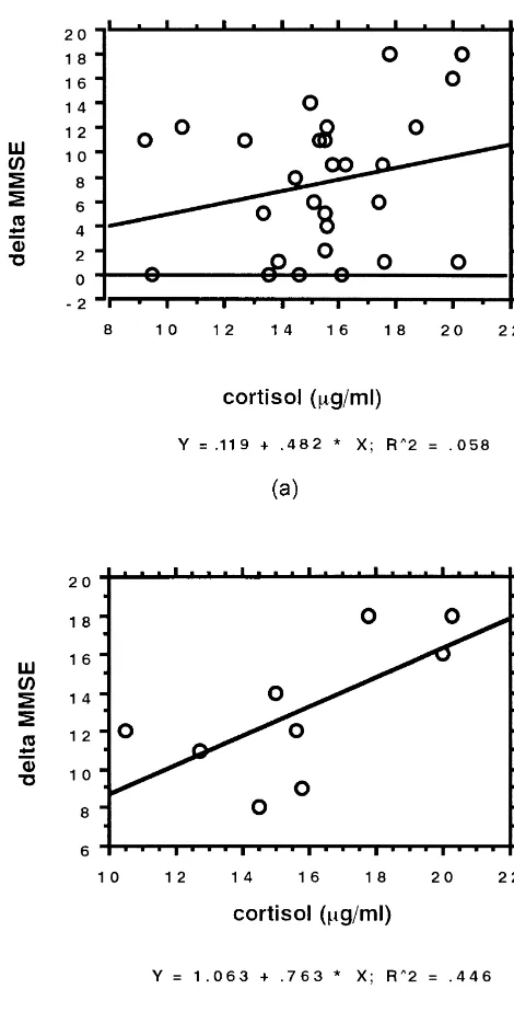
sub-There was no association between the basal cortisol jects. (2) The cortisol levels in a fasting state in AD
level and the changes of MMSE scores during the follow- subjects did not change significantly, while the MMSE
up period in which all subjects were included (Fig. 1A). scores declined significantly over a 40-month period. (3)
However, when only the mildly demented subjects with In AD subjects in relatively early stages of the disease
MMSE scores of more than 14 at baseline were analyzed, a (MMSE$14) who had higher cortisol levels, an
acceler-significant correlation was observed between the changes ated progression of the disease was observed.
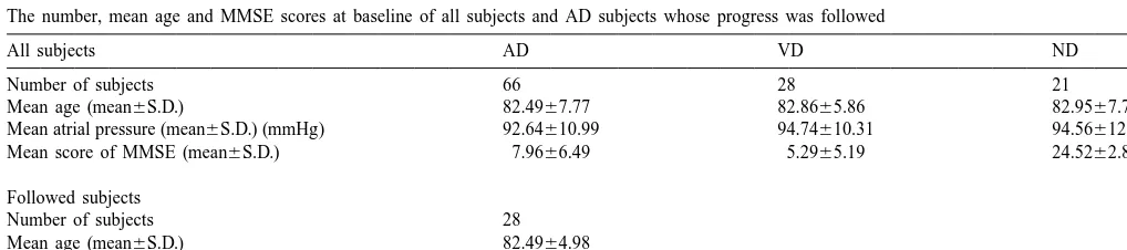
Table 1
The number, mean age and MMSE scores at baseline of all subjects and AD subjects whose progress was followed
All subjects AD VD ND
Number of subjects 66 28 21
Mean age (mean6S.D.) 82.4967.77 82.8665.86 82.9567.77
Mean atrial pressure (mean6S.D.) (mmHg) 92.64610.99 94.74610.31 94.56612.09
Mean score of MMSE (mean6S.D.) 7.9666.49 5.2965.19 24.5262.86
Followed subjects
Number of subjects 28
Mean age (mean6S.D.) 82.4964.98
H. Umegaki et al. / Brain Research 881 (2000) 241 –243 243
[2] I.N. Ferrier, J. Pascual, B.G. Charlton, C. Wright, A. Leake, H.W. Griffiths, A.F. Fairbairn, J.A. Edwardson, Cortisol, ACTH, and dexamethasone concentrations in a psychogeriatric population, Biol. Psychiat. 23 (1988) 252–260.
[3] M.F. Folstein, S.E. Folstein, P.R. McHugh, Mini-Mental State: a practical method for grading the cognitive state of patients for the clinician, J. Psychiatr. Res. 12 (1975) 189–198.
[4] W.R. Markesbery, Neuripathological criteria for the diagnosis of Alzheimer’s disease, Neurobiol. Aging 18 (1997) S13–19. [5] F. Masugi, T. Ogihara, K. Sakaguchi, A. Otuka, Y. Tsuchiya, S.
Morimoto, Y. Kumahara, S. Saeki, M. Nishida, High plasma levels of cortisol in patients with senile dementia of the Alzheimer’s type, Methods Find. Exp. Clin. 11 (11) (1989) 707–710.
[6] A.H. Miller, G. Sastry, A.J. Speranza, B.A. Lawlor, R.C. Mohs, T.M. Ryan, S.M. Gabriel, M. Serby, J. Schmeidler, K.L. Davis, Lack of association between cortisol hypersecretion and nonsuppres-sion on the DST in patients with Alzheimer’s disease, Am. J. Psychiatry 151 (1994) 267–270.
[7] R.M. Sapolsky, L. Krey, B.S. McEwen, The adrenocortical stress-response in the aged male rats: Impairment of recovery from stress, Exp. Gerontol. 18 (1983) 55–64.
[8] R.M. Sapolsky, L. Krey, B.S. McEwen, Corticosterone receptors decline in a site-specific manner in the aged rat brain, Brain Res. 289 (1983) 235–240.
[9] R.M. Sapolsky, L. Krey, B.S. McEwen, Prolonged glucocorticoid exposure reduces hippocampal neuron number: Implications for aging, J. Neurosci. 5 (1985) 1221–1227.
[10] R.M. Sapolsky, B.S. McEwen, Stress, glucocorticoids and their role in degenerative changes in the aging hippocampus, in: T. Crook, R. Bartus, S. Ferris et al. (Eds.), Treatment Development Strategies for Alzheimer’s Disease, Mark Powley Associates, New Canaan, CT, 1986, pp. 151–172.
[11] G.R.J. Swanwick, M. Kirby, I. Bruce, F. Buggy, R.F. Coen, D. Coakley, B.A. Lawlor, Hypothalamic–pituitary–adrenal axis dysfunction in Alzheimer’s disease: Lack of association between longitudinal and cross-sectional findings, Am. J. Psychiatry 155 (1998) 287–289.
[12] G.J. Tucker, M. Popkin, E.D. Caine, M. Folstein, G.L. Gottlieb, I. Grant, B. Liptzin, Delirium, Dementia, and Amnesic and Other Cognitive Disorders, Diagnostic and Statistical Manual of Mental Disorders, 4th edition, American Psychiatric Association, Washing-ton D.C, 1984.
[13] H. Umegaki, H. Ikari, H. Nakahata, J. Yoshimura, H. Endo, T. Yamamoto, A. Iguchi, Low plasma epinephrine in elderly female subjects with dementia of Alzheimer’s type, Brain Res. 858 (2000) 67–70.
[14] B. Vitiello, R.C. Veith, S.E. Molchan, R.A. Martinez, B.A. Lawlor, J. Fig. 1. (A) Simple regression analysis showed no correlation between Radcliffe, J.L. Hill, T. Sunderland, Autonomic dysfunction in plasma cortisol levels and changes in MMSE scores during the follow-up patients with dementia of the Alzheimer type, Biol. Psychiatry 34 period when analyzed in all the subjects (P50.2433). (B) Simple (1993) 428–433.
regression analysis showed a significant correlation between plasma [15] M.F. Weiner, S. Vobach, K. Olsson, D. Svetlik, R.C. Risser, Cortisol cortisol levels and changes in MMSE scores during the follow-up period secretion and Alzheimer’s disease progression, Biol. Psychiatry 42 when analyzed in subjects with relatively mild dementia (MMSE$14) (1997) 1030–1038.
(P50.0307).
References