Gonadoblastoma Arising in Undifferentiated Gonadal
Tissue within Dysgenetic Gonads
Martine Cools, Hans Stoop, Anne-Marie F. Kersemaekers, Stenvert L. S. Drop, Katja P. Wolffenbuttel, Jean-Pierre Bourguignon, Jolanta Slowikowska-Hilczer, Krzysztof Kula, Sultana M. H. Faradz,
J. Wolter Oosterhuis, and Leendert H. J. Looijenga
Department of Pathology and Daniel den Hoed, Josephine Nefkens Institute, (M.C., H.S., A.-M.F.K., J.W.O., L.H.J.L.), and Departments of Pediatric Endocrinology (S.L.S.D.) and Pediatric Urology (K.P.W.), Erasmus Medical Center, University Medical Center, 3000 DR Rotterdam, The Netherlands; Department of Pediatric Endocrinology (J.-P.B.), University Hospital of Lie`ge, B-4000 Lie`ge, Belgium; Department of Andrology and Reproductive Endocrinology (J.S.-H., K.K.), Medical
University of Lodz, 90-419 Lodz, Poland; and Molecular and Cytogenetics Unit (S.M.H.F.), Medical Biotechnology Laboratory, Medical Faculty of Diponegoro University, 50232 Semarang, Indonesia
Purpose:The purpose of the study was to define the histological origin of gonadoblastomas, allowing the identification of high-risk patients.
Experimental Design: Sixty paraffin-embedded gonadectomy or gonadal biopsy samples of 43 patients with gonadal dysgenesis were selected from our archives. We studied the morphology and immu-nohistochemical properties of the germ cells in 40 samples without neoplastic transformation and compared these findings with the mor-phological and immunohistochemical characteristics of 20 samples containing gonadoblastoma/dysgerminoma.
Results:The overall incidence of germ cell tumors in our patient series was 35%. In dysgenetic gonads without germ cell neoplasia, besides the presence of areas with testicular and/or ovarian differ-entiation, areas of undifferentiated gonadal tissue were identified in 13 of 40 samples (32.5%). A subpopulation of germ cells within these undifferentiated areas stained positive for octamer binding
transcrip-tion factor (OCT)3/4, the stem cell factor receptor, placentlike al-kaline phosphatase, and testis-specific protein-Y encoded. Gonado-blastoma germ cells display identical staining results. Moreover, in gonads containing gonadoblastoma, adjacent to this lesion, areas of undifferentiated gonadal tissue with identical immunohistochemical characteristics were identified in 10 of 20 samples (50%). No adjacent tissue was available in five cases, whereas in the five remaining cases, it consisted of streak tissue. In three cases, an accumulation of OCT3/ 4-positive germ cells in the proximity of the malignant lesions was found, suggesting clonal expansion and final organization into go-nadoblastoma nests.
Conclusions:Based on these observations, we hypothesize that go-nadoblastomas originate from surviving OCT3/4-positive germ cells in areas of undifferentiated gonadal tissue within the dysgenetic gonad. Supportive evidence was obtained that carcinoma in situ
arises in regions with testicular differentiation.(J Clin Endocrinol Metab91: 2404 –2413, 2006)
G
ONADOBLASTOMA IS HISTOLOGICALLY defined as a tumor composed of two principal cell types: large germ cells similar to those of seminoma, and small cells resembling immature Sertoli and granulosa cells; elements resembling Leydig or lutein-like cells may also be present (1). This premalignant lesion of the dysgenetic gonad is the coun-terpart of the more frequent carcinomain situ(CIS) lesion, which is found in well-differentiated testicular tissue (2). Gonadal dysgenesis is defined as an incomplete or defective formation of the gonads, resulting from a disturbed process of migration of the germ cells and/or their correct organi-zation in the fetal gonadal ridge. It is caused by structural or numerical anomalies of the sex chromosomes or mutationsin one of the genes involved in the formation of the urogenital ridge and sex determination of the bipotential gonad. Neo-plastic transformation of germ cells in dysgenetic gonads (the formation of gonadoblastoma and/or an invasive germ cell tumor) occurs, according to literature data in 20 –30% of cases and is associated with the presence of (part of) the Y chro-mosome in the patients’ karyotype (3, 4). It is usually diag-nosed at a young age (3, 5, 6). Therefore, early gonadectomy, often combined with gender reassignment and genital sur-gery, is mostly advocated (3, 6, 7). This safe but radical approach results definitely in infertility and lifelong depen-dence on hormonal replacement therapy in all patients. However, genital surgery and early gender assignment pro-cedures have become controversial (8 –11). Hormonal sub-stitutes are sometimes considered as unphysiological, com-pared with endogenous hormone production. Advances in surgical techniques now allow rearing an individual born with ambiguous genitals as a male, preferably with his go-nads positioned into the scrotum (9, 12). The incidence of malignancy in true hermaphroditism is estimated at 2–10% (4, 13) and is thus considerably lower than in other diagnostic groups. Interestingly, preservation of gonadal function has been described mainly in this specific group (13, 14) but also in some other patients with gonadal dysgenesis (15, 16). First Published Online April 11, 2006
Abbreviations: AMH, Anti-Mu¨llerian hormone; CIS, carcinomain situ; c-KIT, stem cell factor receptor; DAB, diaminobenzidine; GA, ges-tational age; HE, hematoxylin-eosin; OCT, octamer binding transcrip-tion factor involved in the regulatranscrip-tion of pluripotency, also referred to as POU domain class 5 transcription factor 1; PLAP, placental-like alkaline phosphatase; TSPY, testis-specific protein-Y encoded; UGT, undiffer-entiated gonadal tissue; VASA, human homolog of the mousevasagene.
JCEM is published monthly by The Endocrine Society (http://www. endo-society.org), the foremost professional society serving the en-docrine community.
These observations led the French group of Josso and co-workers (14) to propose a more conservative approach re-garding gonadectomy in true hermaphroditism.
Dysgenetic gonads containing a germ cell tumor have been examined in more detail in several patient series (5–7, 15, 17). These combined data reveal that the gonad of origin is con-sidered as a dysgenetic testis in 19.8% of cases and a streak in 26.1%, and that it could not be determined in 54.1% of cases. The impossibility to predict (e.g.from a gonadal bi-opsy) which gonad is prone to neoplastic transformation hampers the application of a more conservative approach relative to gonadectomy on a wider scale.
To gain insight into the nature of the gonads in which gonadoblastoma and invasive tumors may arise, we studied the histological and immunohistochemical properties of 40 dysgenetic gonads, removed as a prophylactic measure and in which no malignancy was detected on routine patholog-ical examination, and compared them with the histologpatholog-ical and immunohistochemical characteristics of 20 gonads con-taining gonadoblastoma and/or dysgerminoma lesions. Im-munohistochemistry was performed with the antibodies oc-tamer binding transcription factor (OCT) 3/4, c-KIT (the stem cell factor receptor), and placental-like alkaline phos-phatase (PLAP), which are normally expressed in primordial germ cells/gonocytes and are well-established markers for the diagnosis of CIS and gonadoblastoma (18 –22). Further-more, the expression of testis-specific protein-Y encoded (TSPY), encoded by the TSPY gene and the main candidate gene responsible for the development of gonadoblastoma (19 –21, 23, 24) and the human homolog of the mousevasa gene (VASA), a general marker for germ cells (20, 25), was examined (Table 1). Sertoli/granulosa cells were examined for their expression of anti-Mu¨llerian hormone (AMH).
Patients and Methods
Tissue samples
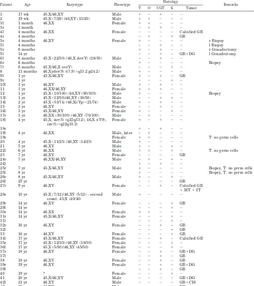
Forty-five patients with gonadal dysgenesis were retrieved from our archives. Samples of 20 dysgenetic gonads containing gonadoblastoma and/or dysgerminoma in 16 different patients and 40 dysgenetic gonads without apparent malignancy in 27 different patients were selected; three samples in two patients were excluded due to bad preservation of material. Patient and tissue characteristics are summarized in Table 2. Two fetal samples (Table 2, patients 1 and 2) obtained after induced abortion were included. Patient 5 underwent a bilateral gonadal biopsy at 4 months and a right gonadectomy 2 months later. A left gonadectomy was performed at 14 yr. Patient 26 underwent a right gonadectomy at 8 yr and a left gonadectomy at 25 yr.
Use of tissues for scientific reasons was approved by an institutional review board. The samples were used according to the Code for Proper Secondary Use of Human Tissue in The Netherlands, as developed by the Dutch Federation of Medical Scientific Societies (version 2002) (26). Samples originating from collaborating centers in Belgium, Poland, and Indonesia were treated in accordance with the above mentioned as well as local medical ethical guidelines.
Immunohistochemical staining
Tissue material was fixed in 10% formalin or Bouin’s fixative, ac-cording to local fixation procedures. Slices of 3–5m thickness were
prepared.
The antibodies used for immunohistochemistry and a schematic rep-resentation of the applied protocols are represented in Table 1. Slides were incubated with the primary antibodies at appropriate dilutions, staining was performed using 3,3⬘ -diaminobenzidine-tetrahydrochlo-ride dehydrate (DAB)/H2O2or New Fuchsin (Fluka Chemica,
Stein-heim, Germany)/Naphtol ASMX (Sigma Aldrich, Zwijndrecht, The Netherlands) phosphate and counterstaining with hematoxylin. As pos-itive controls, a normal adult male gonad for VASA, a seminoma sample for PLAP, c-KIT, TSPY and OCT3/4 and a male fetus, 8 wk gestational age (GA) for AMH were included.
Double-staining experiments were performed using the same detec-tion methods but with different substrates: Fast Blue/Naphtol ASMX phosphate (F3378 and N500; Sigma, Steinheim, Germany) for a blue
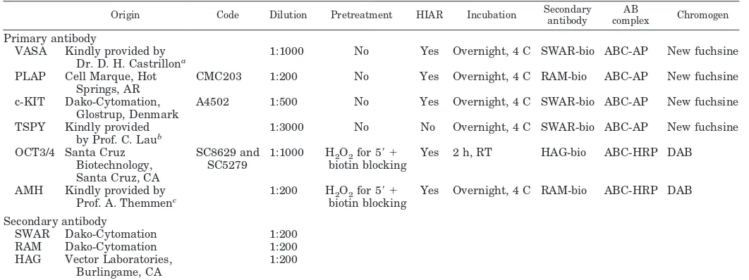
TABLE 1. Schematic representation of origin and protocols used for the different antibodies
Origin Code Dilution Pretreatment HIAR Incubation Secondaryantibody complexAB Chromogen
Primary antibody
VASA Kindly provided by Dr. D. H. Castrillona
1:1000 No Yes Overnight, 4 C SWAR-bio ABC-AP New fuchsine
PLAP Cell Marque, Hot Springs, AR
CMC203 1:200 No Yes Overnight, 4 C RAM-bio ABC-AP New fuchsine
c-KIT Dako-Cytomation, Glostrup, Denmark
A4502 1:500 No Yes Overnight, 4 C SWAR-bio ABC-AP New fuchsine
TSPY Kindly provided by Prof. C. Laub
1:3000 No No Overnight, 4 C SWAR-bio ABC-AP New fuchsine
OCT3/4 Santa Cruz
Yes 2 h, RT HAG-bio ABC-HRP DAB
AMH Kindly provided by Prof. A. Themmenc
1:200 H2O2for 5⬘ ⫹
biotin blocking
Yes Overnight, 4 C RAM-bio ABC-HRP DAB
Secondary antibody
HIAR, Heat-induced antigen retrieval (40); SWAR, swine antirabbit antibody; RAM, rabbit antimouse antibody; HAG, horse antigoat antibody; bio, biotin labeled; AB complex, streptavidin-biotin complex; ABC-AP, streptavidin-biotin-alkaline phosphatase complex; ABC-HRP, streptavidin-biotin-horseradish peroxidase complex; RT, room temperature.
aDepartment of Pathology, University of Texas, Southwestern Medical Center, Dallas, Texas.
TABLE 2. Overview of patient and tissue characteristics
Patient Age Karyotype Phenotype Histology Remarks
T O UGT S Tumor
1 17 wk 45,X/46,XY Male ⫹ ⫺ ⫹ ⫺ ⫺
2 19 wk 45,X (7/20) /46,XY (13/20) Male ⫹ ⫹ ⫺ ⫺ ⫺
3l 1 month 46,XX Female ⫹ ⫹ ⫺ ⫺ ⫺
3r 1 month ⫺ ⫹ ⫹ ⫺ ⫺
4l 4 months 46,XX Female ⫺ ⫺ ⫺ ⫺ Calcified GB
4r 4 months ⫺ ⫹ ⫺ ⫹ GB
5r 4 months 46,XY Female ⫹ ⫺ ⫺ ⫺ ⫺ r Biopsy
5l 4 months ⫺ ⫺ ⫹ ⫺ ⫺ l Biopsy
5r 6 months ⫹ ⫺ ⫹ ⫺ ⫺ r Gonadectomy
5l 14 yr ⫺ ⫺ ⫹ ⫺ GB⫹DG l Gonadectomy
6l 6 months 45,X (22/50) /46,X der(Y) (28/50) Male ⫺ ⫺ ⫹ ⫺ ⫺
6r 6 months ⫹ ⫺ ⫺ ⫺ ⫺ Biopsy
7l 6 months 45,X/46,X iso(Y) Male ⫺ ⫺ ⫺ ⫹ ⫺
8 11 months 46,Xyder(9) t(7;9) (q13.2;p24.2) Male ⫹ ⫺ ⫺ ⫺ ⫺
9l 1 yr 45,X/46,XY Female ⫹ ⫺ ⫹ ⫺ GB
9r 1 yr ⫺ ⫺ ⫺ ⫹ ⫺
10l 1 yr 46,XY Male ⫹ ⫺ ⫹ ⫺ ⫺
11 1 yr 46,XX/46,XY Female ⫹ ⫹ ⫹ ⫺ ⫺
12 1 yr 45,X (10/100) /46,XY (90/100) Male ⫹ ⫺ ⫺ ⫺ ⫺ Biopsy
13l 1 yr 45,X (12/50)/46,XY (38/50) Male ⫺ ⫺ ⫹ ⫹ ⫺
14l 2 yr 45,X (53/74) /46,Xi(Yp) (21/74) Male ⫹ ⫺ ⫹ ⫺ ⫺
15 2 yr 46,XY Female ⫺ ⫺ ⫹ ⫺ ⫺
16l 3 yr 45,X/46,XY Female ⫹ ⫺ ⫺ ⫹ ⫺
17r 3 yr 46,XX (30/100) /46,XY (70/100) Male ⫺ ⫹ ⫺ ⫺ ⫺
18l 4 yr 45,X, inv(5) (q22q33.2); 46,X i(Y9), inv(5) (q22q33.2)
Female ⫹ ⫺ ⫹ ⫺ ⫺
18r ⫹ ⫺ ⫹ ⫺ ⫺
19l 4 yr 46,XX Male, later ⫺ ⫹ ⫺ ⫺ ⫺
19r Female ⫹ ⫹ ⫺ ⫺ ⫺ T: no germ cells
20 4 yr 45,X (11/25) /46,XY (14/25) Male ⫹ ⫺ ⫺ ⫺ ⫺
21 5 yr 46,XY Male ⫹ ⫺ ⫺ ⫹ ⫺
22l 6 yr 46,XX Male ⫹ ⫹ ⫺ ⫹ ⫺ T: no germ cells
23 7 yr 46,XY Female ⫺ ⫺ ⫺ ⫺ GB
24r 7 yr 46,XX/46,XY Male ⫺ ⫹ ⫺ ⫹ ⫺
24l ⫹ ⫺ ⫹ ⫺ ⫺
25r 7 yr 45,X/46,XY Male ⫹ ⫺ ⫺ ⫺ ⫺ Biopsy, T: no germ cells
25l 8 yr ⫹ ⫺ ⫺ ⫺ ⫺ Biopsy, T: no germ cells
26r 8 yr 45,X/46,XY Male ⫹ ⫺ ⫺ ⫹ ⫺
26l 25 yr ⫺ ⫺ ⫺ ⫺ GB
27r 9 yr 46,XY Female ⫺ ⫺ ⫹ ⫺ Calcified GB
⫹MT⫹IT 28r 10 yr 45,X (7/12)/46,XY (5/12) - second
count: 45,X (40/40)
Male ⫹ ⫺ ⫺ ⫺ ⫺
29r 14 yr 46,XY Female ⫺ ⫺ ⫺ ⫹ GB
29l 14 yr ⫺ ⫺ ⫺ ⫹ ⫺
30r 14 yr 46,XX Female ⫹ ⫹ ⫺ ⫺ ⫺
31r 14 yr 45,X/46,XY Female ⫺ ⫺ ⫺ ⫹ ⫺
31l ⫺ ⫺ ⫺ ⫹ ⫺
32r 16 yr 46,XY Female ⫺ ⫺ ⫹ ⫺ GB
32l ⫺ ⫺ ⫺ ⫹ GB
33 16 yr 46,XY Female ⫺ ⫺ ⫺ ⫹ GB
34l 17 yr 45,X/46,XY Female ⫺ ⫺ ⫹ ⫺ Calcified GB
35r 17 yr 45,X (12/30) /46,XY (18/30) Female ⫺ ⫺ ⫺ ⫹ ⫺
36l 17 yr 45,X (5/50)/46,XY (45/50) Female ⫺ ⫺ ⫺ ⫹ ⫺
37r 18 yr 46,XY Female ⫺ ⫺ ⫹ ⫺ GB⫹DG
37l ⫺ ⫺ ⫹ ⫺ GB
38 18 yr 46,XY Female ⫺ ⫺ ⫹ ⫺ GB
39r 19 yr 46,XY Female ⫺ ⫺ ⫹ ⫺ GB⫹DG
39l ⫺ ⫺ ⫹ ⫺ GB
40 19 yr ? Female ⫺ ⫺ ⫺ ⫹ ⫺
41 20 yr 45,X/46,XY Male ⫺ ⫺ ⫺ ⫺ GB⫹DG
42l 21 yr 46,XY Male ⫺ ⫺ ⫺ ⫺ GB⫹CIS
43 25 yr 46,XY Male ⫺ ⫺ ⫺ ⫹ DG
staining and 3-amino-9-ethyl-carbazole (A.5754 and D4254, Sigma)/ H2O2for a red staining, without counterstaining. To reduce background
signal, endogenous peroxidase activity and endogenous biotin were blocked using 3% H2O2(5 min) and a blocking kit for endogenous biotin
(Vector Laboratories, Burlingame CA). For a correct interpretation of histology, hematoxylin-eosin (HE) staining was performed on parallel slides.
Interpretation of results
General morphology and interpretation of the staining results were assessed by two observers with experience in germ cell pathology (M.C. and J.W.O.). Part of the results obtained in five gonadoblastoma samples were reported previously (21).
Statistical analysis
Results were analyzed using the SPSS (SPSS 11.0 for Windows; SPSS Inc., Chicago, IL) statistical program (Fisher exact test for 2⫻2 tables).
Results
Morphology and staining results in dysgenetic gonads without neoplastic transformation
Four gonadal differentiation patterns were recognized in various combinations (Tables 2 and 3 and Fig. 1).
First, tissue containing seminiferous tubules was encoun-tered in 24 of 40 gonads (60%) and was considered as a testicular differentiation pattern. The tubules often displayed
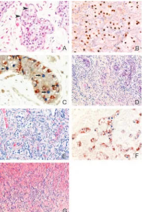
FIG. 1. Differentiation patterns encountered in dysgenetic gonads without neoplastic transformation: A, Patient 30r. Dysgenetic testicular tissue, containing germ cells ( arrow-heads). HE staining,⫻400. B, Patient 18r. Testicular tis-sue. OCT3/4-positive cells are located in the center of the tubule but are also found on the basal lamina. OCT3/4 staining,⫻200. C, Same patient. A subpopulation of germ cells (recognized by the VASA staining) expresses OCT3/4. OCT3/4-positive cells are located in the center of the tubule (arrows) but are also found on the basal lamina ( arrow-heads). OCT3/4 (blue)-VASA (red) double staining,⫻400. D, Patient 19r. Testicular (left part) and ovarian (right part) differentiation; the testicular tissue is devoid of germ cells; ova are enclosed in primordial and primary follicles. HE staining,⫻200. E, Patient 14l. Undifferentiated gonadal tissue. Germ cells align in clusters together with Sertoli/ granulosa cells or lay isolated in fibrous stroma. HE stain-ing,⫻200. F, Same patient. A subpopulation of germ cells within the UGT area expresses OCT3/4. OCT3/4 (blue )-VASA (red) double staining,⫻200. G, Patient 13l. Streak. Remnants of cords that have lost their germ cells and have undergone a fibromatous reaction are recognizable. HE staining,⫻200.
TABLE 3. Summary of staining results in the encountered gonadal differentiation patterns and in gonadoblastoma (GB)/dysgerminoma (DG)
Testis UGT Ovary Streak GB DG
OCT3/4 ⫹ ⫹ ⫺ ⫺ ⫹ ⫹
PLAP ⫹ ⫹ ⫺ ⫺ ⫹ ⫹
c-KIT ⫹ ⫹ ⫹ ⫺ ⫹ ⫹
TSPY ⫹⫹⫹ ⫹⫹⫹ ⫺ ⫺ ⫹⫹⫹ ⫹/⫺
VASA ⫹ ⫹ ⫹ ⫺ ⫹ ⫹/⫺
abnormalities such as a thin basal lamina, shape irregularity, increased intertubular spaces, and branching structures, re-flecting their dysgenetic nature (Fig. 1A). Germ cells were found in 20 of 24 samples (83%) and were easily recognized by positive staining for VASA and TSPY. The TSPY staining was consistently very intense, compared with normal adult and age-matched controls as well as normal fetal testicular samples (our personal observations). Staining of germ cells for OCT3/4, c-KIT, and PLAP was performed in 18 of 24 samples; in the remaining six, it could not be performed due to limited material. A subpopulation of germ cells stained positive for the three markers in 14 of 18 samples (78%). In all cases, positive cells were found centrally in the tubule, pointing at a delay in maturation, but seven of 14 patients (50%) also displayed positive germ cells on the basal lamina (Fig. 1B). OCT3/4-VASA double-staining experiments con-firmed these findings. In general, OCT3/4 expression was lost as cells became positive for VASA, yet sometimes OCT3/ 4-VASA coexpression was observed within a single cell (Fig. 1C). Regardless of their dysgenetic aspect, in all samples the majority of the tubules displayed positive staining for AMH in Sertoli cells.
Second, an ovarian differentiation pattern was defined as gonadal tissue containing germ cells enclosed in primordial and eventually growing follicles, comparable with the ova-ries of female neonates (Fig. 1D). It was encountered in 10 of 40 gonads (25%). Ova homogeneously expressed VASA and c-KIT, whereas OCT3/4-, PLAP-, or TSPY-positive ova were never found. A unique situation was found in patient 3l: follicles, containing VASA-positive and TSPY-negative ova, were enclosed in seminiferous tubules, containing VASA-positive, TSPY-positive spermatogonia (Fig. 2).
A third differentiation pattern, further referred to as un-differentiated gonadal tissue (UGT), consisted of gonadal tissue containing germ cells not enclosed in seminiferous tubules or follicles but organized together with Sertoli/gran-ulosa cells in cord-like structures or residing without appar-ent organization in a background of fibrous stroma (Fig. 1E) or a combination of these two. This pattern, although present
in 13 of 40 gonads (32.5%), was mostly not mentioned in the original pathology reports, or alternatively, it was referred to as a streak. Expression of markers in the germ cells within UGT was similar to that found in testicular tissue. Germ cells could easily be identified by their expression of VASA and abundant expression of TSPY. Due to limited material, OCT3/4, PLAP, and c-KIT staining could be performed in 11 of 13 samples. In nine of them (82%), a subpopulation of germ cells within UGT stained positive for these three markers. OCT3/4-VASA double staining confirmed these results (Fig. 1F). Sertoli/granulosa cells within UGT differed from their apparently more differentiated counterparts in testicular tu-bules: in most samples, AMH expression was totally absent, although sporadic weakly positive Sertoli/granulosa cells were found in two samples.
The fourth pattern, consisting of fibrous stroma devoid of germ cells, was referred to as streak. It was found in 14 of 40 dysgenetic gonads (35%) in an age-related manner: four of 25 cases until 4 yr of age contained streak tissue (15%), whereas it was found in 10 of 15 samples older than 4 yr (66%) (P⫽ 0.002). Cord-like structures were often recognizable and might represent UGT that has lost its germ cells and has undergone a fibromatous involution (Fig. 1G). AMH expres-sion was never observed in this tissue type.
Morphology and staining results in dysgenetic gonads containing gonadoblastoma and/or dysgerminoma (Tables 2 and 3 and Fig. 3)
Typical gonadoblastoma nests were found in 16 of 20 sam-ples, four of them also containing an invasive dysgerminoma component and one combined with CIS. Three samples con-sisted of large calcifications (the so-called burnt-out gonado-blastoma), one of them in combination with mature and im-mature teratoma. In one sample only dysgerminoma was identified. Germ cells within gonadoblastoma stained positive for OCT3/4, c-KIT, PLAP, and TSPY. TSPY was abundantly expressed in every gonadoblastoma sample, but the expression decreased in the invasive tumor components. The expression of
OCT3/4, c-KIT, and PLAP was variable: in some gonadoblas-toma samples, all the germ cells stained positive, whereas in others, only a subpopulation of germ cells expressed these markers; however, in the dysgerminoma samples, these mark-ers were homogenously present. VASA expression within the gonadoblastoma samples was variable: in 12 of 15 examined samples, germ cells were positive; the remaining three samples were negative for VASA.
Nonneoplastic gonadal tissue adjacent to the neoplastic lesions was available in 15 of 20 samples (75%). The four dif-ferentiation patterns described above were encountered: a streak was found in five of 15 cases (33%), one in combination with ovarian tissue, and UGT was present in 10 of 15 cases (67%), one in combination with testicular tissue (Fig. 3, A–C). Again, in UGT, germ cells stained positive for TSPY with a remarkably strong intensity. In all samples containing UGT, OCT3/4-, c-KIT-, and PLAP-positive germ cells were found (Fig. 3D). In patients 23 and 39 (bilaterally), in the proximity of
germ cells was found, suggesting clonal expansion and final organization into gonadoblastoma nests (Fig. 3E).
These findings were confirmed in the OCT3/4-TSPY dou-ble-staining results (Fig. 3, F and G).
Discussion
The overall incidence of germ cell tumors in 45 patients with gonadal dysgenesis retrieved from our database was 35% (16 of 45 patients), with four bilateral cases. Invasive germ cell tumors were found in six of 45 patients (13%).
We studied the histological and immunohistochemical characteristics in dysgenetic gonads that have (not yet) un-dergone neoplastic transformation and compared these find-ings with the properties of the gonadal tissue adjacent to and within gonadoblastoma samples. Given the rarity of the syn-drome, a relatively large patient series could be collected. However, due to limited material, some experiments were FIG. 3. A, Patient 37r. UGT with isolated germ cells (
In dysgenetic gonads, basically four patterns of gonadal differentiation (with each pattern displaying a broad spec-trum of abnormalities in morphology, number, and organi-zation of the germ cells) were found: testicular and ovarian tissue, streak, and UGT. The frequent finding of UGT was unexpected because it had not been mentioned in the original pathology reports, nor is it routinely described in literature, although some histological descriptions might suggest the presence of UGT (17, 27, 28). UGT clearly differed from the other patterns: in contrast to streak tissue, it does contain germ cells, but these are organized in neither seminiferous tubules nor follicles. In contrast, these germ cells reside apparently ran-domly distributed in a background of stromal cells or align in clusters, in close contact with Sertoli/granulosa cells. Without close observation and the use of specific markers, the germ cells within UGT are easily overlooked, hence its frequent classifi-cation as a streak. Alternatively, due to their presence in ovar-ian-type stroma, and although they are not organized in folli-cles, the germ cells are sometimes misinterpreted as residing in ovarian tissue. However, their correct identification is of crucial importance, as is illustrated in patient 5. In this girl, a bilateral biopsy was performed at 4 months, followed by a gonadectomy of the right testis because of its discordance with the sex of rearing. The left gonad, which was considered as ovarian tissue, was left in place. At the age of 14 yr, she developed a gonado-blastoma, already having progressed to a dysgerminoma. A reevaluation of the available biopsy material of the left gonad unequivocally demonstrated germ cells and cord-like struc-tures (Fig. 4, A–C). A similar case is found in the literature (6). A significantly increased incidence of streak tissue was found in patients older than 4 yr, compared with younger patients (P⫽0.002). Cord-like structures were often recog-nized, suggesting a loss of germ cells in UGT, analogous to the germ cell loss that is observed in dysgenetic testicular or ovarian tissue [e.g. the testicular tissue in hermaphrodites (13) or the testes of patients with undervirilization syn-dromes (19)]. This finding is in accordance with previous reports (7).
The fate of the germ cells during normal human gonadal
development was described by Gondos (29) and is largely analogous to the gonadal development and differentiation process analyzed in detail in mice (30). At their arrival in the genital ridge around 5 wk GA, the germ cells and pre-Sertoli/ granulosa cells lay intermingled without specific organiza-tion in the undifferentiated gonad. The first sign of sexual differentiation (the formation of primitive cords) coincides with SRY expression, around wk 6 GA. In the absence of SRY, no changes occur in the undifferentiated gonad until the 12th week, when the germ cells enter meiosis. As stated above, germ cells within UGT either lay randomly in fibrous stroma or line up together with Sertoli/granulosa cells in cord-like structures. It is conceivable that the first pattern represents the undifferentiated state of the gonad, in which no accurate SRY expression has taken place but in which under the in-fluence of unknown (male) characteristics, meiosis and pro-gression along the default pathway are inhibited. The second pattern might represent early sex cords, blocked in their progression toward seminiferous tubules (Fig. 5). The dif-ferential staining results for AMH in testicular tubules and cord-like structures support this hypothesis.
Germ cells were stained with the markers TSPY, OCT3/4, c-KIT, PLAP, and VASA. TSPY expression was never en-countered in ova enclosed in follicles. However, it was abun-dantly expressed in germ cells within dysgenetic testes and UGT, compared with the intensity of the TSPY staining in fetal and age-matched normal gonads (19, 20, 31) (and our personal observations), thereby suggesting an up-regulation of TSPY when germ cells reside in an unfavorable environ-ment. This is in line with previous observations in gonado-blastoma (21). TSPY is thought to be related to the premeiotic proliferation of spermatogonia, although its function is not fully clarified (23). Evidence is growing that TSPY is the main candidate for the hypothetical gene in the gonadoblastoma region on the Y chromosome leading to the development of gonadoblastoma (21, 24, 32). Abnormal TSPY expression has also been related to the development of CIS in undervirilized patients (19).
OCT3/4, c-KIT, and PLAP are well-established markers
for the diagnosis of various germ cell tumors and are nor-mally expressed during fetal gonadal development (20, 33). Maturation delay of germ cells, which has been described in intersex conditions and patients with chromosomal anom-alies, is characterized by a prolonged expression of these markers and is considered to be a risk factor for malignant transformation (2, 19, 31, 34, 35). In the present series, these three markers revealed similar staining patterns; however, OCT3/4, resulting in a well-circumscribed and intense nu-clear staining, was the most stable marker and was easiest for interpretation. OCT3/4, a transcription factor regulating plu-ripotency of embryonal stem cells and essential for the sur-vival of migratory primordial germ cells (36), is consistently expressed in specific germ cell tumors and might play a pathogenetic role in their development (37, 38). In line with previously reported results (33), OCT3/4 expression was never found in ovarian follicles. In contrast, within testicular tissue and UGT of dysgenetic gonads without gonadoblas-toma and tissue adjacent to gonadoblasgonadoblas-toma, a subpopula-tion of germ cells expressed OCT3/4. Gonadoblastoma and dysgerminoma also consistently expressed this marker (Figs. 1 and 3). In the proximity of a gonadoblastoma lesion, an
gesting clonal expansion within UGT toward gonadoblas-toma formation (Fig. 3E). Previously we demonstrated that OCT3/4-positive prespermatogonia located centrally in the seminiferous tubule reflect a state of maturation delay, whereas OCT3/4-positive cells on the basal lamina are prone to malignant transformation (19). In the present study, the latter pattern was encountered in seven samples. It is tempt-ing to speculate that these patients were at high risk for developing CIS, eventually progressing to invasiveness within the dysgenetic testis if a gonadectomy had not been performed.
Based on our results, a model for the development of gonadoblastoma and CIS in the dysgenetic gonad containing Y chromosome material (i.e.theTSPYgene) emerges: a dys-genetic gonad may consist of different parts with various degrees of differentiation (ovarian tissue, in which the germ cells evolve along the default pathway of meiosis). These germ cells have lost OCT3/4 expression and do not express TSPY; therefore, they cannot give rise to a malignant germ cell tumor. Testicular tissue, showing variable degrees of dysgenesis, contains germ cells that abundantly express TSPY, possibly in an attempt to survive and proliferate in an FIG. 5. Model for the development of UGT, gonadoblastoma, and CIS within the dysgenetic gonad.Upper panel, In the developing embryo, germ cells migrate from the yolk sac into the bipotential gonad and intermingle with pre-Sertoli/granulosa cells.Middle panel(from therightto the
OCT3/4 expression, even after having reached the spermato-gonial niche due to a block in their maturation. Over time, most of these germ cells will die; however, some, possibly due to abundant TSPY levels and prolonged OCT3/4 ex-pression, will survive and proliferate, eventually leading to clonal expansion and CIS formation. In UGT, a similar pro-cess takes place, ending in a streak in case all the remaining germ cells die or alternatively in the development of go-nadoblastoma (Fig. 5). If the latter evolution takes place more rapidly than the progression toward CIS, this would explain the more frequent finding of gonadoblastoma rather than CIS in dysgenetic gonads containing both UGT and testicular tissue. Alternatively, it is conceivable that immature germ cells, blocked in their maturation but residing in UGT, which is their natural environment during early embryonic life, have increased survival chances, compared with immature germ cells residing in more differentiated testicular tissue. Our findings are not disconcordant with the hypothesis that gonadoblastomas originate from germ cells developing along the female pathway but failing to complete the meiotic prophase and organize in primordial follicles (39).
In conclusion, extrapolation to a clinical situation would suggest that a gonadal biopsy revealing the presence of UGT or testicular tissue with OCT3/4-positive cells on the basal lamina contains a high risk for germ cell tumors and should imperatively lead to gonadectomy. Ovarian tissue can safely be left in place; testicular tissue displaying maturation delay of germ cells can be left in situ, given that its localization allows adequate follow-up. A streak is not functional, mak-ing its preservation controversial. Evidently the decision with regard to gonadectomy is not only based on a patho-logical analysis of biopsy material but also must be inspired by the combined interpretation of the patients’ karyotype, internal genitalia, phenotype, gender, and psychological well-being. The value, safety, and applicability of this model should be thoroughly tested in large patient series and mul-ticenter studies before it may contribute to a more conser-vative approach regarding gonadectomy in selected patients with gonadal dysgenesis.
Acknowledgments
The authors thank Friedemann Honecker (Department of Oncology/ Hematology, University Medical Center Hamburg-Eppendorf, Ham-burg, Germany) for critical reading of the manuscript; Michel Molier (Department of Pathology, Erasmus Medical Center Rotterdam, Rotter-dam, The Netherlands) for performing some of the staining experiments; and Cecile Janssens (Center for Clinical Decision Sciences, Department of Public Health, Erasmus Medical Center Rotterdam, Rotterdam, The Netherlands) for support with statistical analysis.
Received November 23, 2005. Accepted March 30, 2006.
Address all correspondence and requests for reprints to: Leendert H. J. Looijenga, Department of Pathology, Erasmus Medical Center, University Medical Center Rotterdam, Josephine Nefkens Institute, Room 430b, P.O. Box 1738, 3000 DR Rotterdam, The Netherlands.
This study was supported by the ESPE Research Fellowship, spon-sored by Novo Nordisk A/S (to M.C.), and by the Dutch Cancer Society (including Grant DDHK 2002-2682, to A.-M.F.K., L.H.J.L., J.W.O., and K.P.W.).
References
1. Woodward PJ, Heidenreich A, Looijenga H, Oosterhuis JW, McLeod DG, Møller H, Manivel JC, Mostofi FK, Heilemariam S, Parkinson MC, Grigor
K, True L, Jacobsen GK, Oliver TD, Talerman A, Kaplan GW, Ulbright TM, Sestermenn IA, Rushton MG, Michael H, Reuter VE2004 World Health Organization Classification of Tumours. In: Eble JN, Sauter G, Epstein JI, Sesterhenn IA, eds. Pathology and genetics of tumours of the urinary system and male genital organs. Lyon, France: IARC Press; 217–278
2. Oosterhuis JW, Looijenga LH2005 Testicular germ-cell tumours in a broader perspective. Nat Rev Cancer 5:210 –222
3. Manuel M, Katayama PK, Jones Jr HW1976 The age of occurrence of gonadal tumors in intersex patients with a Y chromosome. Am J Obstet Gynecol 124:293–300
4. Verp MS, Simpson JL1987 Abnormal sexual differentiation and neoplasia. Cancer Genet Cytogenet 25:191–218
5. Slowikowska-Hilczer J, Szarras-Czapnik M, Kula K2001 Testicular pathol-ogy in 46 XY dysgenetic male pseudohermaphroditism: an approach to patho-genesis of testis cancer. J Androl 22:781–792
6. Donahoe PK, Crawford JD, Hendren WH1979 Mixed gonadal dysgenesis, pathogenesis, and management. J Pediatr Surg 14:287–300
7. Robboy SJ, Miller T, Donahoe PK, Jahre C, Welch WR, Haseltine FP, Miller WA, Atkins L, Crawford JD1982 Dysgenesis of testicular and streak gonads in the syndrome of mixed gonadal dysgenesis: perspective derived from a clinicopathologic analysis of twenty-one cases. Hum Pathol 13:700 –716 8. Slijper FM, Drop SL, Molenaar JC, de Muinck Keizer-Schrama SM1998
Long-term psychological evaluation of intersex children. Arch Sex Behav 27: 125–144
9. Crone J, Amann G, Gheradini R, Kirchlechner V, Fekete CN2002 Manage-ment of 46, XY partial gonadal dysgenesis—revisited. Wien Klin Wochenschr 114:462– 467
10. Creighton S, Minto C2001 Managing intersex. BMJ 323:1264 –1265 11. Reiner WG1999 Assignment of sex in neonates with ambiguous genitalia.
Curr Opin Pediatr 11:363–365
12. Sievert KD2003 Vaginal and penile reconstruction. Curr Opin Urol 13:489 – 494
13. van Niekerk WA1976 True hermaphroditism: an analytic review with a report of 3 new cases. Am J Obstet Gynecol 126:890 –907
14. Nihoul-Fekete C, Lortat-Jacob S, Cachin O, Josso N1984 Preservation of gonadal function in true hermaphroditism. J Pediatr Surg 19:50 –55 15. Scully RE1970 Gonadoblastoma. A review of 74 cases. Cancer 25:1340 –1356 16. Pratt-Thomas HR, Cooper JM1976 Gonadoblastoma with tubal pregnancy.
Am J Clin Pathol 65:121–125
17. Krasna IH, Lee ML, Smilow P, Sciorra L, Eierman L1992 Risk of malignancy in bilateral streak gonads: the role of the Y chromosome. J Pediatr Surg 27:1376 –1380
18. de Jong J, Stoop H, Dohle GR, Bangma CH, Kliffen M, van Esser JW, van den Bent M, Kros JM, Oosterhuis J, Looijenga LH2005 Diagnostic value of OCT3/4 for pre-invasive and invasive testicular germ cell tumours. J Pathol 206:242–249
19. Cools M, van Aerde K, Kersemaekers AMF, Boter M, Drop SLS, Wolffen-buttel KP, Steyerberg EW, Oosterhuis JW, Looijenga LHJ2005 Morpholog-ical and immunohistochemMorpholog-ical differences between gonadal maturation delay and early germ cell neoplasia in patients with undervirilisation syndromes. J Clin Endocrinol Metab 90:5295–5303
20. Honecker F, Stoop H, de Krijger RR, Chris Lau YF, Bokemeyer C, Looijenga LH2004 Pathobiological implications of the expression of markers of testicular carcinomain situby fetal germ cells. J Pathol 203:849 – 857
21. Kersemaekers AM, Honecker F, Stoop H, Cools M, Molier M, Wolffenbuttel K, Bokemeyer C, Li Y, Lau YF, Oosterhuis JW, Looijenga LH2005 Identifi-cation of germ cells at risk for neoplastic transformation in gonadoblastoma. Hum Pathol 36:512–521
22. Rajpert-De Meyts E, Skakkebaek NE1994 Expression of the c-kit protein product in carcinoma-in situand invasive testicular germ cell tumours. Int J Androl 17:85–92
23. Schnieders F, Dork T, Arnemann J, Vogel T, Werner M, Schmidtke J1996 Testis-specific protein, Y-encoded (TSPY) expression in testicular tissues. Hum Mol Genet 5:1801–1807
24. Lau Y, Chou P, Iezzoni J, Alonzo J, Komuves L2000 Expression of a candidate gene for the gonadoblastoma locus in gonadoblastoma and testicular semi-noma. Cytogenet Cell Genet 91:160 –164
25. Castrillon DH, Quade BJ, Wang TY, Quigley C, Crum CP2000 The human VASA gene is specifically expressed in the germ cell lineage. Proc Natl Acad Sci USA 97:9585–9590
26. Oosterhuis JW, Coebergh JW, van Veen EB 2003 Tumour banks: well-guarded treasures in the interest of patients. Nat Rev Cancer 3:73–77 27. Damjanov I, Klauber G1980 Microscopic gonadoblastoma in dysgenetic
gonad of an infant: an ultrastructural study. Urology 15:605– 609
28. Jorgensen N, Muller J, Jaubert F, Clausen OP, Skakkebaek NE1997 Heter-ogeneity of gonadoblastoma germ cells: similarities with immature germ cells, spermatogonia and testicular carcinomain situcells. Histopathology 30:177– 186
29. Gondos B1985 Development of the reproductive organs. Ann Clin Lab Sci 15:363–373
Wolffenbuttel KP, Bokemeyer C, Lau C, Drop SLS, Looijenga LHJ2006 Maturation delay of germ cells in trisomy 21 fetuses results in increased risk for the development of testicular germ cell tumors. Hum Pathol 37:101–111 32. Page DC1987 Hypothesis: a Y-chromosomal gene causes gonadoblastoma in
dysgenetic gonads. Development 101(Suppl):151–155
33. Stoop H, Honecker F, Cools M, de Krijger R, Bokemeyer C, Looijenga LH 2005 Differentiation and development of human female germ cells during prenatal gonadogenesis: an immunohistochemical study. Hum Reprod 20: 1466 –1476
34. Rajpert-De Meyts ER, Jorgensen N, Muller J, Skakkebaek NE1996 Pro-longed expression of the c-kit receptor in germ cells of intersex fetal testes. J Pathol 178:166 –169
35. Rajpert-De Meyts E, Hanstein R, Jorgensen N, Graem N, Vogt PH, Skak-kebaek NE2004 Developmental expression of POU5F1 (OCT-3/4) in normal and dysgenetic human gonads. Hum Reprod 19:1338 –1344
36. Kehler J, Tolkunova E, Koschorz B, Pesce M, Gentile L, Boiani M, Lomeli
H, Nagy A, McLaughlin KJ, Scholer HR, Tomilin A2004 Oct4 is required for primordial germ cell survival. EMBO Rep 5:1078 –1083
37. Looijenga LH, Stoop H, de Leeuw HP, de Gouveia Brazao CA, Gillis AJ, van Roozendaal KE, van Zoelen EJ, Weber RF, Wolffenbuttel KP, van Dekken H, Honecker F, Bokemeyer C, Perlman EJ, Schneider DT, Kononen J, Sauter G, Oosterhuis JW2003 POU5F1 (OCT3/4) identifies cells with pluripotent potential in human germ cell tumors. Cancer Res 63:2244 –2250
38. Gidekel S, Pizov G, Bergman Y, Pikarsky E2003 Oct-3/4 is a dose-dependent oncogenic fate determinant. Cancer Cell 4:361–370
39. Pauls K, Franke FE, Buttner R, Zhou H2005 Gonadoblastoma: evidence for a stepwise progression to dysgerminoma in a dysgenetic ovary. Virchows Arch 447:603– 609
40. Shi SR, Key ME, Kalra KL1991 Antigen retrieval in formalin-fixed, paraffin-embedded tissues: an enhancement method for immunohistochemical staining based on microwave oven heating of tissue sections. J Histochem Cytochem 39:741–748