CASE REPORT: MAMMARY GLAND HYPERPLASIA IN A DOMESTIC SHORT HAIR CAT
1*WiwikMisacoYuniarti, 2*HardanyPrimarizky
1,2 Department of Veterinary Clinic, Faculty of Veterinary Medicine,Universitas Airlangga,
Jl.Muryorejo, Kampus C, Surabaya-Indonesia, 60115
ABSTRACT
Mammary gland hyperplasia also called as benign mammary hypertrophy, mammary fibroadenomatosis, and fibroglandular mammary hypertrophy. It is progesterone-dependent enlargement of one or more mammary glands. Clinical signs that appeared are localized or diffuse enlargement of one or more mammary glands. The visible masses are firm and no pain observed. No concurrent sign of systemic illness. The differential diagnosis of mammary gland are mastitis and mammary neoplasia. The treatment for the case of mammary hypertrophy was due to high endogenous progesterone regresses when progesterone fall at the end of gestation or pregnancy. Hypertrophy that occurs due to administration of exogenous progesterone’s regresses when the medications was withdrawn. The surgery procedure which is ovariohysterectomy must be considered and the cats should not intended for breeding.Dexamethasone as an anti-inflammatory drug was used to overcome the inflammatory reaction that occurs due to the open wounds lesion when performing mammocentesis or wounds that caused by licked activity. Dexamethasone will bind to glucocorticoid receptors in the cell of mammary gland with very high affinity. This may stimulate the anti-inflammatory process, so the proliferation of cell may be reduced.
Keywords: Cat, mammary gland hyperplasia, progesterone, dexamethasone.
*Correspondence: Department of Veterinary Clinic, Faculty of Veterinary Medicine, Universitas
Airlangga, Jl. Mulyorejo, Kampus C, Surabaya– Indonesia, 60115. Email: wiwikmisaco@yahoo.com
INTRODUCTION
A cat has brought to Animal Teaching Hospital at the Faculty of Veterinary Medicine Universitas Airlangga with signalmen as female Medium Persian, named “Kim Kim”, age 1,5 years old and has brownish-orange colored.
Anamnesis
Physical Examination
Physical examination was done and its revealed normal body temperature at 38,8°C, pulse rate on 144 beat/minutes, respiratory rate 42 beat/minutes, 2,7 kg of body weight. Normal body condition also observed but the cat has hair loss. Upon auscultation, ronchi sounds of respiration was heard and abdominal palpation revealed enlargement of the last mammary glands without any open wound, with well-defined margin, soft consistency and no pain reaction when palpated.
Diagnostic Work Up
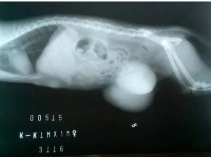
Radiograph of abdominal position revealed the appearance of last mammary glands was enlarged bilaterally and the density has increased with size 10 cm and 15 cm in diameter. The first and second mammary gland also started to increase in size and density.From the results of mammo-centesis obtained no liquid aspirated.
Figure 1 .The radiograph result showed there was enlargement and increased radiopacity of the mammary glands
Differential Diagnosis
2. Mammary tumor(consistency slightly hard and gradually observed).Cause of the mammary tumor so far is unknown. Mammary gland tumors often has receptors for the female cat’s hormone (estrogen and progesterone). This means that the hormone has a chance to trigger tumor growth. Due to that condition, a case of mammary gland tumors are not commonly found in dogs and cats that have been ovariohysterectomized (OH) and at a very young age.Otherwise, the administration of synthetic progesterone to prevent pregnancy or for any other treatment, increases the risk of mammary gland tumors.The tumor mass usually placed in the chest or lower abdomen. These masses are usually found in the mammary glands near the mammary teats, but sometimes it’s located quite far from the mammary gland and was not related to the mammary glands. These masses rather hard, swollen gradually and sometimes ruptured with foul-smell secrete oozed out (Birchard and Sherding, 2005; Sorenmo, 2011).
3. Galactostasis (occurred on postpartum and accompanied with pain).
Galactostasis is an abnormal delays or difficulties in the milk let-down process which can lead to the accumulation in the mammary gland. Usually it happens due to the anatomical structures of cats that can inhibit the release of breast milk. It can also happen if after parturition, a kitten dies or sudden weaning kittens, and when the kittens cannot breastfeed completely (Johnston et al., 2001). Mammary glands in the inguinal area often experienced an oedematus and enlargement of mammary glands (Linde-Forsberg, 2005). The skin in the inguinal area was tightened and the animals showed signs of discomfort and pain. The milk becomes difficult to come out or even not able to. Galactostasis can develop into mastitis, due to the accumulation of milk in the mammary gland which is a good medium for bacterial growth.
4. Mammary neoplasia (occurred in adults).
Mammary neoplasia can occured in adult cats at the age of 10 to 12 years. The risk of mammary neoplasia in the female cat increases with age, especially in animals that are not sterilized. Sterilization influence on the development of mammary carcinoma in cats is less clear, but there is evidence to suggest that sterilization can prevent the development of mammary tumors (Moore, 2006).
Diagnosis and Prognosis
Based on the signalment, anamnesis, physical examination and other investigations, the diagnosis of Kim Kim was Mammary Glands Hyperplasia. The prognosis of this disease is fair (dubious)
Treatment
Therapy was given to the cat Kim-Kim is an oral prescription drugs which contains antibiotic (Amoxicillin 20 mg/kg bb twice a day), anti-inflammatory (Dexamethasone 0,02 mg/kg bb twice a a day) and multivitamins (1/4 tablet twice a day) for 5 days to see its progress.
DISCUSSION
Feline mammary glands hyperplasia, or fibroadenomatous hyperplasia, is a benign, often drastic enlargement of the mammary glands typically seen in younger cats.Mammary glands hyperplasia is a benign condition where’s the tissue of the mammary glands are growed rapidly within 1-2 weeks after estrus or 2-5 weeks after progesterone hormone therapy. This case often affected on mammary glands and occurred in un-neutered young female cats. Sometimes it can also reported in a spayed male or female cats. Enlargement of the mammary glands in this hyperplasia case is reversible. The mass volume of the mammarymay decreased after luteolysis or after the end of exogenous progesterone hormone activity (Buritica et al., 2010). Mammary glands hyperplasia might cause by high levels of progesterone hormone that stimulates the mammary glands to proliferate though the lactation time had not started.
The pathogenesis of mammary glands hyperplasia still remains unclear. The activity of the sexual hormone with the mammary glands are closely linked.Shortly, the development and the growth of mammary glands were influenced byprogesterone which mediated by progesterone receptor in the stroma and epithelial cells. The local activation of progesterone triggers a specificcascade of progesterone that would stimulates the mammary glands to conducted a proliferation. In physiological conditions, analteration of the estrogen and progesterone cycle will make either stimulation or suppression of the progesterone cycle activation. Those pathway might responded to impaired progesterone stimulation and contributed to development of mammary gland hyperplasia and neoplastic activity (Linde-Forsberg, 2005).
It is possible that the response can be attributed to two factors: 1. the extreme sensitivity of cat’s mammary gland for therapy using corticosteroids hormone; 2. the fact that the glands are usually very thin-small when the cats are not pregnant or breastfeeding. In recent studies, there are two progesterone receptor isoforms (A and B) have been demonstrated in tissue samples from fibro-epithelial hyperplasia, with a dominant expression in epithelium duct.
Kim-Kim is a young female cat that has not been neutered. Before the enlargement of mammary glands are noticed by the owner, Kim-Kim was got a birth-control injection using Depoprogestin® which is an exogenous progesterone. The high number of exogenous progesterone will stimulate high proliferation of the mammary glands. It would stop if the activity of the hormone progesterone decreased. Based on the anamnesis with the client, it mentioned that the onset of mammary glands enlargement with Depoprogestin® injection was around 4 weeks. This is similar with the characteristics of mammary glands hyperplasia that growth fast and affected to un-neutered young female cats.
It may be able to use a broad spectrum antibiotic such as Amoxicillin or using drugs of anti-inflammatory drugs in short period. Until end of 90's decade, ovariectomy or ovariohysterectomy is considered as a suitable therapy. Excision of the ovary usually caused regression on mammary tissue within 3 - 4 weeks. Mastectomy was not recommended as first approach for treatment of mammary glands hyperplasia. An aggressive mastectomy often caused some complications and only recommended when other options have failed (Tilley and Smith, 2015). In the case where the hyperplasia is due to synthetic progesterone administration, the use of the drug is simply discontinued. In either case, after removing the source of progesterone, the mammary glands generally return back to normal within a few weeks. Some of the recent studies showed that using Aglepristone or progesterone receptor blockers (Alizine®, Virbac, France) could be succeed. Aglepristone is a competitive molecule that binds to progesterone receptor without activated cascade hormone reponse in target tissue. This drug bound the progesterone receptor with 9 times affinities, and the withdrawal time is 6 days if given once a day in dose of 20 mg/kg or twice a day at 10 mg/kg (Enginler and Senunver, 2011).
CONCLUSSION
In this case the therapy that been used is dexamethasone oral continued with injection subcutaneously. Dexamethasone is a corticosteroid anti-inflammatory drugs. This medicine was used to resolve the inflammatory reaction which occurred due to an open wound after conducted mammocentesis or because of the swollen mammary glands that licked by the cats. In addition, this medicine is a corticosteroid drug that can bind to the glucocorticoid receptor in mammary gland cells with very high affinity. Glucocorticoid receptor with Dexametasone bond can stimulated anti-inflammatory process, so the proliferation can be reduced. Besides that, cold water compression can be made if necessary. Supportive injection therapy with Biosol® can provide the stability condition of the patient.
REFERENCES
Birchard, SJ. and Sherding, RG. 2005. Saunder’s Manual of Small Animal Practice. 3rd Edition. WB
Saunders Co. USA. 114.
Buriticá EF, Echeverry DF, Lozada AF. 2010 Hiperplasia fibroepitelial mamaria feli‐na: reporte de uncaso. Rev Ces Med Vet Zootec. 5 (1):70-76.
Enginler SÖ, Şenünver A. 2011. The Effects of Progesterone Hormone Applications Used for Suppression of Estrus on Mammary Glands in Queens. Kafkas Üniversitesi Veteriner Fakültesi Dergisi 17 (2): 277-284.
Johnston, SD., Kustritz, MVR, Olson, PNS. 2001. Canine and Feline Theriogenology. 215.
Leidinger E, Hooijberg E, Sick K, Reinelt B, Kirtz G. 2011. Fibroepithelial hyperplasia in an entire male cat: cytologic and histopathological features. Tierarztl Prax Ausg K Kleintiere Heimtiere 39(3): 198-202.
Moore, A. 2006. O-Oncology : Advances in the Treatment of Mammary Neoplasia. Proceeding of the WSAVA World Congress/FECAVA/CSAVA. pp.
562-565.http://ivis.org/proceedings/wsava/2006/lecture20/Moore2.pdf?LA=1>.
Sorenmo, KU. 2011. Mammary gland tumors in cats: Risk factors, clinical presentation, treatments and outcome. Proceedings of the 36th World Small Animal Veterinary Congress‐ 230. Insights from Veterinary Medicine Congress, Jeju (Korea), 14 to 17 October. OC-I10: 764– 767.
Tilley, LP.and Smith, FWK. 2015. The 5 Minute Veterinary Consult Canine and Feline. 6th Ed. William and Wilkins Co, USA. 839.
Yuniarti, W.M. and Lukiswanto, B.S. 2014. Mastitis pada Kucing Mona. Vet Medika J Klin Vet. 2 (2). http://journal.unair.ac.id/JKV@case-study-article-6884-media-85-category-5.html