Intervertebral Disc Degeneration and Low Back Pain: Molecular Mechanisms
and Stem Cell Therapy
Anna Meiliana
1,2,, Nurrani Mustika Dewi
1,2, Andi Wijaya
1,21Postgraduate Program in Clinical Pharmacy, Padjadjaran University, Jl. Eijkman No.38, Bandung, Indonesia 2Prodia Clinical Laboratory, Jl. Cisangkuy No.2, Bandung, Indonesia
Corresponding author. E-mail: [email protected]
Received date: Jan 19, 2018; Revised date: Apr 8, 2018; Accepted date: Apr 18, 2018
B
ACKGROUND:
Low back pain (LBP) mostly
caused by disc degeneration, reflects to a
tremendous of health care system and economy.
More knowledge about these underlying pathologies
will improve the opportunities that may represent critical
therapeutic targets.
CONTeNT:
Basic research is advancing the understanding
of the pathogenesis and management of LBP at the
molecular and genetic levels. Cytokines such as matrix
metalloproteinases, phospholipase A2, nitric oxide, and
tumor necrosis factor-α are thought to contribute to the
development of LBP. Mesenchymal stem cells (MSCs)
transplant to cartilage-like cells and secrete extracellular
matrix and encourage nucleus pulposus (NP) cell activity
Abstract
R E V I E W A R T I C L E
inhibiting NP cell apoptosis, together with some chemical
mediators such as cytokines and growth factors become
a safe and effective new strategy for intervertebral disc
degeneration (IDD) treatment and regeneration.
SUMMARy:
IDD occurs where there is a loss of homeostatic
balance with a predominantly catabolic metabolic profile. A
basic understanding of the molecular changes occurring in
the degenerating disc is important for practicing clinicians
to help them to inform patients to alter lifestyle choices,
identify beneficial or harmful supplements, or offer new
biologic, genetic, or stem cell therapies.
KeywORDS:
low back pain, intervertebral disc,
degeneration, nucleus pulposus, annulus fibrosus,
extracellular matrix, genetic, stem cells
Indones Biomed J. 2018; 10(1): 1-15
Introduction
The lumbar intervertebral discs (IVDs) play important roles
for the support and mobility of spine.(1-3) These remarkable
tissues is able to maintain stability under a large variety of
loading conditions, while still permitting intersegmental
motion.(1,4) Disc herniation and IVD degeneration (IDD)
are two of the most common causes of low back pain (LBP)
which is targeted for intervention.(5) Disc degeneration
(DD) is a multifactorial process characterized by cellular
and biochemical alternations in disc tissue which result
in structural failure.(6) While DD is a part of normal
aging, a significant number of people with indications of
DD on magnetic resonance imaging (MRI) are actually
asymptomatic, with no history of pain or disability.(7,8)
The risk of back pain is increasing with the severity of
DD.(9,10) Biological changes like proteoglycan and water
loss is not really related with back pain, but back pain is
more related to structural alternations, such as endplate
defects and annulus height loss.(11-15) Most closely linked
to pain are radial fissures in the annulus, whether or not they
cause disc herniation.(14-17)
inflammatory-like reactions, so that they can signal pain
after minimal mechanical stimulation in animal experiments
and in pain-provocation studies on humans.(21,22) The
different knowledge of pain-sensitization processes may
explain why some degenerated discs are painful, whereas
the others are not.(23) More insights into the pathogenesis
of DD may establish new paradigms for early or differential
diagnostics of degeneration using new techniques such as
systemic biomarkers. Research on the mechanobiology of
disease also enriches the development of therapeutics for
disc repair, with potential to reduce pain and disability
associated with DD.(24) More recently, some studies
about the application of mesenchymal stem cells (MSC)
from many source for example bone marrow, synovial
membrane and adipose tissues showed a promosing results
for IDD.(25-27)
The Lumbar IVD, Structure and
Functions
The healthy IVD is composed of some concentrically
arranged layers of fibrocartilage which surround and restrain
an amorphous, well-hydrated, inner core of proteoglycan
gel.(1-3) Strongly bound to the vertebral bodies and
cartilaginous vertebral endplates, the composite make-up
of the IVD creates a hydraulic system that can absorb and
transmit various combinations of compression, shear, and
tensile forces.(6,28,29) The healthy disc creates a “spacer
effect” which maintains sufficient vertical distance between
the vertebrae (disc height) to provide ligamentous tension,
alignment of the facet joints, and adequate space for the
passage of neurovascular structures within the vertebral
foramina.(1) The IVDs lie between the vertebral bodies,
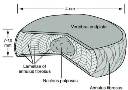
linking them together (Figure 1). They are the main joints of
the spinal column and occupy one-third of its height.
The foremost function of the IVD is mechanical:
it transfers loads, dissipates energy and facilitates joint
mobility. The IVDs are complex structures that consist of
a thick outer ring of fibrous cartilage termed the annulus
fibrosus (AF), which surrounds a more gelatinous core
known as the nucleus pulposus (NP). The NP is
sandwiched inferiorly and superiorly by cartilage endplates.
(30) The NP and the AF structures act synergistically to
distribute and transmit loads between the vertebral bodies
(Figure 2).(31,32) The central NP containing collagen fibers
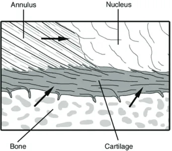
and elastin fibers (Figure 3).(30) The vertebral endplate
containing hyaline cartilage bonded to the perforated
cortical bone of the vertebral body and collagen fibers of
the annulus and the nucleus (Figure 4).(30) When the disc
is compressed, hydrostatic pressure is generated within the
NP, which is constrained peripherally by the AF, generating
tensile circumferential stresses within the lamellar structure.
(31,32) Compressive loads are also supported directly by the
inner AF, which is rich in proteoglycans.(33,34) The
angle-ply structure and nonlinear properties of the AF facilitate
both joint mobility and stability in multiple modalities,
including bending and rotation, and combinations thereof.
(35-38) They provide flexibility to this, allowing bending,
flexion, and torsion.(39,40)
Cells in the anulus are elongated parallel to the
collagen fibers, rather like fibroblasts. Cells in the nucleus
are initially notochordal but are gradually replaced during
Figure 1. A line drawing of the spinal segment consisting of two vertebral bodies and a normal IVD sandwiched between them. (30) (Adapted with permission from John Wiley & Sons).Figure 3. The central NP containing collagen fibers and elastin fibers. The solidified portion of the NP is surrounded by gel-like NP.(30) (Adapted with permission from John Wiley & Sons).
Figure 4. The organization of the vertebral endplate containing hyaline cartilage bonded to the perforated cortical bone of
the vertebral body and collagen fibers of the annulus and the
nucleus. Arrows indicate routes for nutrient transport from blood vessels into the central portion of the disc.(30) (Adapted with permission from John Wiley & Sons).
childhood by rounded cells resembling the chondrocytes of
articular cartilage. Anulus cells synthesize mostly collagen
type I in response to deformation, whereas nucleus cells
respond to hydrostatic pressure by synthesizing mostly
proteoglycans and fine collagen type II fibrils. Cell density
declines during growth (41), and in the adult is extremely
low, especially in the nucleus (42,43). In adult discs, blood
vessels are normally restricted to the outmost layers of
the anulus. Metabolite transport is done by both diffusion,
which is important for small molecules, and by bulk fluid
flow, which is important for large molecules.(42,44)
Low oxygen tension in the center of a disc could causes
anaerobic metabolism, which results in a high concentration
of lactic acid and low pH.(42)
In vitro
experiments indicate
that a chronic lack of oxygen causes nucleus cells to
become quiescent, meanwhile a chronic lack of glucose
can kill them.(45) Deficiencies in metabolite transport are
known to limit both the density and metabolic activity of
disc cells.(42) As a result, discs have limited capability to
recover from any metabolic or mechanical injury. During
growth and the process of aging, it is normal for endplate
permeability and also disc metabolite transport to decrease,
but it increase in the presence of DD and following endplate
damage.(46)
Degradative enzymes, such as matrix
metalloproteinases (MMPs) and a disintegrin and
metalloproteinase (ADAM), are produced by disc cells to
synthesize their matrix and break down the existing matrix.
(47-53) Molecular markers of matrix turnover are naturally
found most abundant during growth, but usually decline
thereafter.(54) The major structural alternations to the disc
occur during fetal and juvenile growth, when the nucleus
changes in consistency from a translucent fluid to a soft
amorphous tissue, caused mainly by an increase in collagen
content.(6,55) Collagen turnover time in articular cartilage
is approximately 100 years (56) and could be even longer
in the disc. Meanwhile the proteoglycan turnover is faster,
possibly around 20 years (57), and some regeneration of
NP is possible in young animals (58). Injuries that affect
the inner annulus or endplate decompress the nucleus and
healing processes are then overtaken by severe degenerative
changes.(59,60)
Molecular Mechanisms of IDD
Figure 5. The classification of internal disc disruption from grade 0 to grade 5, based on the Modified Dallas Classification.
(30) (Adapted with permission from John Wiley & Sons, Inc).
in the disc are pentosidine and carboxymethyl-lysine.
Pentosidine crosslinks collagen molecules and increases
collagen stiffness as well as decreasing the synthesis of
matrix proteins and proteoglycans.(62,64) Additionally,
notochordal cells, the cells that persist in the NP, which
are of notochordal origin, are greatly affected by oxidative
stressors and activate both intrinsic and extrinsic pathways
of apoptosis.(65) Without aging, there is reduced catabolic
activity of NP cells and decreased NP cell number.(66)
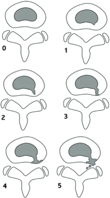
Structural disruption of the IVD is manifested by a
loss of the hydrostatic capacity of the nucleus that occurs
when its surrounding connective tissues cannot provide
adequate restraint.(6) This may occur following an injury
to, or disruption of, the vertebral endplate and/or annulus.
(67,68) Structural changes often initially occur in small
and localized regions of the IVD.(69) Figure 5 shows the
classification of internal disc disruption from grade 0 to
grade 5, based on the Modified Dallas Classification.
Over time, tissue disruption can spread diffusely
throughout the disc and cause a reduction in stiffness and
loss of fluid pressure, also some various combinations of
bulging, herniation, and decreased disc height.(6,70,71)
The most significant biochemical change to occur in DD is
loss of proteoglycan.(72) The aggrecan molecules become
degraded, with smaller fragments being able to leach
from the tissue more readily than larger portions.(30)
Decreasing aggrecan content in the NP leads to reduced
hydration (73), leading in turn to impaired mechanical
function (74,75). A less hydrated, more fibrous NP is unable
to evenly distribute compressive forces between the vertebral
bodies. The forces are instead transferred non-uniformly
to the surrounding AF (28), which can result in altered AF
mechanical properties (76,77) and progressive structural
deterioration, including the formation of circumferential
and radial tears (78). On occasion, radial tears can progress
to a posterior radial bulge or herniation of NP material (78),
resulting in painful symptoms. Decreased disc height is also
commonly associated with advanced DD (12) and results in
painful compression of surrounding structures.
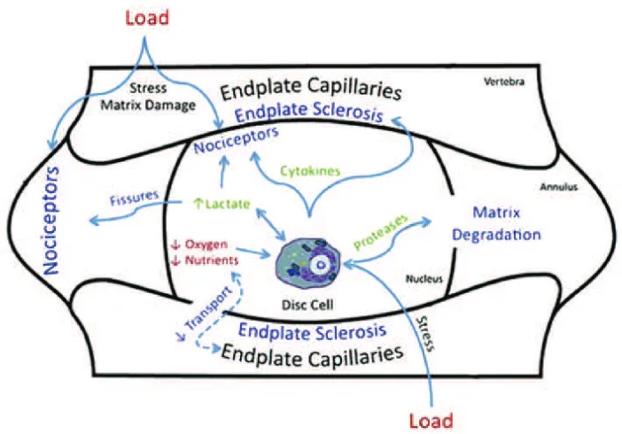
The vertebral endplate is believed to play a critical
role in the transport of nutrients into the IVD and in the
removal of waste products.(42,79,80) One of the main
cause of DD is thought to be failure of the nutrient supply
to the disc cells.(81) Just like all cell types, the cells of the
disc need nutrients, such as glucose and oxygen, to remain
alive and active. The activity of disc cells is very sensitive
to extracellular oxygen and pH in
in vitro
experiment, with
matrix synthesis rates falling steeply at acidic pH and at low
oxygen concentrations and the cells do not survive prolonged
exposure to low pH or glucose concentrations.(45,82,83)
Decrease in nutrient supply that leads to a lowering of
oxygen tension or of pH, which arising from raised lactic
acid concentrations, thus could cause the ability of disc cells
to synthesize and maintain the disc’s extracellular matrix
and could ultimately lead to DD. Thus, endplate disruption
can lead to impaired diffusion (46), disruption in nutrient
supply (45), and/or cell death within the IVD, resulting from
excessive tissue loading.(29,84,85)
Abnormal mechanical loads are also thought to provide
a pathway to DD. For many decades, it was suggested
that a major cause of back problems is injury, often
work-related, that causes structural damage. It is believed that
such an injury initiates a pathway that leads to DD and
finally to clinical symptoms and back pain.(86) Although
LBP Pathophysiology
Significant numbers of recent works assume that
the factors that lead to DD may have important genetic
components. Several studies have reported a strong familial
predisposition for DD and herniation.(92-94) Findings from
two different twin studies conducted during the past decade
showed heritability exceeding 60%.(95,96) MRI in identical
twins were very similar with respect to the spinal columns
and the patterns of DD.(97) Genes associated with DD have
been identified. Individuals with a polymorphism in the
aggrecan gene were found to be at risk for early DD. Studies
of transgenic mice have demonstrated that mutations in
structural matrix molecules such as aggrecan (98), collagen
II (99) and collagen IX (100) can lead to DD. Mutations in
genes other than those of structural matrix macromolecules
have also been associated with DD.(101-103)
There is increasing evidence supporting the role of the
inflammatory cytokine interleukin (IL)-1 in the processes
which leads to degeneration.(104-110) During this process,
there is an increase in the production of the IL-1 agonists
(IL-1α and IL-1β) and their active receptor IL-1 receptor
I (IL-1RI), without a concordant increase in the natural
inhibitor, IL-1 receptor antagonist (IL-1Ra) or the decoy
receptor IL-1RII within the cells of the NP and inner AF. IL-1
induces the expression of a number of MMPs and ADAM
with thrombospondin motifs (ADAMTS) family members
(109,111), and reduces the expression of normal matrix
genes.(109) Neurotrophic factor expression is modulated by
IL-1 (110), and has been linked to induction of senescence
in articular chondrocytes (112-113) and fibroblasts (114), all
of which are features associated with IDD.(115-118)
The aforementioned evidence shows that many
different influences are at work in old and DD, including
genetic inheritance, impaired metabolite transport, altered
levels of enzyme activity, cell senescence and death, changes
in matrix macromolecules and water content, structural
failure, and neurovascular ingrowth.(6)
LBP is related to ageing, mechanical stresses (119) and
genetic predisposition (120), and it is attributed to DD
in around 40% of patients (121,122). It appears that
alteration in biomechanical properties of the disc structure,
sensitization of nerve endings by release of chemical
mediators, and neurovascular ingrowth into the degenerated
discs all may contribute to the development of pain. The
loss of disc structure also alters the loading response and
alignment of the rest of the spinal column, including that of
the facet joints, ligaments, and paraspinal muscles, which
eventually may become additional pain generators.(123)
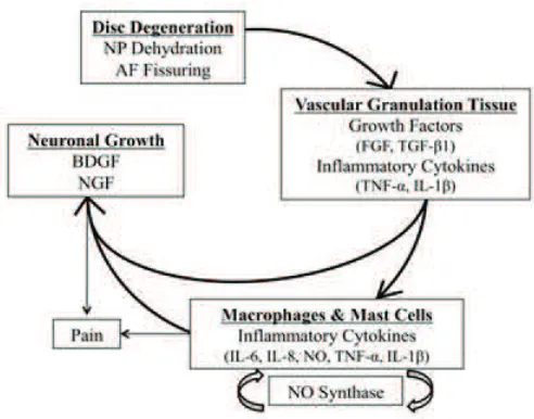
The development of pain might be the result of the
presence of macrophage and mast cells that propagates the
inflammatory cascade. Macrophages increase the levels of
multiple inflammatory mediators, especially 6 and
IL-8, nitric oxide, tumor necrosis factor (TNF)-
a
and IL-1
b
.
(124) The concentration levels of the said cytokines have
correlated with pain intensity, and persistent activation of
sensory fibers upregulates nitric oxide synthase, therewith
increasing the level of nitric oxide, suggesting a possible
positive feedback loop of pain generation.(125)
The onset of discogenic pain is characterized
by nerve fiber ingrowth into an otherwise aneural
tissue (Figure 6).(20,126-128) The interplay between
inflammatory cytokines and neurotrophins, produced by disc
cells and infiltrating immunocytes as well as neurotrophin
receptors and their modulators may guide this process.
Then neuronal tissue will develop after the development
of vascularized granulation tissue. Degenerated disc
cells secrete brain-derived growth factor (BDNF), which
promotes neuronal development.(129) The release of
proinflammatory cytokines IL-1
b
and TNF-
a
from the
surrounding tissues also upregulates nerve growth factor
(NGF) and expression of its receptors on the disc tissue.
(130) Progressively small nerve fibers form along with
the granulation tissue.(131) NGF promotes the collateral
sprouting of additional peripheral sensory nerves into
The recent standard of care for LBP due to degenerative
disc changes includes non-operative approaches, such as
pain management, and operative approaches. The
non-operative management aims primarily in symptomatic pain
relief, while permitting possible endogenous recovery such
as resolution of herniation (148,149) or the repairment
of structural damage (150,151). The main target of
non-operative LBP management is analgesia. It is accomplished
by a combination of nonsteroidal anti-inflammatory drugs
Gene Therapy for IDD
the inner AF and the NP, increases nerve survival, and
increases the action and sensitivity of nociceptive sensory
neurons.(18-20,130,132-135)
Of note, nerve fibers that innervate disc tissue are
categorized as nociceptive and thought to be derived from
the dorsal root ganglia. They express acetylcholinesterase,
protein gene product (PGP) 9.5, substance P (SP), BDNF,
transient receptor potential cation channel subfamily
V member 1 (TrpV1), calcitonin gene related peptide
(CGRP), and neurofilament protein (NFP).(136-140) These
relationships suggest a direct linkage between inflammatory
cytokines, neurotrophins and nociception.(141) Mechanical
stimuli which are normally innocuous to disc nociceptors
can, in certain circumstances, generate an amplified
response which has been termed ‘peripheral sensitization’.
This may explain why some degenerative discs are painful
and others not. There is growing evidence that these pain
receptors in painful disc are peripherally sensitized by the
activity of sympathetic efferent which may initiate a pain
impulse in response to ischemia, pressure changes or in
amatory irritation.(142,243)
Knowing why nerves and blood vessels grow into AF
may lead to effective strategies to hinder the process, or
render it less painful. AF may reflects a microenvironment
of low mechanical stress within a tissue which normally
exhibits a fluid pressure many times greater than systolic
blood pressure.(144,145) Normally, fluid pressure will
extend from the nucleus into the inner and middle annulus
(28), where it would be expected to collapse any blood
vessel. Reduced pressure within a fissure is able to provide
a route for ingrowth of blood vessels and accompanying
nerves. In addition, proteoglycans can inhibit the growth
of nerves (146) and blood vessels
in vitro
(147), and any
loss of proteoglycans from within an annulus fissure may
increase their attractiveness to ingrowing vessels.(23)
(NSAIDs) and physical therapy to strengthen core muscles
among other programs. Surgical management may start
with epidural injections of local anesthetic, steroids, or
a combination of both prior to more invasive surgical
approach.(24)
The current treatment options for IDD and the pathology
associated with it are not the underlying pathophysiology.
(152-155) With advances in molecular and cellular biology,
researchers have start to characterize the pathophysiological
pathways associated with DD and thus provided targets for
potential biological treatments to augment or reverse the
course of IDD.(156) Although the certain pathophysiology
of DD still has not completely understood, however it is
known to be affected by the interaction between various
genetic, biologic and biomechanical factors.(95,157-159)
The hallmark of DD is the progress loss of proteoglycans
which coincides with decreases in oxygen tension, free
radial accumulation, decreased pH, and the increased
activity of aberrant proteolytic enzymes.(73,160,161)
With the loss of proteoglycans the NP cannot maintain
normal physiologic hydrostatic pressure, resulting in in the
dehydration of the disc.(162) There is also a progressive
fibrosis of the NP as the ratio between type I to type
II collagen increases.(163) The NP and AF lose their
morphological diversity as the degeneration happens, which
ultimately distracts the finely balance biomechanics of the
disc and spine as whole.(162,164)
Numerous risk factors, such as age, abnormal physical
loading, and genetics, may lead to the development of
IDD (Figure 7).(165) The homeostasis of IVD tissues is
biologically regulated by the active maintenance of a balance
between the anabolism and catabolism of disc cells. This is
achieved through a complex and precise coordination of a
variety of substances, including cytokines, growth factors,
enzymes and enzyme inhibitors, in a paracrine or/and
autocrine fashion.(166,167) The latest therapeutic strategies
for DD have included some efforts in upregulating the
production of key matrix proteins (
e.g.,
aggrecan), or
downregulating the catabolic events induced by the
Figure 7. Numerous risk factors that may cause to the development of IDD.(165) (Adapted with permission from Elsevier).
(177) The stimulation of matrix synthesis by cytokines or
growth factors changes IVD homeostasis by shifting cellular
metabolism to its anabolic state.(166) It was demonstrated
that the rate of synthesis of proteoglycans by IVD cells
increases several-fold following the addition of transforming
growth factor (TGF)-
a
and epidermal growth factor (EGF).
(178,179) Insulin-like growth factor (IGF)-1 also stimulates
IVD cell proliferation and matrix synthesis
in vitro
.(180,181)
Members of the bone morphogenic protein (BMP) family,
osteogenic protein (OP)-1 (182) and BMP-2 (183), have
both been found to enhance the propylene glycol metabolism
of IVD cells. OP-1 strongly stimulates the production and
formation of the extracellular matrix by rabbit IVD cells
(182), as well as by human IVD cells
in vitro
(184). OP-1
was also found to be effective in the replenishment of a
matrix rich in proteoglycans and collagens after depletion
of the extracellular matrix following exposure of IVD cells
to IL-1 or chondroitinase ABC.(185,186) BMP-2 is known
to facilitates the expression of the chondrogenic phenotype
by human IVD cells, increases proteoglycan synthesis and
up-regulates the expression of aggrecan, collagen type I,
and collagen type II mRNA, compared to untreated control
levels.(187) Both recombinant human BMP (rhBMP)-2 and
-12 increased human NP cell proteoglycan and collagen
synthesis while having minimal effects on AF cells.(188)
Another member of the BMP family, namely growth
and differentiation factor-5 (GDF-5), was also found to
stimulate propylene glycol and type II collagen expression
in mouse IVD cells.(189) Moreover, the recombinant
human GDF-5 (rhGDF-5) enhances cell proliferation and
matrix synthesis and accumulation by both bovine NP and
AF cells.(190) Some epidemiologic studies highlighted that
DD may be caused to a large degree by hereditary factors
with apparently a relatively minor effects of environmental
and behavioral risk factors (191-195), which indicated
that genetic factors might play an important role in the
pathogenesis of IDD.
Recently, Mayer,
et al.
, reviewed the literature and
found that the genetic polymorphisms of 21 genes have been
associated with IDD, including vitamin D receptor (VDR),
GDF5, aggrecan, collagen types I, IX, and XI, fibronectin,
provides a mechanism for the sustained production of the
desired therapeutic product.(156) The role of gene therapy
in the treatment of LBP has been extensively evaluated
to prevent DD disease, regenerate degenerated IVD, and
promote spinal fusion.(123)
Beside the dependence of sustained expression of the
therapeutic gene, the success of gene therapy also lie on the
efficiency of the genetic material transfer to the host cell.
With very little exceptions naked plasmid DNA alone is
not an effective means of gene transfer. Therefore the use
of vectors is necessary to facilitate the transfer of genetic
information to host cell. There are some types of vectors,
which later classified into either viral or nonviral vectors
(include liposomes, gene guns, DNA-ligand complexes,
and microbubble enhanced ultrasound). Liposomes are
phospholipid vesicles which deliver the genetic material into
the cell by fusing with the host’s cellular membrane. Viral
vectors use the natural ability of viruses to infect host cells
and thus transfer the viral genetic information into the host.
Viral vectors are very efficient at transducing the desired
genetic material to the host cell, even into slowly dividing
senescent cellular populations like those of the IVD. Viral
vectors which is used for the gene therapy applications
include adenovirus, adeno-associated virus, herpes
simplex virus, lentivirus, retrovirus and also pox virus.
Each viral vector is associated with specific advantages
and disadvantages. Therefore proper selection of vector is
critical to successful gene therapy.(156)
In addition to the selection of the appropriate gene
and vector, another notable consideration with gene therapy
applications is the delivery strategy utilized. There are
currently two basic strategies for the delivery of exogenous
therapeutic genes into target cells. The
in vivo
strategy
involves the direct transfer of the gene-vector complex to
the targeted cellular population within the living host. The
ex vivo
strategy differs significantly as the targeted cells
are isolated and removed from the living host. These cells
are then cultured with transduction of the therapeutic gene
occurring
in vitro
. The final step includes the re-implantation
of the genetically altered cells back into the host.(156)
Stem Cell Therapy for IDD
One of the available sources for cell-based repair of the
disc that recently as been developed is MSC.(197-199)
MSCs are a heterogeneous population of multipotent cells
capable to differentiate along the chondrogenic, osteogenic,
and adipogenic lineages but not the hematopoietic lineage.
Many different sources of MSCs have been identified and
studied, for example bone marrow, synovial membrane and
adipose tissues.(25-27)
Studies with MSC have been particularly promising.
Co-culture of MSC with NP cells stimulates both NP
cells proliferation and MSC differentiation toward the
chondrogenic lineage.(200-203) Increased production of
cytokines, particularly TGF-
b
favors these transformations.
(203-205) The NP contains MSC that are similar to the
MSC recovered from bone marrow (206), and studies in
animal models of DD have shown that MSC injected
in the NP area not only survive for months but also
proliferate in canine (207,208), porcine (209), and rabbit
models (210). Moreover, the transplanted MSC induced
production of extracellular matrix proteins, including
aggrecan and other proteoglycans, and types I and II
collagens.(207,209,210)
A major limitation of using stem cells as a therapy for
IDD is an appropriate delivery method that will not cause
further injury to the IVD. The most direct route is an injection
into the affected IVD, ensuring a localized therapeutic
effect. However, in vivo studies suggest that needle injection
into the IVD may cause further degeneration.(211-213) In
fact, many studies use needle puncture as a model system
to study DD in animals.(214-216) Recently, a population
of stem cells isolated from human umbilical cord blood,
multipotential stem cells (MPSCs), was reported to exhibit
expanded multipotency with the ability to differentiate into
cells of mesoderm, endoderm and ectoderm lineage.(217)
Importantly, these cells were reported to home to sites of
injury (218), and engraftment at the injured site following
an intravenous injection of these cells. Contrasting to
direct injection, intravenous injection neither improved the
degeneration status, nor preserve disc height, however, both
delivery methods increased glycosaminoglycan (GAG)
protein and Acan gene expression relative to controls,
suggesting possible paracrine effects.(219)
and consistent quality of regenerative effect. In a clinical
setting, injection of MSCs has the benefit of minimizing
invasiveness of secondary surgery in comparison with
installation of mechanical device which also requires
tertiary surgery to remove device after treatment.(216)
All the combined evidences support the application
of bone marrow MSCs for regeneration of IVD and that
long-term survival of injected cells in the hypoxic disc
environment is feasible. In addition to MSC long-term
survival
in vivo
, immediate and trophic effects are of great
importance in supporting MSC differentiation into disc cells,
contributing to immediate disc repairing. Therefore, future
studies can also focus on the methods that support MSC
differentiation as adjuvants. It should also be remembered
that the trophic effects from MSCs injected into the IVD
could potentially contribute to activate endogenous disc or
stem cells to enhance the regenerative efficiency.
Extending the concept of stem cell therapy further,
investigators have exploited the use of allogenic stem cells.
This has the added advantage of off-the-shelf availability.
Moreover, as the cause of DD is thought to be multifactorial,
the use of allogenic stem cells could eliminate potential
autogenic precipitating factors such as genetic predisposition
(222-225), or the diminished potency of stem cells due to
natural aging (197). In fact, IVD is suggested to be
immune-privileged due to its avascular nature. A study, showing
allogenic NP cell transplantation did not elicit lymphocyte
infiltration, is consistent with this notion.(197) The problem
of immune rejection is likely to be even less for allogenic
MSCs, since MSCs are capable of escaping alloantigen
recognition.(194,197)
Adipose-tissue-derived stromal cell (ADSC) show
potential for restoring degenerative discs and may prove
effective in the treatment of IVD. The results of ADSC
implantation studies in a DD model were promising,
indicating that ADSCs could maintain their viability and
proliferate within the rat IVD.(194)
Notochordal cells are the developmental origin of
the NP. Yet they are not expressed in adult human IVD.
Induced pluripotent stem cells (iPSCs) have demonstrated
their ability to differentiate into various cell types. In IVD
applications, mouse and human iPSCs have been shown
to differentiate into NP-like cells expressing notochordal
markers and assumed the possibility that they may be used
as a novel cell source for cellular therapy.(200) Notochordal
cells have been observed to substantially stimulate
biosynthetic activity of NP cells through factors secreted
into conditioned medium.(200) These findings support
the notion that molecular agents secreted by notochordal
cells constitute a promising alternative for disc repair.(24)
Results of stem cell studies in IVD are developing and, if
delivery obstacles can be overcome, may offer alternative
future treatment strategies.
DD progresses with age and involves a shift in the metabolic
productivity of the IVD. The degenerative and inflammatory
changes occurring as the disc degenerates promote
increased neural and vascular ingrowth into the disc,
potentially accounting for the painful discomfort patients
experience with DD. Treatments which utilize inherent
growth potential, through growth factors or stem cells, can
stimulate tissue repair but may also provide advantages by
mitigating inflammation. By knowing the mechanism of
IVD contributes an essential piece of the repair puzzle, lead
to an optimum integrated management of LBP for new and
refined concepts in pathophysiology, earlier detection of
disease, and improved developments in tissue engineering
for treatment.
Conclusion
References
1. Bogduk N, Twomey LT. Clinical Anatomy of the Lumbar Spine. New York: Churchill-Living-stone; 1987.
2. Humzah MD, Soames RW. Human intervertebral disc: structure and function. Anat Rec. 1988; 220: 337-56.
3. Lundon K, Bolton K. Structure and function of the lumbar intervertebral disk in health, aging, and pathologic conditions. J Orthop Sports Phys Ther. 2001; 31: 291-306.
4. Alexander LA, Hancock E, Agouris I, Smith FW, MacSween A. The response of the nucleus pulposus of the lumbar intervertebral discs to functionally loaded positions. Spine. 2007; 32: 1508-12. 5. Deyo RA, Weinstein JN. Low back pain. N Engl J Med. 2001; 344:
363-70.
6. Adams MA, Roughley PJ. What is intervertebral disc degeneration, and what causes it? Spine. 2006; 31: 2151-61.
7. Boden SD, McCowin PR, Davis DO, Dina TS, Mark AS, Wiesel S. Abnormal magnetic-resonance scans of the cervical spine in asymptomatic subjects. A prospective investigation. J Bone Joint Surg Am. 1990; 72: 1178-84.
8. Jensen MC, Brant-Zawadzki MN, Obuchowski N, Modic MT, Malkasian D, Ross JS. Magnetic resonance imaging of the lumbar spine in people without back pain. N Engl J Med. 1994; 331: 69-73. 9. Cheung KM, Karppinen J, Chan D, Ho DWH, Song YQ, Sham
P, et al. Prevalence and pattern of lumbar magnetic resonance imaging changes in a population study of one thousand forty-three individuals. Spine. 2009; 34: 934-40.
10. De Schepper EIT, Damen J, van Meurs JBJ, Ginai AZ, Popham M, Hofman A, et al. The association between lumbar disc degeneration
and low back pain: the influence of age, gender, and individual
11. Hamanishi C, Kawabata T, Yosii T, Tanaka S. Schmorl’s nodes on magnetic resonance imaging. Their incidence and clinical relevance. Spine. 1994; 19: 450-3.
12. Videman T, Battie MC, Gibbons LE, Maravilla K, Manninen H, Kaprio J. Associations between back pain history and lumbar MRI
findings. Spine. 2003; 28: 582-8.
13. Boden SD, Davis DO, Dina TS, Patronas NJ, Wiesel SW. Abnormal magnetic-resonance scans of the lumbar spine in asymptomatic subjects. A prospective investigation. J Bone Joint Surg. 1990; 72: 403-8.
14. Boos N, Rieder R, Schade V, Spratt KF, Semmer N, Aebi M. The diagnostic accuracy of magnetic resonance imaging, work perception, and psychosocial factors in identifying symptomatic disc herniations. Spine. 1995; 20: 2613-25.
15. Moneta GB, Videman T, Kaivanto K, Aprill C, Spivey M, Vanharanta H, et al. Reported pain during lumbar discography as a function of anular ruptures and disc degeneration. A re-analysis of 833 discograms. Spine. 1994; 19: 1968-74.
16. Videman T, Nurminen M. The occurrence of anular tears and their relation to lifetime back pain history: a cadaveric study using barium sulfate discography. Spine 2004; 29: 2668-76.
17. Peng B, Hou S, Wu W, Zhang C, Yang Y. The pathogenesis and clinical
significance of a high-intensity zone (HIZ) of lumbar intervertebral
disc on MR imaging in the patient with discogenic low back pain. Eur Spine J. 2006; 15: 583-7.
18. Freemont AJ, Peacock TE, Goupille P, Hoyland J, O’Brien J, Jayson M, et al. Nerve ingrowth into diseased intervertebral disc in chronic back pain. Lancet. 1997; 350: 178-81.
19. Coppes MH, Marani E, Thomeer RTWM, Groen GJ. Innervation of “painful” lumbar discs. Spine. 1997; 22: 2342-9.
20. Freemont AJ, Watkins A, Le Maitre C, Baird P, Jeziorska M, Knight MTN, et al. Nerve growth factor expression and innervation of the painful intervertebral disc. J Pathol. 2002; 197: 286-92.
21. Olmarker K. Puncture of a lumbar intervertebral disc induces changes in spontaneous pain behavior: an experimental study in rats. Spine. 2008; 33: 850-5.
22. Kuslich SD, Ulstrom CL, Michael CJ. The tissue origin of low back pain and sciatica: a report of pain response to tissue stimulation during operations on the lumbar spine using local anesthesia. Orthop Clin North Am. 1991; 22: 181-7.
23. Stefanakis M, Al-Bassi M, Harding I, Pollintine P, Dolan P, Tartlon J,
et al. Annulus fissures are mechanically and chemically conducive
to the ingrowth of nerves and blood vessels. Spine. 2012; 37: 1883-91.
24. Weber KT, Jacobsen TD, Maidhof R, Virojanapa J, Overby C, Bloom O, et al. Developments in intervertebral disc disease research: pathophysiology, mechanobiology, and therapeutics. Curr Rev Musculoscelet Med. 2015; 8: 18-31.
25. Leung VYL, Chan D, Cheung KMC. Regeneration of intervertebral disc by mesenchymal stem cells: potentials, limitations, and future direction. Eur Spine J. 2006; 15: S406-13.
26. Jeong JH, Lee JH, Jin ES, Min JK, Jeon SR, Choi KH. Regeneration of intervertebral discs in a rat disc degeneration model by implanted adipose-tissue-derived stromal cells. Acta Neurochir. 2010; 152: 1771-7.
27. Le Maitre CL, Baird P, Freemont AJ, Hoyland JA. An in vitro study investigating the survival and phenotype of mesenchymal stem cells following injection into nucleus pulposus tissue. Arthritis Res Ther. 2009; 11: R20. doi: 10.1186/ar2611.
28. Adams MA, McNally DS, Dolan P. ‘Stress’ distributions inside intervertebral discs. The effects of age and degeneration. J Bone Joint Surg Br. 1996; 78: 965-72.
29. Hutton WC, Elmer WA, Bryce LM, Kozlowska EE, Boden SD, Kozlowski M. Do the intervertebral disc cells respond to di erent levels of hydrostatic pressure? Clin Biomech. 2001; 16: 728-34. 30. Raj PP. Intervertebral disc:
anatomy-physiology-pathophysiology-treatment. Pain Pract. 2008; 8: 18-44.
31. Johannessen W, Cloyd JM, O’Connell GD, Vresilovic EJ, Elliott DM. Trans-endplate nucleotomy increases deformation and creep response in axial loading. Ann Biomed Eng. 2006; 34: 687-96.
32. OʼConnell GD, Johannessen W, Vresilovic EJ, Elliott DM. Human
internal disc strains in axial compression measured noninvasively using magnetic resonance imaging. Spine. 2007; 32: 2860-8.
33. Roughley PJ, Melching LI, Heathfield TF, Pearce RH, Mort JS. The
structure and degradation of aggrecan in human intervertebral disc. Eur Spine J. 2006; 15: S326-32.
34. Vresilovic EJ, Johannessen W, Elliott DM. Disc mechanics with trans-endplate partial nucleotomy are not fully restored following cyclic compressive loading and unloaded recovery. J Biomech Eng. 2006; 128: 823-9.
35. Heuer F, Schmidt H, Wilke HJ. Stepwise reduction of functional spinal structures increase disc bulge and surface strains. J. Biomech. 2008; 41: 1953-60.
36. Guerin HL, Elliott DM. Quantifying the contributions of structure to
annulus fibrosus mechanical function using a nonlinear, anisotropic,
hyperelastic model. J Orthop Res. 2007; 25: 508-16.
37. Schmidt H, Kettler A, Heuer F, Simon U, Claes L, Wilke HJ.
Intradiscal pressure, shear strain, and fiber strain in the intervertebral
disc under combined loading. Spine. 2007; 32: 748-55.
38. Smith LJ, Nerurkar NL, Choi KS, Harfe BD, Elliott DM. Degeneration and regeneration of the intervertebral disc: lessons from development. Dis Mod Mech. 2011; 4: 31-41.
39. Twomey LT, Taylor JR. Age changes in lumbar vertebrae and intervertebral discs. Clin Orthop. 1987; 224: 97-104.
40. Roberts S, Menage J, Urban JPG. Biochemical and structural properties of the cartilage end-plate and its relation to the intervertebral disc. Spine. 1989; 14: 166-74.
41. Nerlich AG, Weiler C, Weissbach S, Schaaf R, Bachmeier BE, Paesold G, et al. Age-associated changes in the cell density of the human lumbar intervertebral disc. In: The 51st Annual Meeting of the Orthopaedic Research Society; Feb 20-23, 2005; Washington, DC.
42. Urban JP, Smith S, Fairbank JC. Nutrition of the intervertebral disc. Spine. 2004; 29: 2700-9.
43. Setton LA, Chen J. Cell mechanics and mechanobiology in the intervertebral disc. Spine. 2004; 29: 2710-23.
44. Ferguson SJ, Ito K, Nolte LP. Fluid flow and convective transport of
solutes within the intervertebral disc. J Biomech. 2004; 37: 213-21. 45. Horner HA, Urban JP. Effect of nutrient supply on the viability of cells from the nucleus pulposus of the intervertebral disc. Spine. 2001; 26: 2543-9.
46. Rajasekaran S, Naresh Babu J, Arun R, Armstrong BRW, Shetty AP, Murugan S. A study of diffusion in human lumbar discs. Spine. 2004; 29: 2654-67.
47. Visse R, Nagase H. Matrix metalloproteinases and tissue inhibitors of metalloproteinases: structure, function, and biochemistry. Circ Res. 2003; 92: 827-39.
48. Mott JD, Werb Z. Regulation of matrix biology by matrix metalloproteinases. Curr Opin Cell Biol. 2004; 16: 558-64. 49. Duffy MJ, Lynn DJ, Lloyd AT, O'Shea CM. The ADAMs family
of proteins: from basic studies to potential clinical applications. Thromb Haemost. 2003; 89: 622-31.
69. Johnson WE, Roberts S. Human intervertebral disc cell morphology and cytoskeletal composition: a preliminary study of regional variations in health and disease. J Anat. 2003; 203: 605-12. 70. Kirkaldy-Willis WH, Hill RJ. A more precise diagnosis for low-back
pain. Spine. 1979; 4: 102-9.
71. Aoki Y, Ohtori S, Takahashi K, Ino H, Takahashi Y, Chiba T, et al.
Innervation of the lumbar intervertebral disc by nerve growth
factor-dependent neurons related to inflammatory pain. Spine.
2004; 29: 1077-81.
72. Lyons G, Eisenstein SM, Sweet MB. Biochemical changes in intervertebral disc degeneration. Biochim Biophys Acta. 1981; 673: 443-53.
73. Buckwalter JA. Aging and degeneration of the human intervertebral disc. Spine. 1995; 20: 1307-14.
74. Boxberger JI, Sen S, Yerramalli CS, Elliott DM. Nucleus pulposus glycosaminoglycan content is correlated with axial mechanics in rat lumbar motion segments. J Orthop Res. 2006; 24: 1906-15. 75. Costi JJ, Stokes IA, Gardner-Morse MG, Iatridis JC.
Frequency-dependent behavior of the intervertebral disc in response to each of
six degree of freedom dynamic loading: solid phase and fluid phase
contributions. Spine. 2008; 33: 1731-8.
76. Acaroglu ER, Iatridis JC, Setton LA, Foster RJ, Mow VC, Weidenbaum M. Degeneration and aging affect the tensile behavior
of human lumbar anulus fibrosus. Spine. 1995; 20: 2690-701.
77. O’Connell GD, Guerin HL, Elliott DM. Theoretical and experimental
evaluation of human annulus fibrosus degeneration. J Biomech Eng.
2009; 131. 111007. doi: 10.1115/1.3212104.
78. Vernon-Roberts B. Disc pathology and disease states. In: Ghosh P, editor. The Biology of the Intervertebral Disc. Boca Raton: CRC Press; 1988. p. 73-119.
79. Moore RJ. The vertebral endplate: disc degeneration, disc regeneration. Eur Spine J. 2006; 15 (Suppl 3): S333-7.
80. Roberts S, Urban JP, Evans H, Eisenstein SM. Transport properties of the human cartilage endplate in relation to its composition and
calcification. Spine. 1996; 21: 415-20.
81. Nachemson A, Lewin T, Maroudas A, Freeman MAF. In vitro
diffusion of dye through the end-plates and annulus fibrosus of
human lumbar intervertebral discs. Acta Orthop Scand. 1970; 41: 589-607.
82. Ishihara H, Urban JP. Effects of low oxygen concentrations and metabolic inhibitors on proteoglycan and protein synthesis rates in the intervertebral disc. J Orthop Res. 1999; 17: 829-35.
83. Ohshima H, Urban JPG. Effect of lactate concentrations and pH on matrix synthesis rates in the intervertebral disc. Spine. 1992; 17: 1079-82.
84. Lotz JC, Chin JR. Intervertebral disc cell death is dependent on the magnitude and duration of spinal loading. Spine. 2000; 25: 1477-83.
85. Race A, Broom ND, Robertson P. Effect of loading rate and hydration on the mechanical properties of the disc. Spine. 2000; 25: 662-9.
86. Allan DB, Waddell G. An historical perspective on low back pain and disability. Acta Orthop Scand Suppl. 1989; 234: 1-23.
87. Puustjarvi K, Takala T, Wang W, Tammi M, Helminen H, Inkinen R. Proteoglycans in the interverterbal disc of young dogs following strenuous running exercise. Conn Tiss Res. 1994; 30: 225-40. 88. Iatridis JC, Mente PL, Stokes IA, Aronsson DD, Alini M.
Compression-induced changes in intervertebral disc properties in a rat tail model. Spine. 1999; 24: 996-1002.
89. Lotz JC, Colliou OK, Chin JR, Duncan NA, Lieben-berg E. Compression-induced degeneration of the intervertebral disc: an in
vivo mouse model and finite-element study. Spine. 1998; 23:
2493-506. 51. Porter S, Clark IM, Kevorkian L, Edwards DR. The ADAMTS
metalloproteinases. Biochem J. 2005; 386: 15-27.
52. Roberts S, Caterson B, Menage J, Evans EH, Jaffray DC, Eisenstein SM. Matrix metalloproteinases and aggrecanase: Their role in disorders of the human intervertebral disc. Spine. 2000; 25: 3005-13.
53. Goupille P, Jayson MIV, Valat JP, Freemont AJ. Matrix metalloproteinases: The clue to intervertebral disc degeneration? Spine. 1998; 23: 1612-26.
54. Antoniou J, Steffen T, Nelson F, Winterbottom N, Hollander AP, Poole RA, et al. The human lumbar intervertebral disc: evidence for changes in the biosynthesis and denaturation of the extracellular matrix with growth, maturation, ageing, and degeneration. J Clin Invest. 1996; 98: 996-1003.
55. Urban JP, Roberts S, Ralphs JR. The nucleus of the intervertebral disc from development to degeneration. Am Zool. 2000; 40: 53-61. 56. Verzijl N, DeGroot J, Thorpe SR, Bank RA, Shaw JN, Lyons TJ, et
al. Effect of collagen turnover on the accumulation of advanced glycation end products. J Biol Chem. 2000; 275: 39027-31. 57. Roughley PJ. Biology of intervertebral disc aging and degeneration:
Involvement of the extracellular matrix. Spine. 2004; 29: 2691-9. 58. Bradford DS, Oegema TR Jr, Cooper KM, Wakano K, Chao
EY. Chymopapain, chemonucleolysis, and nucleus pulposus regeneration. A biochemical and biomechanical study. Spine. 1984; 9: 135-47.
59. Adams MA, Freeman BJ, Morrison HP, Nelson IW, Dolan P. Mechanical initiation of intervertebral disc degeneration. Spine. 2000; 25: 1625-36.
60. Kaigle AM, Holm SH, Hansson TH. Kinematic behavior of the porcine lumbar spine: A chronic lesion model. Spine. 1997; 22: 2796-806.
61. Nerlich AG, Bachmeier BE, Schleicher E, Rohrbach H, Paesold G, Boos N. Immunomorphological analysis of RAGE receptor expression and NF-kappaB activation in tissue samples from normal and degenerated intervertebral discs of various ages. Ann NY Acad Sci. 2007; 1096: 239-48.
62. Bank RA, Bayliss MT, Lafeber FP, Maroudas A, Tekoppele JM.
Ageing and zonal variation in post-translational modification of
collagen in normal human articular cartilage. The age-related increase in non-enzymatic glycation affects biomechanical properties of cartilage. Biochem J. 1998; 330: 345-51.
63. Sivan SS, Tsitron E, Wachtel E, Roughley P, Sakkee N, van der Ham F, et al. Age-related accumulation of pentosidine in aggrecan and collagen from normal and degenerate human intervertebral discs. Biochem J. 2006; 399: 29-35.
64. DeGroot J, Verzijl N, Bank RA, Lafeber FP, Bijisma JW, TeKoppele JM. Age-related decrease in proteoglycan synthesis of human articular chondrocytes: the role of nonenzymatic glycation. Arthritis Rheum. 1999; 42: 1003-9.
65. Kim KW, Ha KY, Lee JS, Rhyu KW, An HS, Woo YK. The apoptotic effect of oxidative stress and antiapoptotic effect of caspase inhibitors on rat notochordal cells. Spine. 2007; 32: 2443-8. 66. Kadow T, Sowa G, Vo N, Kang JD. Molecular basis of intervertebral
disc degeneration and herniations: what are the important translational questions? Clin Orthop Relat Res. 2015; 473: 1903-12. 67. An HS, Anderson PA, Haughton VM, Iatridis JC, Kang JD, Lotz JC,
et al. Introduction: disc degeneration: summary. Spine. 2004; 29: 2677-8.
90. Osti OL, Vernon-Roberts B, Fraser RD. Anulus tears and intervertebral disc degeneration. An experimental study using an animal model. Spine. 1990; 15: 762-7.
91. Lipson SJ, Muir H. Experimental intervertebral disc degeneration: morphologic and proteoglycan changes over time. Arthritis Rheum. 1981; 24: 12-21.
92. Heikkilä JK, Koskenvuo M, Heliövaara M, Kurppa K, Riihimäki H, Heikkilä K, et al. Genetic and environmental factors in sciatica. Evidence from a nationwide panel of 9365 adult twin pairs. Ann Med. 1989; 21: 393-8.
93. Matsui H, Kanamori M, Ishihara H, Yudoh K, Naruse Y, Tsuji H. Familial predisposition for lumbar degenerative disc disease. A case-control study. Spine. 1998; 23: 1029-34.
94. Varlotta GP, Brown MD, Kelsey JL, Golden AL. Familial predisposition for herniation of a lumbar disc in patients who are less than twenty-one years old. J Bone Joint Surg Am. 1991; 73: 124-8.
95. Sambrook PN, MacGregor AJ, Spector TD. Genetic influences
on cervical and lumbar disc degeneration: a magnetic resonance imaging study in twins. Arthritis Rheum. 1999; 42: 366-72. 96. Battie MC, Videman T, Gibbons LE, Fisher LD, Manninen H, Gill K.
Determinants of lumbar disc degeneration. A study relating lifetime
exposures and magnetic resonance imaging findings in identical
twins. Spine. 1995; 20: 2601-12.
97. Battie MC, Haynor DR, Fisher LD, Gill K, Gibbons LE, Videman T.
Similarities in degenerative findings on magnetic resonance images
of the lumbar spines of identical twins. J Bone Joint Surg Am. 1995; 77: 1662-70.
98. Watanabe H, Nakata K, Kimata K, Nakanishi I, Yamada Y. Dwarfism
and age-associated spinal degeneration of heterozygote cmd mice defective in aggrecan. Proc Natl Acad Sci USA. 1997; 94: 6943-7. 99. Li SW, Prockop DJ, Helminen H, Fassler R, Lapvetelainen T, Kiraly
K, et al. Transgenic mice with targeted inactivation of the Col2 alpha 1 gene for collagen II develop a skeleton with membranous and periosteal bone but no endochondral bone. Genes Dev. 1995; 9: 2821-30.
100. Kimura T, Nakata K, Tsumaki N, Miyamoto S, Matsui Y, Ebara S, et al. Progressive degeneration of articular cartilage and intervertebral discs. An experimental study in transgenic mice bearing a type IX collagen mutation. Int Orthop. 1996; 20: 177-81.
101. Kawaguchi Y, Kanamori M, Ishihara H, Ohmori K, Matsui H, Kimura T. The association of lumbar disc disease with vitamin-D receptor gene polymorphism. J Bone Joint Surg Am. 2002: 84-A: 2022-8. 102. Videman T, Gibbons LE, Battie MC, Maravilla K, Vanninen E,
Leppävuori J, et al. The relative roles of intragenic polymorphisms of the vitamin D receptor gene in lumbar spine degeneration and bone density. Spine. 2001; 26: E7-12.
103. Jones G, White C, Sambrook P, Eisman J. Allelic variation in the vitamin D receptor, lifestyle factors and lumbar spinal degenerative disease. Ann Rheum Dis. 1998; 57: 94-9.
104. Hoyland JA, Le MC, Freemont AJ. Investigation of the role of IL-1 and TNF in matrix degradation in the intervertebral disc. Rheumatology. 2008; 47: 809-14.
105. Sandell LJ, Xing X, Franz C, Davies S, Chang LW, Patra D. Exuberant expression of chemokine genes by adult human articular chondrocytes in response to IL-1beta. Osteoarthritis Cartilage. 2008; 16: 1560-71.
106. Le Maitre CL, Hoyland JA, Freemont AJ. Interleukin-1 receptor antagonist delivered directly and by gene therapy inhibits matrix degradation in the intact degenerate human intervertebral disc: an in situ zymographic and gene therapy study. Arthritis Res Ther. 2007; 9: R83. doi: 10.1186/ar2282.
107. Le Maitre CL, Hoyland JA, Freemont AJ. Catabolic cytokine expression in degenerate and herniated human intervertebral discs:
IL-1beta and TNFalpha expression profile. Arthritis Res Ther. 2007;
9: R77. doi: 10.1186/ar2275.
108. Le Maitre CL, Freemont AJ, Hoyland JA. A preliminary in vitro study into the use of the inhibition of intervertebral disc degeneration. Int J Exp Pathol 2006; 87: 17-28.
109. Le Maitre CL, Freemont AJ, Hoyland JA. The role of interleukin-1 in the intervertebral disc degeneration. Arthritis Res Ther. 2005; 7: R732-45.
110. Purmessur D, Freemont AJ, Holyland AJ. Expression and regulation of neurotrophins in the nondegenerate and degenerate human intervertebral disc. Arthritis Res Ther. 2008; 10: R99. doi: 10.1186/ ar2487.
111. Pockert AJ, Richardson SM, Le Maitre CL, Lyon M, Deakin JA, Buttle DJ, et al. Modified expression of the ADAMTS enzymes and
tissue inhibitor of metalloproteinases 3 during human intervertebral disc degeneration. Arthritis Rheum. 2009; 60: 482-91.
112. Dai SM, Shan ZZ, Nakamura H, Masuko-Hongo K, Kato T, Nishioka K, et al. Catabolic stress induces features of chondrocyte senescence through overexpression of caveolin 1: possible involvement of caveolin 1-induced down-regulation of articular chondrocytes in the pathogenesis of osteoarthritis. Arthritis Rheum. 2006; 54: 818-31. 113. Yudoh K, Shi Y, Karasawa R. Angiogenic growth factors inhibit
chondrocyte ageing in osteoarthritis: potential involvement of catabolic stress-induced overexpression of caveolin-1 in cellular ageing. Int J Rheum Dis. 2009; 12: 90-9.
114. Dumont P, Balbeur L, Remacle J, Toussaint O. Appearance of biomarkers of in vitro ageing after successive stimulation of WI-38
fibroblasts with IL-1alpha and TNF-alpha: senescence associated
beta-galactosidase activity and morphotype transition. J Anat. 2000; 197: 529-37.
115. Roberts S, Evans EH, Kletsas D, Jaffray DC, Eisenstein SM. Senescence in human intervertebral discs. Eur Spine J. 2006; 15: 312-6.
116. Gruber HE, Ingram JA, Norton HJ, Hanley EN. Senescence in cells of the aging and degenerating intervertebral disc: immunolocalization of senescence-associated beta-galactosidase in human and sand rat discs. Spine. 2007; 32: 321-7.
117. Heathfield SK, Le Maitre CL, Hoyland JA. Caveolin-1 expression
and stress-induced premature senescence in human intervertebral disc degeneration. Arthritis Res Ther. 2008; 10: R87. doi: 10.1186/ ar2468.
118. Le Maitre CL, Freemont AJ, Hoyland JA. Accelerated cellular senescence in degenerate intervertebral discs: A possible role in the pathogenesis of intervertebral disc degeneration. Arthritis Res Ther. 2007; 9: R45. doi: 10.1186/ar2198.
119. Adams MA. Biomechanics of back pain. Acupunct Med. 2004; 22: 178-88.
120. Patel AA, Spiker WR, Daubs M, Brodke D, Cannon-Albright LA. Evidence for an inherited predisposition to lumbar disc disease. J Bone Joint Surg Am. 2011; 93: 225-9.
121. Rodrigues-Pinto R, Richardson SM, Hoyland JA. Identifcation of novel nucleus pulposus markers: Interspecies variations and implications for cell-based therapies for intervertebral disc degeneration. Bone Joint Res. 2013; 2: 169-78.
122. Capossela S, Schlafli P, Bertolo A, Janner T, Stadler BM, Potzel T, et al. Degenerated human intervertebral discs contain autoantibodies against extracellular matrix proteins. Eur Cell Mater. 2014; 27: 251-63.
124. Kim HJ, Studer RK, Sowa GA, Vo NV, Kang JD. Activated
macrophage-like THP-1 cells modulate annulus fibrosus cell production of inflammatory mediators in response to cytokines.
Spine. 2008; 33: 2253-9.
125. Koch A, Zacharowski K, Boehm O, Stevens M, Lipfert P, von Giesen HJ, et al. Nitric oxide and pro-inflammatory cytokines correlate
with pain intensity in chronic pain patients. Inflamm Res. 2007; 56:
32-7.
126. Peng B, Wu W, Hou S, Li P, Zhang C, Yang Y. The pathogenesis of discogenic low back pain. J Bone Joint Surg Br. 2005; 87: 62-7.
127. Vernon-Roberts B, Moore RJ, Fraser RD. The natural history of age-related disc degeneration: the pathology and sequelae of tears. Spine. 2007; 32: 2797-804.
128. Melrose J, Roberts S, Smith S, Menage J, Ghosh P. Increased nerve and blood vessel ingrowth associated with proteoglycan depletion in an ovine anular lesion model of experimental disc degeneration. Spine. 2002; 27: 1278-85.
129. Freeman BJ, Fraser RD, Cain CM, Hall DJ, Chapple DC. A randomized, doubleblind, controlled trial: intradiscal electrothermal therapy versus placebo for the treatment of chronic discogenic low back pain. Spine. 2005; 30: 2369-77.
130. Abe Y, Akeda K, An HS, Aoki Y, Pichika R, Muehleman C, et al.
Proinflammatory cytokines stimulate the expression of nerve
growth factor by human intervertebral disc cells. Spine. 2007; 32: 635-42.
131. Peng B, Hao J, Hou S, Wu W, Jiang D, Fu X, et al. Possible pathogenesis of painful intervertebral disc degeneration. Spine. 2006; 31: 560-6.
132. Aoki Y, Takahashi Y, Ohtori S, Moriya H, Takahashi K. Distribution and immunocytochemical characterization of dorsal root ganglion neurons innervating the lumbar intervertebral disc in rats: a review. Life Sci. 2004; 74: 2627-42.
133. Diamond J, Coughlin M, Macintyre L, Holmes M, Visheau B. Evidence that endogenous beta nerve growth factor is responsible for the collateral sprouting, but not the regeneration, of nociceptive axons in adult rats. Proc Natl Acad Sci USA. 1987; 84: 6596-600. 134. Lewin GR, Ritter AM, Mendell LM. Nerve growth factor-induced
hyperalgesia in the neonatal and adult rat. J Neurosci. 1993; 13: 2136-48.
135. Woolf CJ, Ma QP, Allchorne A, Poole S. Peripheral cell types contributing to the hyperalgesic action of nerve growth factor in
inflammation. J Neurosci. 1996; 16: 2716-23.
136. Ohtori S, Takahashi K, Moriya H. Existence of brain-derived neurotrophic factor and vanilloid receptor subtype 1 immunoreactive sensory DRG neurons innervating L5/6 intervertebral discs in rats. J Orthop Sci. 2003; 8: 84-7.
137. Ashton IK, Roberts S, Jaffray DC, Polak JM, Eisenstein SM. Neuropeptides in the human intervertebral disc. J Orthop Res. 1994; 12: 186-92.
138. Brown MF, Hukkanen MVJ, McCarthy ID, Redfern DRM, Batten JJ, Crock HV, et al. Sensory and sympathetic innervation of the vertebral endplate in patients with degenerative disc disease. J Bone Joint Surg Br. 1997; 79: 147-53.
139. Ohtori S, Takahashi K, Chiba T, Yamagata M, Sameda H, Moriya H. Substance P and calcitonin gene-related peptide immunoreactive sensory DRG neurons innervating the lumbar intervertebral discs in rats. Ann Anat. 2002; 184: 235-40.
140. García-Cosamalón J, Del Valle ME, Calavia MG, García-Suárez O, López-Muñiz A, Otero J, et al. Intervertebral disc, sensory nerves and neurotrophins: who is who in discogenic pain? J Anat. 2010; 217: 1-15.
141. Risbud MV, Shapiro IM. Role of cytokines in intervertebral disc degeneration: pain and disc-content. Nat Rev Rheumatol. 2014; 10: 44-56.
142. Edgar MA. The nerve supply of the lumbar intervertebral disc. J Bone Joint Surg Br. 2007; 89: 1135-9.
143. Peng BG. Pathophysiology, diagnosis, and treatment of discogenic low back pain. World J Orthop. 2013; 4: 42-52.
144. Sato K, Kikuchi S, Yonezawa T. In vivo intradiscal pressure measurement in healthy individuals and in patients with ongoing back problems. Spine. 1999; 24: 2468-74.
145. Nachemson AL. Disc pressure measurements. Spine. 1981; 6: 93-7. 146. Johnson WEB, Caterson B, Eisenstein SM, Hynds DL, Snow DM,
Roberts S. Human intervertebral disc aggrecan inhibits nerve growth in vitro. Arthritis Rheum. 2002; 46: 2658-64.
147. Johnson WEB, Caterson B, Eisenstein SM, Roberts S. Human intervertebral disc aggrecan inhibits endothelial cell adhesion and cell migration in vitro. Spine. 2005; 30: 1139-47.
148. Guinto Jr FC, Hashim H, Stumer M. CT demonstration of disk regression after conservative therapy. AJNR Am J Neuroradiol. 1984; 5: 632-3.
149. Keskil S, Ayberk G, Evliyaoglu C, Kizartici T, Yucel E, Anbarci H. Spontaneous resolution of “protruded” lumbar discs. Minim Invasive Neurosurg. 2004; 47: 226-9.
150. Hasue M, Fujiwara M. Epidemiologic and clinical studies of long- term prognosis of low-back pain and sciatica. Spine. 1979; 4: 150-5.
151. Komori H, Shinomiya K, Nakai O, Yamaura I, Takeda S, Furuya K. The natural history of herniated nucleus pulposus with radiculopathy. Spine. 1996; 21: 225-9.
152. Artificial intervertebral disc arthroplasty for treatment of degenerative
disc disease of the cervical spine. Technol Eval Cent Asses Program Exec Summ. 2009; 24: 1-4.
153. Bono CM, Lee CK. Critical analysis of trends in fusion for
degenerative disc disease over the past 20 years: influence of
technique on fusion rate and clinical outcome. Spine. 2004; 29: 455-63.
154. Hwang SL, Hwang YF, Lieu AS, Lin CL, Kuo TH, Su YF, et al.
Outcome analyses of interbody titanium cage fusio used in the anterior discectomy for cervical degenerative disc disease. J Spinal Disord Tech. 2005; 18: 326-31.
155. Karasek M, Bogduk N. Twelve-month follow-up of a controlled trial of intradiscal thermal anuloplasty for back pain due to internal disc disruption. Spine. 2000; 25: 2601-7.
156. Woods BJ, Vo N, Sowa G, Kang JD. Gene therapy for intervertebral disc degeneration. Orthop Clin N Am. 2011; 42: 563-74.
157. Kelsey JL, Githens PB, Walter SD, Southwick WO, Weil U, Holford TR, et al. An epidemiological study of acute prolapsed cervical intervertebral disc. J Bone Joint Surg Am. 1984; 66: 907-14. 158. Noponen-Hietala N, Kyllonen E, Mannikko M, Ilkko E, Karppinen J,
Ott J, et al. Sequence variations in the collagen IX and XI genes are associated with degenerative lumbar spinal stenosis. Ann Rheum Dis. 2003; 62: 1208-14.
159. Pluijm SM, van Essen HW, Bravenboer N, Uitterlinden AG, Smit JH, Pols HA, et al. Collagen type I alpha1 Sp1 polymorphism, osteoporosis, and intervertebral disc degeneration in older men and women. Ann Rheum Dis. 2004; 63: 71-7.
160. Colombini A, Lombardi G, Corsi MM, Banfi G. Pathophysiology of
the human intervertebral disc. Int J Biochem Cell Biol. 2008; 40: 837-42.
161. Urban JP, Roberts S. Degeneration of the intervertebral disc. Arthritis Res Ther. 2003; 5: 120-30.