I n Vitro Antioxidant and Cytotoxicity
Activity of Extract and Fraction
Pyrrosia piloselloides
( L) M.G Price
E.T. Wulandari
1,2*, B. Elya
1, E. Hanani
1, J. A. Pawitan
1. 1
Faculty of Pharmacy, University of Indonesia, Depok, 16424, West Java,
Indonesia,
2
Faculty of Pharmacy,Sanata Dharma University,Yogyakarta, Indonesia,
3
Faculty of Medicine, University of Indonesia, Salemba, Jakarta, Indonesia.
*
Corres.author: teclavion@gmail.com
Phone no.:6285881305915.
Abstract
:
The herbs of Pyrrosia piloselloides (L) M.G Price are well known as traditional medicine in Indonesia for breast cancer . This research was conducted to evaluate the of cytotoxic effect of P. piloselloides(L) M.G Price on MCF-7 Breast cancer cell and in vitro antioxidant activity. P. piloselloides (L) M.G Price herbs macerated successively using n-hexane, dichloromethane and metanol for result n-hexane extract (EHP), dichloromethane extract (EDCMP) and methanol extract (EMP). Cytotoxicity test using 3-(4,5-dimethylthiazol-2-yl)-2,5-diphenyltetrazolium bromide (MTT) assay. Antioxidant activity test using the 1,1 diphenyl-2-picrylhydrazyl (DPPH). The results showed that EDCM has the potential as an antioxidant with the smallest IC50 value that is equal to 12.82 µg/mL and best cytotoxity with IC50 39.54 µg/mL. EDCMP
fractionated using column chromatography with eluent mixture of n-hexane-ethyl acetate and ethyl acetate-methanol with increasing polarity. The results obtained by fractionation of six fractions (FI-FVI EDCMP). Antioxidant activity assay results showed that FIV EDCMP have the greatest antioxidant activity with IC50
value of 28.29 µg/mL. Cytotoxicity activity results that FVIEDCMP have the greatest with IC50 value 28.73
µg/mL.P.piloselloides (L) M.G Price potent for inhibit breast cancer and antioxidant.
Key words:Antioxidant, DPPH, Cytotoxixity, MTT Assay, MCF-7,Pyrrosia piloselloides ( L) M.G Price.
1.Introduction
Aging process and degenerative diseases such as cancer, cardiovascular, blood vessel blockage which includes hiperlipidemic, atherosclerosis, stroke, and high blood pressure as well as the disruption of the body's immune system can be caused by oxidative stress. Oxidative stress is a state of imbalance of oxidants and prooxidant amount in the body. In these conditions, the activity of free radical molecules or reactive oxygen species (ROS) can cause cellular and genetic damage. Dietary deficiencies and any compounds or xenobiotics from food polluted environment will
Plant P. piloselloides (L) MG Price is one of the traditional medicinal plants of Polygonaceae familia. P. piloselloides (L) MG Price are epiphyt plants that can be found in all regions of Tropical Asia. P. piloselloides (L) MG Price not parasitic because it can make their own food. Empirically this plant is used as a breast cancer drug.4 The results of studies that have been carried out it was found that the water extract of leaves of P. nummularifolia
(Sw.) Ching has antiproliferation activity against tumor cells MCM-B2 sustainable by 59.09% at a concentration of 1050 ppm. Antiproliferation activity of aqueous extract of leaves of this plant likely due to the active compounds of flavonoids, saponins, steroids and tannins contained in them.4
2. Material and Methods
2.1 Plant collections
Fresh herbs of P. piloseloides (L) M.G Price collected from Ciawi, West java in March 2012 and were determined by the botanist at Indonesian Institute of sciences, Biologist Research Center, Cibinong. The Fresh herbs sorted, washed, dried in cabinet dryer at 50oC. Then the dried herbs homogenize to fine powder and stored in airtight botlles.
2.2 .Materials
Breast cancer (cell line MCF-7) from research and testing laboratory, Gadjah Mada University, Yogyakarta, Indonesia. Materials for cytotoxicity : Dulbecco’s Modified Eagle Medium
(DMEM ) (Gibco ), Penisilin-streptomisin (Sigma), Foetal Bovine Serum (FBS) 10 % v/v (Gibco), Phosphate Buffer Saline (Merck), Trypsin 0,25% (Sigma), MTT ( Sigma), Fungizone Amphotericin B (Gibco), Reagen Stopper SDS 10% HCl 0,1 N (Merck), DMSO (Merck) materials for extraction and fractination including n-heksan, methanol, dichloromethane, ethyl acetate, methanol technical (Brataco chemika, Indonesia) that have been distilled, n-hexane p.a, ethyl acetate.p.a (Merck, Germany), deminelarized distilled water (Brataco chemika, Indonesia), hydrochloric acid (Univar, USA), thin layer chromatography plate of silica gel 60 F 254 (Merck,Germany), silica gel 60 GF 254 (Merck), DPPH (Sigma Aldric, Singapore)
2.3.Extraction and isolation
Weighed less than 2 kg dry powder macerated with n-hexane solvent. Maceration repeated again with the same solvent until the filtrate gives clear maceration results. Maceration results
obtained filtered and the filtrate was concentrated using a vacuum rotary evaporator at a temperature of approximately 50 oC to obtain a thick n-hexane extract (EHP). Residu n-hexane maceration that macareted again by solvent dichloromethane then filtered and the filtrate maceration result obtained was concentrated by vacuum rotary evaporator at a temperature of approximately 50 oC to obtain a viscous dichloromethane extract (EDCMP), residu from this maceration then macerated with methanol then filtered and the filtrate maceration result obtained was concentrated by vacuum rotary evaporator at a temperature of approximately 50 oC to obtain a viscous methanol extracts (EMP). Then each extract were weighed and calculated for yield of extracts
A total of 30 g of Active extract with IC50
smallest was mixed with silica gel G-60 (70-230 mesh), subjected to the colomn ( 4,5 cm x 75 cm) then eluted with gradient polarity using hexane, n-hexane-ethylacetate, ethyl acetate, ethyl acetate-methanol, methanol ranging from (100,0); (98,2); (96,4) to ratio of (2,98); (0,100). Appropiate fractions (100 mL) were collected. Those fractions showing similar TLC profile by n-hexan:ethyl acetate (4:1) were combined and evaporated yielding some fractions . Each fraction evaluated antioxidant and cytotoxicity activity.
2.4.Assay of Cytotoxic activity
The MCF-7 cell line were cultured stock in DMEM with 10% FBS, 100 µg/mL streptomycin and penicillin (100 IU / ml) and 2 mm glutamine. Cell were incubated in humidified atmosphere of 5% CO2 at 37
o
C. 100 µl cell suspension with 1.5 X 10⁴ cells included in microplate 96 well. EHP/EDCMP/EMP/ fractions of active extract with concentration 39, 78, up to 500 µg/mL with triple replications each cell controls and medium controls . Microplate incubated for 24 hours at 37oC 5% CO2,
the culture medium removed and washed with PBS. Into each well plate added 10 µL of MTT solution (1 mL MTT in 10 ml culture medium) and microplate incubated at 37 oC in 5% CO2. After 4 hours of
stopper reagent added 100 mL of 10% SDS in 0.1 N HCl into each well (to dissolve the purple formazan crystals). Absorbance is read using an ELISA reader at a wavelength of 550 nm.The percentage of cell viability and cell death of extract and fractions on MCF-7 cell line was calculated for each assay by using the formula :
% viability cell = Ods - OD m
% death cell = 100 - % viability Cell
Where ODs = optical density cell with extract or fractions, ODc = optical density cell withaout extract or fractions, ODm = optical density media withaout cell .
Graph percentage of viability cell against logarithm concentration was plotted. The IC50 value were
calculated by using curve in linier equations.
2.5. DPPH Radical Scavenging Activity
Free radical scavenging ability of the extracts was tested by DPPH as described by Blois5 that was modified. The mixture contained 1.0 mL sample at concentration ranging from 0.25, 1.25, to 50 µg/mL, and 1.0 mL DPPH solution of 100 ppm, and 2.0 mL of methanol p.a than homogenized, absorbance of solution was measured at 515 nm after incubation for 30 minutes at 37 oC in a dark tube. The same procedure was also done to quercetin as positive controls at concentrtion of 0.25 to 1.5 µg/mL. The percentage of the inhibition can be calculated by formula:
% inhibition = Ac– As
---X 100 % Ac
Where Ac = Absorbance control As = Absorbance sample
The IC50 value were calculated by using inhibition
curve in linier equations.
3. Results and Discussion
3.1.Cytotoxicity assay
Cytotoxicity assay with MTT is based on tha capacity of mitochondrial dehydrogenase enzym to convert the yellow water soluble substrate MTT into dark blue formazan product which is insoluble in water. The amount of formazan product is directly proportional to the viable cell number in variety of cell types.6-9Cytotoxicity testing of plant extractsP. piloselloides (L) MG Price using MTT assay after 24 h incubation obtained the following results :
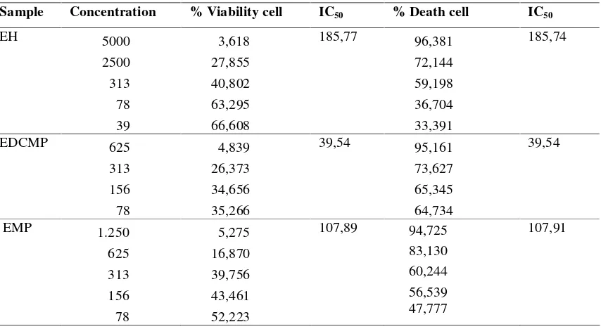
Table I : Cytotoxocity Measurement of extract PyrrosiaPyrrosia piloselloides (L) M.G Price
Sample Concentration % Viability cell IC50 % Death cell IC50
EH 5000
2500 313 78 39
3,618 27,855 40,802 63,295 66,608
185,77 96,381
72,144 59,198 36,704 33,391
185,74
EDCMP 625
313 156 78
4,839 26,373 34,656 35,266
39,54 95,161
73,627 65,345 64,734
39,54
EMP 1.250
625 313 156 78
5,275 16,870 39,756 43,461 52,223
107,89 94,725
83,130 60,244 56,539 47,777
Figure 1. Cytotoxicity activity extract ofPyrrosia piloselloides(L) MG Price
Based on the result in table I, each extract show increase viability cell from higher concentrations to small concentration opposite death cell show
decrease from higher concentrations to small concentrations. IC50are concentration where 50%
death cell or viability cell. Value IC50 each extract can be described in figure 1.
Figure 1 shows that EDCMP have high cytotoxicity because small concentrations of extract ( 39.54 µg/mL) can reduce death cell by 50% . EDMP are active extract then fractionated with chromatography colomn to obtain fractions. Each fractions collect building on similarity chromatogram on TLC, result six fractions, namely FI EDMP, FII EDMP, FIII EDCMP, FIV EDMP, FVEDMP and FVIEDCMP. Each fractions EDCMP evaluated cytotoxicity by MTT method to obtain the following results.
Table II : Cytotoxicity Measurement of Fraction PyrrosiaPyrrosia piloselloides (L) M.G Price Fraction Concentration % viability cell IC50 % inhibition cell IC50
Each fraction have various result % viability cell and death cell but every high concentration causing high % death cell and litlle % viability cell. Value IC50
each fraction can be shown in figure 2.
Figure 2. Cytotoxicity assay fraction of EDCMP
The results of figure 2 shows that FIEDCMP non cytotoxic because value IC50 is very high >
100ug/ml. FVIEDCMP has smallest IC50 with
value is 28.73 µg/mL, this fraction is very cytotoxic. FVIEDCMP can be developed as an alternative treatment for breast-cancer.
3.2. Antioxidant assay
Cancer can be treated with antioxidant compounds to see if the probability P. piloselloides (L) M.G Price also is an antioxidant, each extract from this plant analyzed the antioxidants tested in vitro by DPPH method, the test results can be shown in table III.
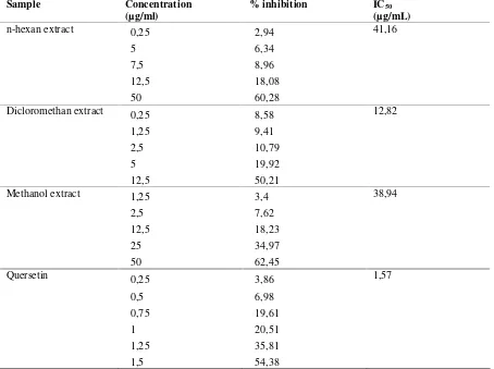
Table III: Antioxidant Measurement of extract PyrrosiaPyrrosia piloselloides (L) M.G Price and Standards Quersetin
Sample Concentration
(µg/ml)
% inhibition IC50
(µg/mL)
n-hexan extract 0,25
5 7,5 12,5 50
2,94 6,34 8,96 18,08 60,28
41,16
Dicloromethan extract 0,25
1,25 2,5 5 12,5
8,58 9,41 10,79 19,92 50,21
12,82
Methanol extract 1,25
2,5 12,5 25 50
3,4 7,62 18,23 34,97 62,45
38,94
Quersetin 0,25
0,5 0,75 1 1,25 1,5
3,86 6,98 19,61 20,51 35,81 54,38
Based on the results in Table III, we obtained that the EHP, EDCMP and EMP are moderately active antioxidant because they have IC50 values below 50 µg/mL.
10
IC50 is concentrated
when extract can inhibit radical ions equal 50%. Value IC50 among three extracts can be described
in figure 3.
Figure 3. Result DPPH assay extractPyrrosia piloselloides (L) M.G Price
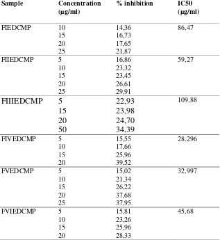
Table IV: Antioxidant Measurement of extract PyrrosiaPyrrosia piloselloides (L) M.G Price and Standards Quersetin
Sample Concentration (µg/ml)
% inhibition IC50 (µg/ml)
FIEDCMP 10
15 20 25
14,36 16,73 17,65 21,87
86,47
FIIEDCMP 5
10 15 20 25
16,86 23,32 23,45 26,61 29,91
59,27
FIIIEDCMP
5
15
20
50
22,93
23,98
24,70
34,39
109,88
FIVEDCMP 5
10 15 20
15,55 17,66 25,96 39,52
28,296
FVEDCMP 5
10 15 20 25
15,02 21,34 26,22 37,68 37,95
32,997
FVIEDCMP 5
10 15 20
15,81 23,26 25,96 28,33
Based on the results in Table IV, the active fractions is FIVEDCMP because it has the smallest IC50.
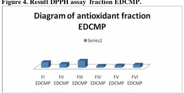
More detail value IC50 on each fractions can
described in figure 4.
Fraction I, II, IV, V and V have activity antioxidants because value IC50 < 100 µg/ml. Fractions V less potent antioxidant opposite another fractions. FIVEEDCMP are active fraction because they have the smalest IC50 (28.29 g/mL). Fractions
FIVEDCMP can be developed as antioxidant drug.
4. Conclusion
. Plants P. piloselloides (L) MG Price can be developed as antioxidant and anticancer , especially
breast cancer because the test results in vitro antioxidant with DPPH method obtain FIVEDCMP as active fraction with IC 50 28.29 µg/mL and
cytotoxicity assay use MTT obtain FVIEDCMP as active fractions with IC50 28.73 µg/mL.
Acknowledgement
This work was supported by funds from the Madya Research Grant–University of Indonesia 2012.
Figure 4. Result DPPH assay fraction EDCMP.
5. References
1. Percival M., Antioxidants. Nut 031, 1/96 Rev. 10/98. Structure Activity Relationship of Cumarin Derivatives on Xanthine Oxidase Inhibiting and Free Radical Scavenging Activities. Biochemical Pharmacology, 1998,75 , 1416-1425.
2. Packer, L.M, T, Yoshikawa, Antioksidant Food Supplement in Human Health, Academic Press, 1999.
3. Bakta IM. Antioksidan dan kanker. Simposium Antioksidan IDI. Bali: IDI Bali; 6 Maret 2004. 4. idiyanti P.M, Aktivitas AntiTumor Ekstrak Air
Daun Sisik Naga (Pyrrosia nummulariforia (SW) Ching) Terhadap Sel Lestari Tumor MCM B2 Secara In Vitro, Nopember 3, http:// duniaveteriner.com/2010/07/aktivitas-anti-tumor-
ekstrak-air-daun-sisik-naga-pyrrosia- nummularifolia-sw-ching-terhadap-sel-lestari-tumor-mcm-b2-secara-in-vitro/print,2011. 5. Bloiss M.S, Antioxidant Determinations by The
Use of Stable Free Radicals, Nature, 1958, 181, 1199-1200.
6. Doyle A, and Griffiths J.B., Cell and Tissue Culture: Laboratory Procedures in Biotech ology, Chichester: Wiley, 2006
7. Freshney, I, Culture of Animal Cell : a Manual of Basic Techniques , fourth Edition, New York, John Wiley & Sons, 1994,
8. Mosmann T, Rapid colorimetric assay for cellular growth and survival:application to proliferation and cytotoxicity assays, .Immunol.Methods ,1983; 5: 55-63
9. King RJB, Cancer biology, 2nd ed. Pearson Education Limited, England, 2002.
10.Young LS and Woodside JV, Antioxidants in health and disease, J Clin Pathol, 2001,54,176-86.