ORIGINAL ARTICLE
Bali Medical Journal (Bali Med J) 2016, Volume 5, Number 3: 484-487 P-ISSN.2089-1180, E-ISSN.2302-2914
484 Open access: www.balimedicaljournal.org and ojs.unud.ac.id/index.php/bmj
CrossMark
Published by DiscoverSys
Association of SDF-1 with metastasis in breast
cancer patient at Sanglah hospital, Bali-Indonesia
Kristanto Yuli Yarso,1,3* Suyatmi,2 Faris Khairuddin Syah,2 Ida Bagus Tjakra Wibawa Manuaba,3 Hakimi,4 Indwiani Astuti,5 Teguh Aryandono6
ABSTRACT
Background: More than 24% breast cancer patients came to Sanglah Teaching Hospital with distant metastasis which cause 90% of cancer related death. Distant metastasis is complex process of interaction between tumor cells and its micro environment involving chemo-attractant cytokines which lead circulating tumor cells toward target organs. One of the most common cytokines involved in metastasis of multiple tumor is SDF-1, produces by target organ or tumor cells it selves. However, only few studies ever evaluate the relationship between its concentrations in tumor tissue with metastasis. Method: A cross sectional analysis study was conducted involving clinical data and paraffin blocks from 46 patients. Samples were grouped into metastasis and non-metastasis group and level of tumor tissue SDF-1 was evaluated by immunohistochemistry method.
Numerical conversion was done using modified “Mirisola” technique and statistical analysis was conducted using SPSS 16 software. Results: The overall median expression of SDF-1 was 4in which the median is 3 in non-metastatic group and 6 in metastatic group. Elevated SDF-1 expression was significantly associated with increase metastatic risk (PR: 1.34; P=0.04; CI 95%: 1.014-1.769). In addition, parenchymal carcinoma cell had significantly higher expression of SDF-1 compared with micro-environmental cell (median SDF-SDF-1 in carcinoma vs microenvironment; 3vs.2; p=0,004). Finally, multivariateanalysis of SDF-1 expression also gave significant result that MBC had significantly higher expression of SDF-1 (p=0.019).
Conclusions: Elevated SDF-1 expressions significantly increase metastatic risk and majority of SDF-1 was produce by tumor parenchyma.
Keywords: Breast Cancer, SDF-1, Metastasis
Cite This Article: Yarso, K., Suyatmi, S., Khairuddin Syah, F., Wibawa Manuaba, I., Hakimi, H., Astuti, I., Aryandono, T. 2016. Association of SDF-1 with metastasis in breast cancer patient at Sanglah hospital, Bali-Indonesia. Bali Medical Journal 5(3): 484-487. DOI:10.15562/bmj.v5i3.320
INTRODUCTION
Breast cancer is the second most prevalent cancer among women worldwide. In United States, the incidence is about 246.660 new cases in 2016, comprising 14.6% of all cancer incidences. It also one of leading cause of death among women in United States with 40.450 deaths in 2016. Breast cancer is also the most common cancer in Europe in 20061-4
. In Indonesia, breast cancer is the second highest malignancy in women ater cervical carci-noma with tendency to increase in number in the upcoming years5
. Sanglah General Hospital itself found new cases of breast cancer approximately 90 cases each year. More than 43% of patients with breast cancer came to Sanglah in advanced stage in which 26% had metastasis.6
he most feared consequences of malignancy are metastasis as its cause majority of cancer related deaths in all cancer types. In case of breast cancer, 90% of deaths are attributed to metastasis. he mech-anism of metastasis is already extensively studied. However, there are still lots of elusive mechanism yet uncovered and new indings arise almost every year. One of new theoretical advancement in meta-static process is interaction between tumour paren-chyma and tumour microenvironment (TME). his
interaction is proved vital as microenvironment plays essential role to determine metastatic location and tumour response to therapy as well as length of survival. 7,8,9,10
CXCL-12 (Chemokine Motif Ligand-12) or Stromal Cell-Derived Factor 1α (SDF-1) is a cyto-kine that produced by Carcinoma Associated Fibroblasts (CAFs). CXCL-12 is a speciic ligand of (Chemokine Motif Receptor 4) CXCR-4 and plays role in the function of organogenesis, regen-eration and tumorigenesis. hey drive cells express-ing CXCR4 close to cell expressexpress-ing SDF-1.11,12,13
he high expression of CXCR4 on breast cancer relates to younger age, larger tumour and lower rates of overalls suvival.14
Expressions of both of these proteins are strongly inluenced by HIF-1α which is mostly expressed by the hypoxic tissue.15
In tumour tissue, hypoxic state begin when tumour mass reach 2 mm3 in volume and 1% O
2 concentration,
indi-cating that metastatic potential begin very early in tumour progression.16,17
he aim of this study is to evaluate the associa-tion between level of tissue SDF-1 and distant meta-static event. Currently, there is no similar research being conducted in Indonesia.
1Department of Surgery, Oncology
Division Medical Faculty of Sebelas Maret University Solo-Indonesia
2Medical Faculty of Sebelas Maret
University Solo-Indonesia
3Department of Surgery, Division
of Oncologic Surgery, Faculty of Medicine Udayana University Bali-Indonesia
4Department of Obstetric
Gynecology, Faculty of Medicine Gadjah Mada University Jogjakarta- Indonesia
5Department of Histology,
Faculty of Medicine Gadjah Mada University Jogjakarta- Indonesia
6Department of Surgery, Division
of Oncologic Surgery, Faculty of Medicine Gadjah Mada University Jogjakarta- Indonesia
*Correspondence to: Kristanto Yuli Yarso, Department of Surgery, Oncology Division Medical Faculty of Sebelas Maret University Solo-Indonesia
485 Published by DiscoverSys | Bali Med J 2016; 5 (3): 484-487 | doi: 10.15562/bmj.v5i3.320
ORIGINAL ARTICLE
MATERIAL AND METHODS
he study was conducted at the Department of Surgery Medical Faculty of Udayana University/ Sanglah General Hospital. his cross sectional study was aimed to determine the relationship of the expression of SDF-1 in tumour tissue with distant metastases. Samples were taken from all parain blocks of breast cancer patients who had biopsy in 2009 to 2011 as well as clinical data from the patient’s medical record. Research data collection began in July 2010. he study was conducted until
suicient sample. he inclusion criteria are biopsy sample of breast cancer patient treated in Sanglah general Hospital. All stages and types of breast cancer are included. Samples with incomplete clini-cal and pathologiclini-cal data, unrepresentative parain blocks, diagnosed other than breast cancer, or if the result of staining could not be interpreted were excluded.
he collected data included basic, clinical and pathological data. Data were divided into 2 groups: metastatic group and non-metastatic group. All samples with complete medical data were examined by using Immunohistochemistry (IHC) method to delineate CXCL12 / SDF-1 expression which were carried out in Laboratory of Pathology Anatomy Medical Faculty of Udayana University/ Sanglah General Hospital. We used rabbit monoclonal anti-SDF-1 antibody (AbCAM) diluted until 1:75. he data were analysed statistically by both bivariate and multivariate analysis. All analysis process was conducted using SPSS 16 for windows sotware.
RESULTS
All samples were obtained from 2009-2011 regis-ter. 247 samples were collected in which 63 meta-static and 184 non-metameta-static cases were collected during the courses of the study. However, only 89 samples had complete medical data. From these samples, 36 samples were classiied as metastasis and 53 were non-metastasis. Only 46 samples were eligible for IHC-stained (21 patients with metas-tasis and 25 patients without metasmetas-tasis). Of all samples, 37 samples were Balinese, 5 patients were Sasak tribe, and 4 patients were Javanese. he mean age of patients was 45.93 with the youngest was 30 years old and the eldest was 62 years. In term of menstrual status, 37 patients (80.4%) were on premenopausal age and 9 patients (19.6%) were already menopause.
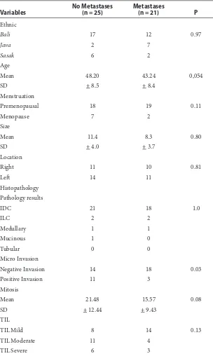
he data about cancer grade and apoptotic were not obtained in this study because some samples were diagnosed other than IDC and apop-totic assessment was not routinely conducted in Laboratory of Pathology Anatomy Medical Faculty of Udayana University/ Sanglah General Hospital. Samples baseline characteristics are described in Table 1.
he result of IHC staining showed that the median value of SDF-1 expression was 4 from all samples. Separating the samples into metastatic and non-metastatic group reveal that metastatic group had higher level of SDF-1 compare to non-met-astatic group (mean SDF-1 expression score 6 vs. 3). Logistic regression analysis showed that SDF-1 signiicantly increased metastatic risk (PR: 1.34; P=0.04; CI 95%:1.014-1.769) (Table 2). Moreover, Table 1 Baseline characteristic of subjects
Variables
No Metastases (n = 25)
Metastases
(n = 21) P
Ethnic
Bali 17 12 0.97
Java 2 7
Sasak 6 2
Age
Mean 48.20 43.24 0,054
SD + 8.5 + 8.4
Menstruation
Premenopausal 18 19 0.11
Menopause 7 2
Size
Mean 11.4 8.3 0.80
SD + 4.0 + 3.7
Location
Right 11 10 0.81
Let 14 11
Histopathology Pathology results
IDC 21 18 1.0
ILC 2 2
Medullary 1 1
Mucinous 1 0
Tubular 0 0
Micro Invasion
Negative Invasion 14 18 0.03
Positive Invasion 11 3
Mitosis
Mean 21.48 15.57 0.08
SD + 12.44 + 9.43
TIL
TIL Mild 8 14 0.13
TIL Moderate 11 4
486 Published by DiscoverSys | Bali Med J 2016; 5 (3): 484-487 | doi: 10.15562/bmj.v5i3.320 ORIGINAL ARTICLE
cancer cells itself appear to produce more SDF-1 compared with micro-environmental cell in both metastatic and non-metastatic groups (Table 3). However, we found no correlation between SDF-1 and mitotic index in this study (Table 4).
Multivariate analysis of the relationship between the occurrences of metastases as dependent vari-ables with several independent varivari-ables using logistic regression analysis showed that SDF-1 was signiicantly associated with metastasis. Other factors that showed similar result were age and mitosis. Table 4 summarize the result of multivar-iate analysis.
DISCUSSION
he relationship between SDF-1 and metastasis have already studied extensively both in labora-tory and clinically. Mego et.al states SDF-1 related with circulating tumour cells and act as chemo-tactic signal.18 It also found that SDF-1 is directly correlated with prognosis and tumour progres-sion.19-23 However, some research found the oppo-site efect of SDF-1 in which it decreases cancer cell motility, hence, preventing metastasis.24
he result of this study, however, conirming that SDF-1 was, in fact, associated with metastasis. It also found that tumour cell itself produce SDF-1 even in greater amount compared with stromal
tissue. his result conirms that cancer cell is capa-ble of producing SDF-1 and not just as its target. However, because we found that SDF-1 production by cancer cell exceeded the stromal tissue, further investigation is needed to conirm this inding.
SDF-1/CXCR4 axis play important role in cancer pathogenesis. It controls wide array of cancer char-acteristic, ranging from maintaining cell prolif-eration to angiogenesis and cell survival.19,20,21,25 SDF-1 also required to maintain steady cancer stem cell population. his is not surprising since SDF-1 normally act as regulator of stem cell population in bone marrow.25
In relation with metastasis, SDF-1 act as chemo-tactic factor that attract circulating tumour cell to homing into target organ, oten with already formed metastatic niche.18
SDF-1 also induces cell mobilisation by increasing the expression of β1-in-tegrin.25
It also induce the expression of several matrix metalloproteinase (MMP) family gene which result in stromal remodelling that favour metastatis.26
hough not directly related, SDF-1 also plays integral role in angiogenesis. Increase vascular supply not only ensure the oxidative status of cancer mass but also provide easy way for cancer cell to enter vascular system since newly formed blood vessel tend to have fragile and permeable structure.27
he aforementioned mechanisms are underly-ing reason why SDF-1 is oten associated with poor prognosis in wide array of cancer.19-23
In adult acute myelogenous leukemic, SDF-1 was related with lower chemotherapeutic response.20
Meanwhile in prostate cancer and malignant glioma, it is asso-ciated with tumour aggressiveness and progres-sion.21,22
Overall, it showed that SDF-1 is strong candidate as prognostic biomarker of prognostic in almost every type of cancer.
CONCLUSION
SDF1 over expression in tumour tissue shown to be associated with increased risk distant metastasis in carcinoma cells and it appeared that tumour cell itself is the major source of SDF-1. However, further prospective studies are needed to determine the relative risk SDF-1 over expression in tumour tissue on the occurrence of distant metastases by controlling other factors such as receptors and subtypes of breast cancer.
REFERENCES
1. Ferlay J, Autier P, Boniol M, et al. 2007. Estimates of the Cancer Incidence and Mortality in Europe in 2006. In: Ann Oncology vol 18 no 3: P 581.
2. American Cancer Society. 2005. Breast cancer facts and ig-ures 2005 - 2006. American Society Inc. Atlanta. Table 2 Logistic regression analysis of association between SDF-1
expression and metastasis
Var B S.E P-Value PR
95.0% C.I. for OR
Lower Upper
Sdf1 0.292 0.142 0.039 1.34 1.014 1.769
Table 3 Comparison of SDF-1 expression between carcinoma cell and tumour microenvironment
SDF-1 Expression
Cell Type
P Carcinoma cell Microenvironment
Median 3 (1-9) 2 (1-9) 0.001
Table 4 Multivariate analysis between several predictors with metastasis
Predictor B P Exp (B)
95.0% CI
Lower Upper
Age -0241 0.016 0786 0647 0955
Size 0483 0.080 1,622 0944 2,786
Mitosis -0266 0.007 0766 0631 0.930
TIL -1471 0.442 0.230 0005 9,770
Invasion 3,279 0.057 26 537 0906 777 524
487 Published by DiscoverSys | Bali Med J 2016; 5 (3): 484-487 | doi: 10.15562/bmj.v5i3.320
ORIGINAL ARTICLE
3. Burstein. H, Harris. J, & Morrow. M. 2008. Section 2: Malignant Tumors of the Breast. In: Devita, Hellman & Rosenberg’s Cancer: Principles & Practice of Oncology, 8th Edition. Lippincott Williams & Wilkins. P 1606
4. Zager. J, Solorzano. C, homas. E, Feig, B, & Babiera. G. 2006.Invasive Breast Cancer. In: MD Anderson Surgical Oncology Handbook, he, 4th Edition. Lippincott Williams
& Wilkins.
5. Tjindarbumi, D. &Mangukusumo, R. 2002. Cancer in Indonesia Present and Future. In: Japan Journal Clinical Oncology vol 32 (Supplement). P 17-21
6. Yarsa K.Y., Sudarsa I., Manuaba I.B.T.W. 2010. Clinical Initial Response of Neoadjuvant Chemotherapy in Triple Negative, HER-2, & Luminal Types of Breast Cancer in Denpasar. DipresentasikanpadaPertemuanIlmiahTahunan PERABOI. November. Medan.
7. Su. J.L., Yen. C.J., Chen. P.S., Chuang. S.E., Hong. C.C., Kuo. I.H., Chen. H.Y., Hung. M.C., Kuo. M.L. 2007. he role of the VEGF-C/ VEGFR-3 axis in Cancer Progression. In: British Journal of Cancer Vol 96. PP 541-545.
8. Alfred. D.C., Mohsin. S.K., Fuqua. S.K., 2001. Histopathological and Biological Evolution of Human Premalignant of Breast Disease. In: Endocrine Related Cancer Vol 8. P 47-61
9. Bodenstine. T &. Welch. D. 2008. Metastasis Suppressors and the Tumor Microenvironment. In: Cancer Microenvironment. Springer Science Business Media. P 1–11
10. Mansel. R, Fodstad. O, & Jiang. W. 2007. Metastasis of breast cancer: an introduction. In: Metastasis Breast Cancer, Cancer Metastasis – Biology and Treatment Vol 11. Springer. Dordrecht. P 1-7
11. Hartmann. T.N., Grabovsky. V., Pasvolsky. R., Shulman. Z., Buss. E.C., Spiegel. A., Nagler A., Lapidot. T., helen. M., & Alon. R. 2008. A crosstalk between intracellular CXCR7 and CXCR4 involved in rapid CXCL12-triggered integ-rin activation but not in chemokine-triggered motility of human T lymphocytes and CD34_ cells. In: Journal of Leukocyte Biology Vol 84, October 2008
12. Ratajczak. M.Z., Surma. E.Z., Kucia. M., Reca1. R., Wojakowski. W., &Ratajczak. J. 2006. he pleiotropic efects of the SDF-1–CXCR4 axis in organogenesis, regen-eration and tumorigenesis. In: Leukemia vol 20. Nature Publishing Group. P 1915–1924
13. Schier AF. 2003. Chemokine signaling: rules of attraction. In: Current Biology vol 13. Elsevier Science Ltd. P 192–194. 14. Kim. J.O, Suh. K.S, Lee. D.O, Sul. H.J, Lee. J.U, & Song. K.S.
2009. Expression of CXCR4 and SDF-1 in Primary Breast Cancer and Metastatic Lymph Nodes. In: Journal of Breast Cancer vol 12 no 4. P 249-256.
15. Ceradini DJ, Kulkarni AR, Callaghan MJ, Tepper OM, Bastidas N, Kleinman ME, Capla JM, Galiano RD, Levine JP, Gurtner GC. 2004. Progenitor cell traicking is regulated by hypoxic gradients through HIF-1 induction of SDF-1. In: Nat Med vol10.P 858–864
16. Burger. J & Kipps. T. 2006 CXCR4: A Key Receptor in the Crosstalk Between Tumor Cells and heir Microenvironment. In: BLOOD, Vol 107, no 5. he American Society of Hematology. P 1761-1767
17. Vaupel. P., & Mayer. A. 2007. Hypoxia in Cancer: Signiicance and Impact on Clinical Outcome. In: Cancer Metastasis Reviews Vol 26, No 2. P 225–239.
18. Mego M, Cholujova D, Minarik G, et.al. CXCR4-SDF-1 interaction potentially mediates traicking of circulating tumor cells in primary breast cancer. BMC Cancer 2016; 16:127
19. Kim J, Takeuchi H, Lam ST, et al. Chemokine receptor CXCR4 expression in colorectal cancer patients increases the risk for recurrence and for poor survival. J Clin Oncol
2005, 23: 2744-53.
20. Sun YX, Fang M, Wang J, Cooper CR, Pienta KJ, Taichman RS. Expression and activation of alpha v beta 3 integrins by SDF-1/CXC12 increases the aggressiveness of prostate cancer cells. Prostate 2007, 67: 61-73.
21. Bian XW, Yang SX, Chen JH, et al. Preferential expres-sion of chemokine receptor CXCR4 by highly malignant human gliomas and its association with poor patient sur-vival. Neurosurgery 2007, 61: 570-8.
22. Rombouts EJ, Pavic B, Lowenberg B, Ploemacher RE. Relation between CXCR-4 expression, Flt3 mutations, and unfavorable prognosis of adult acute myeloid leukemia. Blood 2004, 104: 550-7.
23. Kato M, Kitayama J, Kazama S, Nagawa H. Expression pattern of CXC chemokine receptor-4 is correlated with lymph node metastasis in human invasive ductal carci-noma. Breast Cancer Res 2003;5: R144-50
24. Mirisola V, Zuccarinoa A, Bachmeierb BE, Sormanic MP, Falterd J, Nerlichd A, Pfefera U. 2009. CXCL12/SDF1 expression by breast cancers is an independent prognos-tic marker of disease-free and overall survival. European Journal of Cancer. 45;P 2579 –2587
25. Gelmini, Mangoni M, Serio M, Romagnani P, Lazzeri E. he critical role of SDF-1/CXCR4 axis in cancer and can-cer stem cells metastasis. J. Endocrinol. Invest. 2008;31: 809-819
26. Osman NM, Osman WM. SDF-1 and MMP2 cross talk in cancer cells and tumor microenvironment in non-small cell lung cancer. Egyptian Journal of Chest Diseases and Tuberculosis 2016; 65:517–525
27. Orimo A, Gupta PB, Sgroi DC, et al. Stromal ibroblasts present in invasive human breast carcinomas promote tumor growth and angiogenesis through elevated SDF-1/ CXCL12 secretion. Cell 2005;121: 335-48.