* corresponding author: [email protected]
Cranial unifocal langerhans cell histiocytosis
in a female child: a difficult case with S-100
and CD1a immunonegativity
Hanggoro Tri Rinonce1*,Sagiri Mangunsudirdjo1, Soeripto1, J Bras2
1Department of Anatomical Pathology, Faculty of Medicine, Gadjah Mada University,
Yogyakarta, Indonesia,
2Department of Pathology, Academic Medical Center, Amsterdam University,
The Netherlands
ABSTRACT
A 13-years old female child was carried to Dr. Sardjito General Hospital, Yogyakarta by her mother with chief complaint of a mass on her forehead. Since eight months prior to her visiting, she had a mass on her forehead which became larger slowly without tenderness and had no fever. Clinical examination revealed a well circumscribed mass, 3 cm in diameter, fixed, with rubbery consistency. The skull X-ray revealed a punch out lesion in frontal bone. The head CT scanning revealed a destruction of frontal bone. Clinical diagnosis of dermoid cyst was determined, excision and curettage was performed. Gross examination showed 2.5 cc fragmented tissue, brownish yellow, with rubbery consistency. A diagnosis of benign histiocytosis (Langerhans cell histiocytosis or non-Langerhans cell histiocytosis) of frontal bone was determined based on morphological and immunohistochemical examination. The aim of this presented article was to report a rare case of cranial unifocal Langerhans cell histiocytosis in a female child with S-100 and CD1a immunonegativity, and to discuss how to determine its diagnosis based on literature review.
Key words: Langerhans cell histiocytosis - juvenile xanthogranuloma – reticulohistiocytoma - eosinophilic granuloma – S100 – CD1a
ABSTRAK
Seorang anak perempuan dibawa oleh ibunya ke Rumah Sakit Umum Pusat Dr. Sardjito, Yogyakarta dengan keluhan utama benjolan pada dahi. Sejak 8 bulan sebelum masuk rumah sakit, ia mengeluhkan benjolan pada dahi yang perlahan semakin membesar tanpa disertai nyeri. Ia tidak mengeluhkan demam. Pemeriksaan fisik menunjukkan adanya massa berukuran diameter 3 cm, berbatas tegas, terfiksir, dengan konsistensi kenyal. Pemeriksaan sinar X kepala menunjukkan suatu lesi punch out pada tulang frontal. Pemeriksaan CT scanning kepala menunjukkan suatu destruksi tulang frontal. Diagnosis klinik kista dermoid ditegakkan dan dilakukan eksisi dan kuretase massa. Pemeriksaan makroskopis menunjukkan jaringan pecah belah, 2,5 cc, kuning kecoklatan, dengan konsistensi kenyal. Diagnosis histiositosis jinak (histiositosis sel Langerhans atau histiositosis non sel Langerhans) tulang frontal ditegakkan berdasarkan pemeriksaan morfologik dan immunohistokimia. Tujuan penulisan artikel ini adalah untuk melaporkan suatu kasus jarang histiositosis sel Langerhans kranial unifokal pada seorang anak perempuan dengan immunonegativitas S-100 dan CD1a, dan mendiskusikan bagaimana menegakkan diagnosisnya berdasarkan tinjauan pustaka yang dipaparkan.
INTRODUCTION
Langerhans cell histiocytosis (LCH) is a disorder of uncontrolled pathologic clonal proliferation of dendritic cells with Langerhans cell characteristic.1
LCH is one of the most common forms of histio-cytoses and the one most encountered in the pediatric population.2 The annual incidence in the
pediatric age has been estimated to be in the range of 2 to 5 per million per year.1,3 Males are more
frequently affected than females and age at
presen-tation varies from a few months to 15 years.4
Affected bones in order of frequency are skull, long bones, flat bones (scapula, rib, and mandible), and vertebrae.5,6,7
Morphological diagnosis of LCH is often difficult to be determined because many other lesions have the similar pattern with LCH, including non-LCH (juvenile xanthogranuloma and reticulohistio-cytoma). Because LCH is progressive but non-LCH is always self-limiting, it is important to distinguish these two diseases. Immunohistochemical staining has an important role for differentiating LCH from non-LCH. The diagnosis of LCH is regarded as presumptive when the typical morphological characteristics of Langerhans cell are seen with light microscopy. It is regarded as designated when additional stains (e.g., ATP-ase and S-100) are positive. However, sometimes immunohistochemical
staining is not helpful in differentiating LCH from non-LCH, particularly in small specimens.
A rare case of cranial unifocal LCH in a female child with S-100 and CD1a immunonegativity was reported in this article. How to determine its diagnosis was discussed deeply based on literature review.
CASE
Clinical history and examination
A 13-year old female child was carried to Dr. Sardjito General Hospital, Yogyakarta by her mother with chief complaint of a mass on her forehead. Since eight months prior to her visiting, she had a mass on her forehead which slowly became larger without tenderness. She had no fever. There were also multiple lymphadenopathies in right and left aspect of her neck.
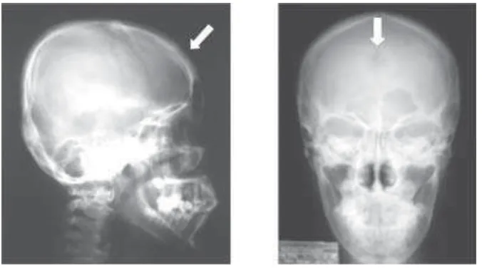
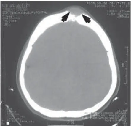
Clinical examination revealed a well circumscribed mass, 3 cm in diameter, fixed, with rubbery consistency on frontal bone. The skull X ray revealed a punch out lesion in frontal bone (FIGURE 1). The head computed tomography (CT) scanning revealed a destruction of frontal bone (FIGURE 2). Laboratory test including complete blood count, electrolyte levels, liver function test, kidney function test, urine analysis, and the chest radiograph were normal, except low level of hemoglobin.
FIGURE 2. Head CT scanning revealed a destruction of frontal bone, pointed by black arrows
Clinical diagnosis of dermoid cyst was determined and excision and curettage were performed. Gross examination showed 2.5 cc fragmented tissue, brownish yellow, with rubbery consistency. One slide was chosen for microscopic examination by routine hematoxylin-eosin staining. A block of paraffin embedded tissue was sent to The Department of Pathology, Amsterdam Medical Center, The Netherlands, for confirmation of the diagnosis and immunohistochemical examination.
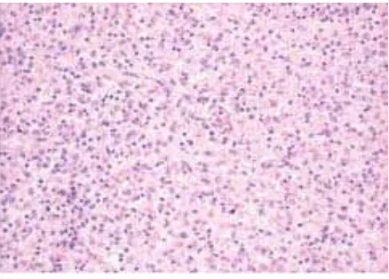
Microscopic examination showed fragments of a cellular lesion, composed of epitheloid and plump spindle shaped cells, admixed with inflammatory cells (granulocytes and lymphocytes). The cytoplasm were abundant, eosinophilic or amphophilic. The nuclei of the epitheloid and plump spindle shaped cells showed little variation in size and vary from round to lobulated. Chromatin pattern varied from granular to usually open with a recognizable nucleolus. Mitosis was not found. Sometimes, cells with 2 or even more nuclei were found. Morphologically, the cells impressed as histiocytic nature, could be LCH or non-LCH (FIGURE 3 and 4).
FIGURE 3.Microscopic feature of presented case showed a cellular lesion, epitheloid (arrow heads) and plump spindle (arrows) shaped cells, admixed with inflammatory cells (HE
staining, 100X)
FIGURE 4. Microscopic feature of presented case showed cells with 2 or even more nuclei, pointed by black arrows
(HE staining, 200X)
FIGURE 5. Positive expression of vimentin showed by cytoplasmic brown staining (100 X)
FIGURE 6. Positive expression of CD68 showed by cytoplasmic brown staining, some positive cells pointed by
black arrows (100 X)
FIGURE 7. Positive expression of HLA-DR showed by cytoplasmic brown staining (100 X)
FIGURE 8. Positive expression of LCA showed by membranous brown staining, some positive cells pointed by
black arrows (100 X)
FIGURE 9. Positive expression of CD31 showed by membranous brown staining, some positive cells pointed by
black arrows (100 X)
FIGURE 11. Negative expression of CD1a repeatedly (100 X)
Clinical examination of her right and left neck revealed multiple well circumscribed mass, mobile, 1-2 cm in diameter, with rubbery consistency. Fine needle aspiration biopsy (FNAB) was performed.
Microscopic examination of smear obtained from FNAB of right and left neck showed clustered and dispersed cells, composed of epitheloid cells. The cytoplasm was abundant with indistinct border. The nuclei showed little variation in size and vary from round to lobulated. Chromatin pattern varied from granular to usually open with a recognizable nucleolus. Between these cells, there were inflammatory cells, particularly lymphocytes (FIGURE 12 and 13)
FIGURE 13. Smear obtained from FNAB showed clustered and dispersed cells, composed of ephitheloid cells (arrows) admixed with inflammatory cell, particularly lymphocytes (arrow heads) (Giemsa staining, 200 X)
PATHOLOGICAL DIAGNOSIS
Based on morphological and immunohisto-chemical examination a diagnosis of benign histiocytosis (Langerhans cell histiocytosis or non-Langerhans cell histiocytosis) in frontal bone was determined. FNAB of right and left neck suggested benign lesion, a granulomatous inflammation.
DISCUSSION
LCH spans a spectrum from the localized, usually benign form (eosinophilic granuloma) through a chronic disseminated form (Hand– Schuller–Christian disease) to the acute form which is often fatal (Letterer–Siwe disease). It is important to distinguish this disease with non-LCH because non-LCH is always self-limiting.
Morphologically, the typical lesion of LCH is composed of an admixture of Langerhans cell histiocytes, intermediate cells and interdigitating cells of a dendritic cell lineage, T-cell lymphocytes, eosinophils, and macrophages. The hallmark cell is the Langerhans cell histiocyte. This cell has abundant eosinophilic to amphophilic cytoplasm and a nucleus that appears reniform, deeply indented, or grooved. The number of eosinophils is quite variable from being abundant with eosinophilic abscesses to sparse or even absent. Occasional giant cells representing fusion of either macrophages or Langerhans cell histiocytes may be seen. The presence of this granulomatous inflammation with occasional giant cells raises the concern for an infectious process, such as tuberculosis in the past and viral infection with an agent that is capable of inducing syncytial
(giant) cells. Necrosis within this granulomatous lesion is not unusual, and again reinforced the suggestion in the past that these lesions represented an infectious process. The lesions vary from an indistinct focus with blending into the adjacent normal tissue to nodular in appearance, depending on tissue types involved. Although present, mitotic activity tends to be low to moderate without atypical mitotic figures. The lesions may take on a more atypical appearance and appear as epithelioid granulomas that lack the typical features of LCH. The lesions may resemble other histiocytic lesions, such as early juvenile xanthogranuloma that lack characteristic of Touton giant cells. Definitive diagnosis for these atypical lesions requires immunocytochemistry and occasionally electron microscopy.10
Morphologically, the presented case composed of cells impressing as histiocytic nature, with sparse eosinophils’ infiltration, could be LCH or non-LCH. In this presented case, immunohistochemical staining is very important to determine the definitive diagnosis.
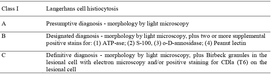
The diagnosis of LCH is regarded as presumptive when the typical morphological characteristics of Langerhans cell are seen with light microscopy. It is regarded as designated when additional stains (e.g., ATP-ase and S-100) are positive. Diagnosis is confirmed if stains for CD1a antigen are positive or when Birbeck granules are
seen with electron microscopy.8 Degrees of
confidence level for the diagnosis of LCH that has established by the Writing Group of the Histiocyte Society showed in TABLE 1.
TABLE 1. Degrees of confidence level for the diagnosis of Langerhans cell histiocytosis11
Class I Langerhans cell histiocytosis
A Presumptive diagnosis - morphology by light microscopy
B Designated diagnosis - morphology by light microscopy, plus two or more supplemental
positive stains for: (1) ATP-ase; (2) S-100, (3) o-D-annosidase; (4) Peanut lectin
C Definitive diagnosis - morphology by light microscopy, plus Birbeck granules in the
Immunohistochemical staining of the presented case showed negative expression of epithelial marker (cytokeratin, CAM5.2, and EMA) which confirmed that the presented case was not epithelial lesion.
The presented case showed expression of vimentin, CD68, HLA-DR, LCA, and CD31. CD68 is lysosomal glycoprotein present in monocytes and
macrophage.12 CD45, also known as leukocyte
common antigen (LCA), is a family of trans-membrane protein tyrosine phosphatases. It is expressed on the surface of all hematopoietic cells except erythroid and megakaryocytic cells.13 The
platelet-endothelial adhesion molecule-1 (PECAM-1) is also known as CD31. It is a 130-kD trans-membrane glycoprotein that is shared by vascular lining cells, megakaryocytes, platelets, and other selected hematopoietic elements, as recognized by monoclonal antibody JC/70. This marker is highly restricted to endothelial neoplasms among all tumors of the soft tissue, and its sensitivity is also excellent.13
Expression of these markers could support the diagnosis of LCH.
However, imunohistochemical staining with S100 and CD1a were negative repeatedly. CD1a is a transmembrane antigen normally expressed in cortical thymocytes, Langerhans cells, and interdigitating dendritic reticulum cells.12,13 In
addition, the expression of CD68 and LCA could also support the diagnosis of non-LCH. It is useful for identifying normal and neoplastic Langerhans cells, where it is considered as sensitive but more specific than S100.26-28
Based on morphological and immunohisto-chemical examination, a diagnosis of benign histiocytosis (LCH or non-LCH) in frontal bone was determined. In that situation, immunostaining could not overcome the diagnostic problem. Unfortunately, electron microscopy study could not be performed because the specimen was not prepared for electron microscopy.
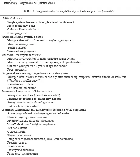
Several clinical classifications and categorization of LCH are used by many practitioners (TABLE 2 and TABLE 3). Clinically the presented case was suffered by 13-years old female child and affected frontal bone (skull) as single lesion, most suitable for clinical diagnosis of eosinophilic granuloma.
If LCH was assumed, the presented case was categorized as unifocal disease. Scintigraphy study to rule out a multifocal disease was not performed because of our limitation. Cervical lymphadeno-pathies were only examined by FNAB. Biopsy of the neck lymph nodes was not performed. FNAB could support the diagnosis granulomatous inflammation, but it could not exclude LCH or NHL of the lymph node. Lymph node involvement in LCH occurs in a number of different clinical situations, as the only site of involvement, so-called primary eosinophilic granuloma of lymph nodes, accompanying or as the presenting manifestation of limited and focal LCH, usually involving lytic bone lesions or cutaneous manifestations, or as part of the disseminated type.29 In the presented case, we
considered that the cervical lymphadenopathies were reactive lesions accompanying unifocal LCH.
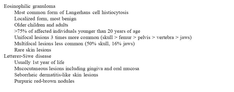
TABLE 2. Clinical types of Langerhans cell histiocytosis29
Eosinophilic granuloma
Most common form of Langerhans cell histiocytosis Localized form, most benign
Older children and adults
>75% of affected individuals younger than 20 years of age
Unifocal lesions 3 times more common (skull > femur > pelvis > vertebra > jaws) Multifocal lesions less common (50% skull, 16% jaws)
Rare skin lesions Letterer-Siwe disease
Usually 1st year of life
Mucocutaneous lesions including gingiva and oral mucosa Seborrheic dermatitis-like skin lesions
Ulcerated painful nodules involving perineal, inguinal, retroauricular, and external auditory canal regions Lung, liver, and spleen involvement
Hand-Schuller-Christian disease Usually 2- to 6-year-old children
Classic triad: osteolytic lesions, exophthalmos, and diabetes insipidus Skin and oral lesions
Congenital self-healing Langerhans cell histiocytosis (reticulohistiocytosis, Hashimoto-Pritzker disease) Pulmonary Langerhans cell histiocytosis
TABLE 3. Categorization by Histiocyte Society for treatment protocols (current)30,31
Unifocal disease
Single system disease with single site of involvement Most commonly bone
Older children and adults Good prognosis
Multifocal single system disease
Multiple sites of involvement in single organ system Most commonly bone
Young children Intermediate prognosis Multifocal multisystem disease
Multiple involved sites in more than one organ system Most commonly bone, skin, liver, spleen, and lymph nodes Children younger than 2 years of age and infants
Poor prognosis
Congenital self-healing Langerhans cell histiocytosis
Multiple skin lesions at birth or shortly after mimicking congenital neuroblastoma or leukemia (‘‘blueberry muffin baby’’)
Neonates and infants Self-healing involution
Pulmonary Langerhans cell histiocytosis Young adult smokers (‘‘smokers malady’’) Indolent progression to pulmonary fibrosis Strong association with malignancies Extremely rare in children
Secondary Langerhans cell histiocytosis associated with neoplasms Acute lymphoblastic and myelogenous leukemias
Chronic myelogenous leukemia Myelodysplastic disorder association Non-Hodgkin and Hodgkin lymphoma Retinoblastoma
Osteosarcoma Thyroid carcinoma
Lung cancer (adenocarcinoma, small cell carcinoma) Prostate cancer
Breast cancer Parathyroid adenoma Pancreatic cystadenoma
A diagnosis of cranial unifocal Langerhans cell histiocytosis (eosinophilic granuloma) was determined based on clinical data and morphological examination of HE-stained specimen only.
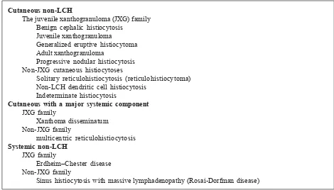
If LCH was excluded, a non-Langerhans cell histiocytosis remained. The non-Langerhans cell histiocytoses (non-LCH) are a diverse group of disorders defined by the accumulation of histiocytes that do not meet the phenotypic criteria for the
diagnosis of Langerhans cells (LCs) 32 and was
classified as listed in TABLE 4. Juvenile xanto-granuloma (JXG) was included as differential diagnosis because JXG was the commonest form of the non-LCH, usually affected young child as solitary lesion.32 1. However, JXG could be ruled
out by negative expression of Factor XIIIa. Immuno-cytochemical features of dendritic cell disorders is listed in TABLE 5.
TABLE 4.Classification of non-Langerhans cell histiocytosis
Cutaneous non-LCH
The juvenile xanthogranuloma (JXG) family Benign cephalic histiocytosis
Juvenile xanthogranuloma
Generalized eruptive histiocytoma Adult xanthogranuloma
Progressive nodular histiocytosis Non-JXG cutaneous histiocytoses
Solitary reticulohistiocytosis (reticulohistiocytoma) Non-LCH dendritic cell histiocytosis
Indeterminate histiocytosis
Cutaneous with a major systemic component JXG family
Xanthoma disseminatum Non-JXG family
multicentric reticulohistiocytosis Systemic non-LCH
JXG family
Erdheim–Chester disease Non-JXG family
Sinus histiocytosis with massive lymphadenopathy (Rosai-Dorfman disease)
Reticulohistiocytoma was included as differential diagnosis of the presented case. A diagnosis of reticulohistiocytoma was still possible, but it was extremely rare in children.33 Leukemia (monocytic/
histiocytic) was also possible. However, it contradicted with negative expression of myeloperoxidase (MPO).
Langerhans cell histiocytosis
Markers important for diagnosis:
CD1a, CD207 (Langerin), S100, Lag antigen Additional markers:
HLA-DR, E-cadherin, peanut agglutinin, CD4, CD31, CD40,CD49d, CD52, CD54, CD80, CD86, CD116 (GM-CSFR),CD209 (DC-SIGN), CCR6, PLAP, NSE, vimentin, IL2-R(CD25), IFN-gamma,
TNF-alpha, acid phosphatase, CD68(weak), LCA (CD45, weak),lysozyme (weak)
Xanthogranuloma family (juvenile xanthogranuloma, Erdheim-Chester disease, xanthoma disseminatum, dermal dendrocytomas)
Factor XIIIa, Fascin, CD68 (PGM1), CD163, CD14,Ki-M1P, CD45
Rosai-Dorfman disease (sinus histiocytosis with massivelymphadenopathy, sinus dendritic cell) CD68, S100, fascin, CD163, cathepsin E, alpha-1-antitrypsin,Si-M9, CD31
Dendritic cell histiocytoma, indeterminant cell type CD1a, S100, fascin, CD45
Dendritic cell histiocytoma, interdigitating dendritic cell type CD1a, S100, Fascin, CD83, CD45
Dendritic cell histiocytoma, follicular dendritic cell type CD21, CD35, Ki-M4, Fascin, S100 variable (±)
In the presented case panel of immuno-histochemical staining was not helpful to determine definitive diagnosis because of CD1a immuno-negativity. Negative expression of S-100 could not support diagnosis of Langerhans cell histiocytosis.
CONCLUSION
A rare case of cranial unifocal Langerhans cell histiocytosis in female child with S-100 and CD1a immunonegativity and discussion how to determine its diagnosis based on literature review was reported. Careful microscopic examination and immunohisto-chemical staining are very important to determine the diagnosis of LCH. In cases in which imunohisto-chemical staining is not helpful to determine the definitive diagnosis, diagnosis only can be determined by clinical and morphological pattern on microscopic examination on HE staining. Thus, clinical and morphological expertise is still very important in diagnosing a difficult case.
ACKNOWLEDGMENT
Authors would like to thank our colleagues from Department of Anatomy Pathology for their support in preparing and review this manuscript.
REFERENCES
1. Laman JD, Leenen PJM, Annels NE, Hogendoorn PCW, Egeler RM. Langerhans- cell histiocytosis ‘insight into DC biology’. Trends Immunol 2003; 24(4):190 –96. 2. Nezelof C, Basset F, Rousseau MF. Histiocytosis X:
histiogenetic arguments for a Langerhans cell origin. Biomedicine 1973; 18: 365–71.
3. Thomas C, Donnadieu J, Emile JF, Brousse N. Langerhans cell histiocytosis. Arch Pediatr 1996; 3:63–9.
4. The French Langerhans’ Cell Histiocytosis Study Group. A multicentre retrospective survey of Langerhans’ cell histiocytosis: 348 cases observed between 1983 and 1993. Arch. Child Dis. 75 (1996) 17-24.
5. Velez-Yanguas MC, Warrier RP. Langerhans cell histiocytosis. Orthop Clin North Am 1996; 27:615- 23. 6. Ghanem I, Tolo VT, et al. Langerhans cell histiocytosis of
bone in children and adolescents. J Pediatr Orthop 2003; 23:124- 30.
7. Bertram C, Determinedrt J, Eggers C. Eosinophilic granuloma of the cervical spine. Spine 2002; 27:1408- 13. 8. Leiberman A, Asher T, Bar Ziv J. Intracranial hypertension due to eosinophilic granuloma of the temporal bone. J Pediatr 1979; 95:275–76.
9. MacCumber MW, Hoffman PN, Wand GS, Epstein JI, Beschorner WE, Green WR. Opthalmic involvement in aggressive histiocytosis X. Opthalmology 1990; 97:22–7. 10. Hicks J, Flaitz CM. Langerhans cell histiocytosis: current insights in a molecular age with emphasis on clinical oral and maxillofacial pathology practice. Oral Surg Oral Med Oral Pathol Oral Radiol Endod 2005;100:S42-66 11. Writing Group of the Histiocyte Society (Chu T, D’Angio
Histiocytosis syndromes in children. Lancet 1:208-209, 1987.
12. Taylor CR, Cote RJ, editors. Immunomicroscopy: a diagnostic tool for the surgical pathologist. 3rd edition.
Philadelphia: Saunders Elsevier, 2006.
13. Dabbs, D, editors. Diagnostic immunohistochemistry. 2nd
edition. Philadelphia: Churchil Livingstone Elsevier, 2006. 14. Jaffe R. The histiocytoses. Clinics Lab Med 1999;
19:135-55.
15. Jaffe R. The other histiocytosis. Pediatr Develop Pathol 2004; 7:2-4.
16. Weitzman S, Jaffe R. Uncommon histiocytic disorders: the non-Langerhans cell histiocytoses. Pediatr Blood Cancer 2005; 44:1-9.
17. Hicks J. Congenital self-healing Langerhans cell histiocytosis: complementary role of immunocytochemical and ultrastructural evaluation. Pathol Case Rev 2002; 7:209-17.
18. Jaffe R, DeVaughn D, Langhoff E. Fascin and the differential diagnosis of childhood histiocytic lesions. Pediatr Develop Pathol 1998; 1:216-21.
19. Chu T, Jaffe R. The normal Langerhans cell and the LCH cell. Br J Cancer 994; 70(Suppl XXIII):S4-10.
20. Favara BE, Jaffe R. The histopathology of Langerhans cell histiocytosis. Br J Cancer 994; 70(Suppl XXIII):S17-23.
21. Chikwava K, Jaffe R. Langerin (CD207) staining in normal pediatric tissues, reactive lymph nodes, and childhood histiocytic disorders. Pediatr Develop Pathol 2004; 7:607-14.
22. McDermott R, Ziylan U, Spehner D, Bausinger H, Lipsker D, Mommaas M, et al. Birbeck granules are subdomains of endosomal recycling compartment in human epidermal Langerhans cells, which form where langerin accumulates. Molecular Biol Cell 2002; 13:317-35.
23. Valladeau J, Clair-Moninot V, Dezutter-Dambuyant C, Pin J-J, Kissenpfennig A, Mattei M-G, Ait-Yahia S, et al.
Identification of mouse langerin/CD207 in Langerhans cells and some dendritic cells of lymphoid tissues. J Immunol 2002; 168:782-92.
24. Valladeau J, Dezutter-Dambuyant C, Saelund S. Langerin/ CD207 sheds light on formation of Birbeck granules and their possible function in Langerhans cells. Immunol Res 2003; 28:93-107.
25. Hunger RE, Sieling PA, Ochoa MT, Sugaya M, Burdick AE, Rea TH, et al. Langerhans cells utilize CD1a and langerin to efficiently present nonpeptide antigens to T cells. J Clin Invest 2004; 113:701-8.
26. Emile JF, Boulland ML, Haioun C, et al. CD5-CD56+ T-cell receptor silent peripheral T-T-cell lymphomas are natural killer cell lymphomas. Blood 1996; 87: 1466.
27. Krenacs L, Tiszalvics L, Krenacs T, et al.
Immunohistho-chemical detection of CD1a antigen in formalin-fixed and paraffin embedded tissue sections with monoclonal antibody 010. J Pathol 1993; 171: 99.
28. Pileri SA, Grogan TM, Harris NL, et al. Tumours of histiocytes and accessory dendritic cells: an immunohisto-chemical approach to classification from the International Lymphoma Study Group based on 61 cases. Histo-pathology 2002; 41: 1.
29. Egeler RM, Nesbit ME. Langerhans cell histiocytosis and other disorders of monocyte-histiocyte lineage. Crit Rev Oncol Hematol 1995; 18: 9-35.
30. Favara B, Feller A, Pauli M, Jaffe E, Weiss L, Arico M, et al. Contemporary classification of histiocytic disorders. Med Pediatr Oncol 1997; 29:157-66.
31. McClain KL. Langerhans cell histiocytosis: what is the orphan telling us? Hematology 2004; 84-7.
32. Weitzman S, Jaffe R. Uncommon histiocytic disorders: the non-Langerhans cell histiocytoses. Pediatr Blood Cancer 2005; 45:256–64.