J.Food Pharm.Sci. 1 (2013) 68-73
Avalaible online at www. jfoodpharmsci.com
Research Article
Differentiation of Bovine and Porcine Gelatin Based on Spectroscopic and
Electrophoretic Analysis
Sandra Hermanto*, La Ode Sumarlin, Widya Fatimah
Department of Chemistry, Faculty of Science and Technology,, Syarif Hidayatullah Jakarta State Islamic University, Indonesia
ARTICLE INFO ABSTRACT
Received 03/07/2012
Received in revised form 11/10/2012 Accepted 17/10/2012
Available online 27/11/2012
1. Introduction
Globalization in all aspects has brought consequences in many local and imported food products either halal or forbidden food easily founded in our society. It is obviously quite worrying, especially since the majority of Indonesian's population was Muslims. The Government of Indonesia (LPPOM MUI, Ministry of Religious Affairs and BPOM RI) has launched a Halal Assurance System as a guidance which manifested in the
form of halal certification for each food product manufacturers. Actually, implementation of the halal assurance system still got many obstacles, one of which is the absence of a method that is truly effective to analyze the presence of non halal substances in food product (Apriantono, 2004).
One of food product which undoubtedly of halal status was gelatin-based food products. Gelatin, a This study was conducted to explore the differentiation of bovine and porcine gelatins before and after pepsin hydrolysis based on peptide pattern from spectroscopic and electrophoretic analysis due to development of the halal food products analysis. In this study, pepsin (EC 3.4.23.1) was used to hydrolyze the two sources of gelatin with consideration to its ability to digest up to 20% of ingested amide bonds by cleaving preferentially after the N-terminal of aromatic amino acids such as phenylalanine, tryptophan, and tyrosine. In this study, we expect to produce the fragment of gelatins with differentiation in relative molecular weights. Gelatins fragments then analyzed by UV-Vis and FTIR spectroscopy to characterize the functional groups on each source of gelatins, followed by SDS-PAGE (Sodium Duodecylsulphate Polyacrylamide Gel Electrophoresis) to identify the molecular weight of the resulting fragments. In UV-Vis spectroscopy, both gelatin source before and after hydrolysis had different absorbance at 229 nm and 240 nm showing the proportion of C=O amida and differences in two-dimensional conformation of the peptide. In terms of FTIR spectra, both gelatin have wavenumber at 3300-3400 cm-1 (NH stretching), 1600 cm-1 (C=O stretching, amida), 1500 cm-1 (C-N stretching), and at 620-767 cm-1 (OCN bending). This indicates that the relative amino acid compositions from two sources of gelatins were relatively different. In contrast, SDS-PAGE analysis does not give a real differentiation, except for porcine gelatin, that fragments which on 2 hour incubation show two peptide fragments with molecular sizes below 36,2 kDa and 28.6 kDa.
Keywords: Gelatin, pepsin, FTIR, UV-Vis, SDS-PAGE
glutinous material commonly used in jellied desserts, merinque, aspics, taffy, marshmallow, fondant, cream and desserts making is obtained by controlled hydrolysis of collagen (Hidaka & Liu, 2002). Gelatin is available in capsule and powder form and it is well-known for its unique properties such as foam stabilizer, gelling agent, binding agent, emulsifier, micro-encapsulation and clarifying agent. Commercially available gelatin generally contains 9–10% moisture. It is not only soluble in water, but also in aqueous solutions of polyhydric alcohols such as glycerol and propylene glycol. Traditionally, gelatins have been extracted mainly from porcine source (Hidaka & Liu, 2002). However, due to special requirement, bovine sources have to some extent replaced porcine sources. Since the source of gelatin are derived from the skin, white connective tissue and bones of animals, its different gelatin profile from different sources through the study of the intra molecular structure. Recent studies have reported the differentiation between bovine and porcine gelatins (Hidaka & Liu, 2002; Nemati, et.al, 2004; Venien & Levieux, 2005). Hidaka and Liu (2002) have shown that bovine and porcine gelatin may be distinguished using a pH drop method after calcium phosphate precipitation. Amino acid analysis (Nemati et al., 2004) and ELISA (Venien & Levieux, 2005) can also differentiate between bovine and porcine gelatins, but both methods need repeated results and experience since the sample preparation is very sensitive and rigid. Fourier transform infrared (FTIR) spectroscopy have been shown to be a very useful technique for determining a range of adulteration problems in food products such as lard content in cakes and chocolates (Che Man et al., 2005), lard in mixture of animal fats (Syahariza et al., 2005), adulteration in jam (Defernez & Wilson, 1995), extra virgin olive oil (Lai et al., 1995), lard in meat balls (Rohman et al., 2011) and maple syrup (Paradkar & Irudayaraj, 2002). It is a powerful analytical technique that provide fast, accurate and environmental friendly tool which have the potential for discriminating spectra between two samples.
IR spectroscopy measures the amount of light absorbed due to molecule vibrations over a range of frequencies of the incident light. The FTIR spectroscopy together with attenuated total reflectance (ATR) or transmission accessories has been used to determine chemical, physicochemical and morphological properties, gelation as well as intermolecular cross-linking study of collagen and proteins (Cao & Xu, 2008; Friess & Lee, 1996; Muyonga et al., 2004; Petibois & Deleris, 2006; Prystupa & Donald, 1996). Infrared spectroscopy is among the most powerful spectroscopic techniques for food analysis since it covers the details on
functional group as well as chemical composition that are contained in the infrared spectrum of specific substances (Guillen and Cabo, 1997).
Hafidz et.al. (2011) mentioned that there are three different amino acid compositions between bovine and porcine gelatin, namely glycine, proline and arginine residues. However these differences need to be examined and combined with the enzymatic hydrolysis treatment to identify the differences in molecular weight by SDS-PAGE (Sodium Duodecil Sulphate Polyacrylamide Gel Electrophoresis) for discriminating of gelatin levels between bovine and porcine gelatins. Previous research has been done by Gomez (2005), which mentions that the gelatin extracted from fish skins by high pressure treatment leads to different molecular weights and different degree of solubility of collagen in each treatment.
In this study, we tried to hydrolyze both sources of gelatin using pepsin enzyme (EC 3.4.23.1). The use of pepsin was based on its ability to digest up to 20% of ingested amide bonds by cleaving preferentially after the N-terminal of aromatic amino acids such as phenylalanine, tryptophan, and tyrosine. Therefore, after being hydrolyzed by pepsin enzyme, we expect to obtain peptide fragments of gelatins which different in molecular weights.
2. Materials and Methods 2.1. Chemical
Powdered porcine gelatin type A (Catalogue No. G8150) and bovine gelatin type B (Catalogue No. G1393) purchased from Sigma-Aldrich, Powdered pepsin (Sigma Aldrich catalog number 76218. NaH2PO4.5H2O and Na2HPO4 for Phosphate buffer pH 6.0, Sample buffer
for electrophoresis
(Tris-HCl,glycerol, bromophenol blue and -merkapto etanol), Running buffer for electrophoresis (Tris-glycine), acrylamide, Bis-acrylamide, SDS (sodium duodecyl
sulphate , Tris base, TEMED N,N,N’,N’-tertamethyl
ethylenediamine), APS (Ammonium persulfate), Coomasie Brilaint blue G250, NaCl (s), Methanol, Acetic acid glacial, SDS-PAGE Protein Standards catalog no. 161-0318 (Biorad).
2.2. Hydrolysis of Gelatin
10,000 rpm. Spectrum of supernatan was measured by UV spectrophotometer and FTIR, while the precipitate was analyzed by SDS-PAGE electrophoresis.
2.3. Analysis of gelatin spectrums by UV Spectrophotometer
Gelatin solutions (type A and type B) prepared by dissolving in deionized water and the mixtures were placed in a sonicator at 50 oC for 10 min until a clear solution was acquired. Subsequently absorbance of the mixtures measured by the UV spectrophotometer Perkin Elmer Lambda 25 with : range 200-400nm and scan speed 10 nm. The resulting spectrum of UV absorption was observed and compared for both types of gelatins. Instrument calibration performed previously with distilled water as a blank solution.
2.4. FTIR measurement for both of gelatins
Perkin Elmer (USA) FTIR spectrophotometer with a DTGS-KBr detector was used during the measurements of ll samples. All spectra were recorded within a range of 4000–650 cm-1 with a 4 cm-1 resolution. Two replicate spectra were obtained from two independent experiments, and the average spectrum was taken for further investigation. All measurements were performed in a dry atmosphere at room temperature (25 ± 0.5 oC). A single beam spectrum was obtained for all samples. These spectrums were subtracted against a background air spectrum and the results were presented in Transmitans (%T) units.
2.5. Electrophoretic analysis of gelatin
Protein patterns of gelatin and gelatin gel samples were determined using sodium dodecyl sulfate polyacrylamide gel electrophoresis (SDS-PAGE) according to the method of Laemmli (1970). The samples (1g) were dissolved in 10 ml of 5% SDS solution (w/v). The mixture was heated at 85oC for 5 minutes in a water bath to dissolve the total proteins. Supernatants were collected after centrifuging at 6000 rpm for 3 min. The supernatants were then mixed with sample buffer (0.5M Tris-HCl, pH 6.8 containing 4% (w/v) SDS and 20% (v/v)
glycerol at the ratio of : v/v . Samples μg protein
were loaded into the polyacrylamide gel made of 10% running gel and 4% stacking gel and subjected to electrophoresis at a constant current of 15mA per gel using a Mini Protean II unit (Bio-Rad Laboratories, Inc., Richmond, CA, USA). After electrophoresis, gel was stained with 0.05% (w/v) Coomassie blue R-250 in 15% (v/v) methanol and 5% (v/v) acetic acid and destained with 30% (v/v) methanol and 10% (v/v) acetic acid. SDS-PAGE Protein Standards catalog no. 161-0318 (Biorad) was used as a standard.
3. Result and Discussion
3.1. Ultraviolet Spectrum Profile of Gelatins
To identify the amino acids contributing to the characteristics of both types of gelatin, we characterize the two source of gelatin using UV spectrophotometer in the range of wavelength 200-400 nm before and after enzymatic hydrolysis. In the wavelength range which has been determined, we expect a difference in the profiles of chromophoric groups of the two gelatin fragments. Chromophore groups which give absorption at 210 - 240 nm indicate the presence of characteristic peptide bond fragments from each of the gelatin.
UV-VIS spectra of gelatins were shown in figure 1, exhibiting the difference in absorbance profiles, having slightly different in the two fragments of gelatin particularly in the wavelength range 210-240 nm. The phenomena indicated that the hydrolysis of gelatin led to changes in the number and location of the peptide carbonyl group in both of gelatins before and after hydrolysis by pepsin. This is appropriate with the results of previous studies by Hafidz, et.al. (2011) reporting that the amino acid composition between bovine skin gelatin and porcine skin gelatin was relatively different, especially for glycine, proline and arginine (table 1).
Fig 1. UV-Vis Spectrum Profile of Gelatins before and after pepsin hydrolysis
(a)Before hydrolysis
Table 1. Amino acid composition of bovine and porcine gelatins (Number residues per 1000 total amino acid residues), as cited from (Hafidz et.al., 2011).
Similarly, for the polar amino acids such as aspartic acid, glutamic acid and arginine with a relatively larger composition on porcine gelatin. Table 1 also shows that both gelatins contain all amino acids except histidine and triptopan, which are not detected in both. The differences in amino acid composition were very influential on the chemical and physical properties of gelatin. Bovine gelatin has isoelectric points between pH 4.8-5, while porcine gelatin has isoelectric points in the pH range of 8.5-9.
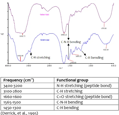
3.2. FTIR Spectrum Profile of Hydrolyzed Gelatins
The characteristic of amino acids in bovine and porcine gelatins before and after pepsin hydrolysis was further identified using FTIR spectroscopy. This analysis aims to identify the functional groups of amino acids making up the source of gelatin. Identification by FTIR spectroscopy was based on the characteristics of functional groups present in gelatin. hydrolysis can be seen in Figure 2.
Frequency (cm-1) Functional group
3400-3200 N-H stretching (peptide bond) 3100-2800 C-H stretching
1660-1600 C=O stretching (peptide bond)
1565-1500 C-N-H bending
1450-1300 C-H bending
(Derrick, et al., 1995)
Fig 2. FTIR profile of hydrolyzed gelatin after pepsin hydrolysis for 1 hour.
Based on Figure 2, it is seen that the FTIR spectra of hydrolyzed gelatin after one hour hydrolysis show that bending), which indicate differences in amino acid composition of the two sources of gelatin. Aliphatic amino acids in porcine gelatin is much larger than the bovine gelatin. These results are consistent with previous studies (Hafidz et.al, 2011) which is the amino acid composition between bovine skin gelatin and porcine skin gelatin was relative differ especially for glycine, proline and arginine residues.
3.3. Polypeptides Pattern of Hydrolyzed Gelatins
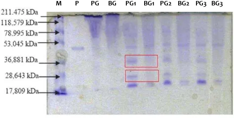
Gelatin, as hydrolyzed collagens, is composed of peptides with wide molecular weight distribution. These peptides were further degraded into peptides with lower molecular weight when digested by pepsin. To identify the different profile of peptide of the gelatins after pepsin hydrolysis, SDS-PAGE electrophoresis was used to differentiate the two sources of hydrolyzed gelatin, characterized by differences in molecular weight pattern. SDS-PAGE electrophoresis results obtained for both sources of gelatin are as showed in Figure 3.
Fig 3. Polypeptide pattern of hydrolyzed gelatin. M : Protein standard, P : pepsin, PG : porcine gelatin, BG : bovine gelatin before and after pepsin hydrolysis for 1, 2 and 3 hours.
significant differences are indicated by the appearance of bands of fragments with molecular weights 36.8 and 28.6 kDa in porcine gelatin gelatin while the bovine gelatin was not found. The next difference seen from the appearance of bands below 28.6 kDa, whereas in bovine gelatin found only one band, while the bovine gelatin found two bands of fragments.
According to Zhang et al. (2009), bovine and porcine collagens type I contain many segments with identical amino acid sequence and some segments containing differential amino acid residues. The peptide corresponding to segments containing differential amino acid residues could be used as marker peptides for differentiation between bovine and porcine gelatins. The amino acid sequence of collagens from different animals is not identical, which is the theoretical foundation of gelatin differentiation based on marker peptide detection. Compared to methods reported in literatures, gelatin differentiation according to the sequences of marker peptides may provide definite information on species origin of gelatin (Guifeng Zhang et al, 2009)
4. Conclusion
Bovine dan porcine gelatins hydrolyzed with pepsin give a characteristics difference of molecular fragments in both gelatin fragments. The analysis using SDS-PAGE electrophoresis showed that there are two bands on the porcine gelatin with molecular weights 36.8 kDa and 28.6 kDa, respectively. Based on UV-VIS and FTIR spectroscopy results, both types of spectra of both gelatins were slightly different in the two fragments of gelatin. This indicates that the hydrolysis of gelatin led to the changes in the number and location of the peptide carbonyl group in both of gelatins.
5. Acknowledgements
We sincerely thank to Research Institute (LEMLIT) of Syarif Hidayatullah, State Islamic University, Jakarta for the research funding. The authors also thank to the
center Integrated Laboratory (PLT) Syarif Hidayatullah, State Islamic University Jakarta for the research facilities.
REFERENCE
Apriantono, A., 2004, Masalah Halal: Kaitan Antara Syar’i, Teknologi dan Sertifikasi, Fakultas Teknologi Pertanian, IPB Bogor.
Che Man, Y. B., & Mirghani, M. E. S. 2001. Detection of lard mixed with body fats of chicken, lamb, and cow by Fourier transform infrared spectroscopy. Journal of American Oil Chemists’ Society, 78, 753–761.
Che Man, Y.B., Z.A. Syahariza, M.E.S. Mirghani, S. Jinap, J. Bakar, 2005, Analysis of potential lard adulteration in chocolate and chocolate products using Fourier transform infrared spectroscopy, Food Chemistry 90, 815–819
Defernez, M., & Wilson, R. H. 1995. Mid-infrared spectroscopy and chemometrics for determining the type of fruit used in jam. Journal of the Science of Food and Agriculture, 67, 461–467.
Gelatin Manufacture Association of Asia Pacific, 2005, About
Gelatin, didownload dari
http://www.gmap-gelatin.com/about_gelatin.html tanggal 1 September 2011.
Gómez-Guillén, M. C., Giménez, B. and Montero, P. 2005. Extraction of gelatin from fish skins by high pressure treatment. Food Hydrocolloids, 19 (5): 923-928.
Guifeng Zhang, Tao Liu, Qian Wang, Li Chen, Jiandu Lei, Jian Luo, Guanghui Ma, Zhiguo Su, 2009, Mass spectrometric detection of marker peptides in tryptic digests of gelatin: A new method to differentiate between bovine and porcine gelatin, Food Hydrocolloids, 23, 2001–2007
Guillen, M.D., and N. Cabo, 1997, Characterization of Edible Oils and Lard by Fourier Transform Infrared Spectroscopy. Relationships Between Composition and Frequency of Concrete Bands in the Fingerprint Region, Journal of the American Oil Chemists’ Society. 74:1281–1286.
Hafidz, R. N., Yaakob, C. M., Amin, I. and Noorfaizan, A., 2011, Chemical and functional properties of bovine and porcine skin gelatin, International Food Research Journal 18: 813-817.
Hans-Hartwig Otto and Tanja Schirmeister, 1997, Cysteine Proteases and Their Inhibitors. Chemical Reviews. 97, 133-171
Hidaka, S. & S. Y. Liu. 2002. Effect of gelatins on calcium phosphate precipitation: a possible application for distinguishing bovine bone gelatin from porcine skin gelatin. Journal of Food Composition and Analysis 16: International Food research Journal 16: 381-389.
Mammalian Gelatins. Food hydrocolloids, 23: 563-576,656.
Kayla, 2010, Marvelous Mixtures Part 2, Perfect Panna Cotta, http://chemistrykitchen.blogspot.com/2010/04/marvelo us-mixtures-part-2-perfect- panna.html didownload 27 Agustus 2011.
Lai, Y. W., Kemsley, E. K., & Wilson, R. H. 1995. Quantitative analysis of potential adulterant of extra virgin olive oil using infrared spectroscopy. Food Chemistry, 53, 95–98. Go´mez-Guille´n, M.C., Gime´nez, P. Montero, B. 2005, Extraction of gelatin from fish skins by high pressure treatment, Journal of Food Hydrocolloids, 19, 923–928. Mahrus Ali, Doni Muhamad Irawan dan Indra Kristiana. 2006.
Isolasi Gelatin dari Limbah Ikan Tuna (Thunnus sp.) dan Ikan Pangkol (Aluterus monoceros) sebagai Alternatif Penyedia Gelatin Halal. Laporan Pekan Kreatifitas Mahasiswa (PKM, 2006).
Nemati, M; Oveisi, M. R.; Abdollahi, H. and Sabzevari, O. 2004. Differentiation of bovine and porcine gelatins using principal component analysis. Journal of Pharmaceutical and Biomedical Analysis 34: 485-492
Paradkar, M. M., Sivakesava, S., & Irudayaraj, J. (2002). Discrimination and classification of adulterants in maple syrup with the use of infrared spectroscopic techniques. Journal of the Science of Food and
Technology, , − .
Rohman, A., Sismindari, Erwanto, Y., and Che Man, Y.B. 2011. Analysis of pork adulteration in beef meatball using Fourier transform infrared (FTIR) spectroscopy. Meat Science 88: 91–95
The Gelatin Manufacturers Institute of America, (GMIA) 2011, Raw Materials, Production & Uses of Gelatin, didownload
dari
http://www.gelatin-gmia.com/html/rawmaterials_app.html, 27 Agiustus 2011.
Venien, A., & Levieux, D. 2005. Differentiation of bovine from porcine gelatins using polyclonal anti-peptide antibodies in indirect and competitive indirect ELISA. Journal of Pharmaceutical and Biomedical Analysis, 39, 418–424.