Cardiovascular reflex
Refleks Cardiovascular
Ikhlas Muhammad Jenie
Bagian Fisiologi Fakultas Kedokteran Universitas Muhammadiyah Yogyakarta
Abstrak
Tekanan darah dan frekuensi denyut jantung merupakan parameter kardiovaskular.
Pengukurannya dalam praktek kedokteran klinis telah menjadi suatu hal yang wajar dilakukan,
sedangkan kepentingannya telah banyak diketahui. Pada dasarnya, parameter kardiovaskular tersebut
merupakan hasil dari refleks. Tulisan ini memaparkan dasar refleks kardiovaskular ditinjau dari
pengaturan melalui saraf, tidak ditinjau dari pengaturan oleh secara mekanis dan maupun faktor
lokal. Refleks kardiovaskular yang dibahas meliputi barorefleks, refleks kardiopulmonar,
khemorefleks, refleks kardiovaskular yang disebabkan oleh stimulasi eksteroreseptor, serta interaksi
antarrefleks kardiovaskular tersebut.
Kata kunci : refleks kardiovaskular, barorefleks, tekanan darah, frekuensi denyut jantung
Abstract
It is familiar to measure cardiovascular parameters, such as blood pressure and pulse
rate, in health care practice. The importance of such measure in clinical practice is out of
question. Those cardiovascular parameters, basically, are products of reflex. In this paper,
we would like to describe the basic tenets of cardiovascular reflex limited to that governed by
autonomic nervous system, but not to that exerted by mechanical and local factors. The
cardiovascular reflexes to be discussed are arterial baroreflex, cardiopulmonary baroreflex,
cardiovascular reflex of stimulation of peripheral chemoreceptors, cardiovascular reflex
associated to stimulation of exteroceptors, and interaction among those reflexes.
Keywords: cardiovascular reflex, baroreflex, blood pressure, heart rate
Introduction
Reflex implies all process develops in an organism in response to stimulation of the receptors with indispensable participation of the central nervous system1. Reflex is produced by the activity of certain structures of the nervous system called reflex arc, which can be represented as follows: receptor Æ afferent nerve fibres Æ central nervous system Æ efferent nerve fibres Æ effector. Thus, reflex arc of any reflex must include: a) a receptor, a nervous structure able to transform energy coming from specific stimulus into a process of nervous
excitation, b) an afferent neuron transmitting impulses from the receptor to the central nervous system, c) a nerve centre with an efferent neuron, d) an efferent nerve fibres extending to certain working organs1.
Vol. 7 No. 1: 43-50, Januari 2007 may produce a reflex excitation of the nerves
that innervates a certain endocrinal gland, which can alter the passage of the hormone of the given gland into the blood. The hormone released by the endocrinal gland will modify the activity of certain effector organs1.
The nature of the reflex depends on two aspects: 1) the state of any structure along the reflex arc, 2) the nature and strength of the stimulus over the receptor1. Cardiovascular parameters, such as blood pressure, heart rate, cardiac output, total peripheral resistance, basically, are products of reflex. Those parameters are actually cardiovascular responses to stimuli. Here, we would like to describe the basic tenets of cardiovascular reflex limited to that governed by autonomic nervous system, but not to that exerted by mechanical and local factors.
Discussion
The reflex arc in the cardiovascular system
1. Receptors
There are two kinds of receptors: interoceptors and exteroceptors. Interoceptors are receptors located within the effector organs, which can be stimulated by agents arising within the organism itself, although some time can be stimulated also by external agents. In the cardiovascular system, interoceptors lie in the heart and blood vessels. Two kinds of interoceptors here are mechanoreceptors and chemoreceptors, which sensitive to mechanical and chemical stimulus, respectively1.
Mechanoreceptors in the cardiovascular system are known as baroreceptors since they respond to stretching usually caused by changes in pressure (baro = pressure). High blood baroreceptors, which located in the adventitial layer of the carotid sinus and aortic arch (thus they are known also as arterial baroreceptors), are sensitive to changes in arterial blood pressure2. Low pressure baroreceptors, which lies on the walls of the right and left atria near the
entrance of the superior and inferior vena cava and pulmonary veins (veno-atrial stretch receptors), on the wall of left ventricles, and in the pulmonary circulation (together known as cardiopulmonary baroreceptors), are sensitive to changes in the blood volume that represented by changes in the central venous pressure, the intra-ventricular pressure, and the pulmonary artery pressure, respectively3. There are also numerous baroreceptors in the vessels of tissues and organs, which continuously maintain tone of blood vessels, responsible for redistribution of the blood among different organs1. Baroreceptors in blood vessels of skeletal muscle are stimulated by local pressure and active muscle tension2.
Peripheral chemoreceptors, which located in the carotid bodies and aortic bodies3, and in small vessels of tissues and organs1, are sensitive to a rise of the partial pressure of CO2, concentration of H+ ions and K--+ ions in the arterial blood, and a decline in the partial pressure of arterial O2. In the blood vessels of skeletal muscle, they are known as metaboloreceptors that activated by an increase in K+ ions, H
2PO4 ions and H+ ions during hard muscular works or exercise2.
Exteroceptors are located outside the cardiovascular system, such as: a) receptors of the skin and mucous membranes of the mouth cavity, upper respiratory tracts, cornea (tactile, thermal, and pain receptors), b) receptors receiving the action of special chemical substances, namely taste and olfactory receptors, c) auditory nerves, d) visual receptors, which stimulated by agents from external environment but can be associated to certain cardiovascular response (conditional reflexes)1.
2 Afferent nerve fibers
a. The glossopharyngeal nerve (the IXth cranial nerve)
b. The vagus nerve (the X cranial nerve) Through with the impulses from baroreceptors in the aortic arch, veno-atrial stretch receptors, the stretch receptors in the pulmonary artery, and chemoreceptors in the aortic bodies is transmitted to cardiovascular centre2. c. The cardiac sympathetic nerves
Through with the impulses from the stretch receptors in the left ventricle is travelled to cardiovascular centre2. d. Small myelinated (group III) and small
unmyelinated nerve fibres (group IV) They carry excitatory input from skeletal muscle2.
3. Cardiovascular centre
The location of cardiovascular centre is in the medulla oblongata. It consists of: a. Sensory centre
It is the nucleus tractus solitarius (NTS), which located on the dorsomedial part of the medulla, is the site of the synapse for the cardiovascular afferents from the baroreceptors, cardio-pulmonary receptors, arterial chemoreceptors, pulmonary stretch receptors, and muscle receptors as well as from the higher centres, namely the hypothalamus, limbic system, and cerebral cortex. NTS evaluates and processes sensory information, which its output is relayed to hypothalamus and cerebellum as well as to various parts of the medulla, e.g. to acceleratory centre and cardiac-inhibitory centre2.
b. Cardiac-acceleratorycentre (CAC) It has intrinsic activity. However, when NTS is stimulated by impulses from peripheral receptors, the cardiac-acceleratory centre will be inhibited. CAC then will depress the spinal centre of the sympathetic nervous system. In turn, heart rate will be decreased (negatively chronotropic effect) and contractility of cardiac muscle be weakened (negatively inotropic effect). Reduced sympathetic tone will cause vasodilatation of most vessels. When NTS is inhibited by impulses from peripheral receptors or higher centres, CAC will be excited and activate the spinal centre of sympathetic nervous system. As a
consequence, heart rate will be increased (positively chronotropic effect) and contractility of the cardiac muscle will be strengthened (positively inotropic effect). Sympathetic tone to most vessels will be increased to cause vasoconstriction except in the skeletal muscle, cardiac muscle, liver, and brain. CAC will also stimulate adrenal medulla to release circulating epinephrine in the blood4.
c. Cardiac-inhibitory centre (CIC) CIC is the nucleus ambiguus of the vagus nerve. When NTS is stimulated, CIC will be activated, and then will increase vagal tone causing a decrease in heart rate. When NTS is inhibited by impulses from peripheral receptors or the higher centres, CIC will be depressed to inhibit the vagal tone. In turn, heart rate will be increased (vagal withdrawal) 4.
As we note, it exists the influence from higher nervous centre such as hypothalamus and cerebral cortex to the cardiovascular centre. Many parts of hypothalamus and cerebral cortex can inhibit or excite CAC5. Under normal condition, cerebral cortex inhibits cardiovascular reflexes exerted by interoceptors. Direct stimulation of the cerebral cortex leads to a decrease in the reflex reaction caused by stimulation of the interoceptors. If the influence of cerebral cortex to the cardiovascular center is blocked, stimulation of interoceptors causes greater changes in cardiovascular reflexes1.
4. Efferent nerve fibres
Vol. 7 No. 1: 43-50, Januari 2007 Acetylcholine released by the postganglionic
parasympathetic nerve terminal binds to muscarinic (M2) receptors in the cell membrane of sinu-atrial node, atrio-ventricular node, and atrial muscle2. The preganglionic sympathetic nerve to the heart arises in the grey matter of the lateral horns of the spinal cord segments T1 to T5. These preganglionic nerves then enter the sympathetic chain and terminate on the stellate ganglion. The postganglionic sympathetic fibres surround subclavian artery form the so-called loop of Vieussens, and enter the rami of the cardiac nerves, where it frequently mixes with the preganglionic parasympathetic fibres of vagus nerves. The postganglionic sympathetic nerve fibres end in the ventricular muscle as well as in the atrial muscle, sinu-atrial node and atrio-ventricular node1. Norepinephrine released by the postganglionic sympathetic fibres terminal binds to the 1 adrenergic receptors either in the cell membrane of sinu-atrial and atrio-ventricular node to increase heart rate (positively chronotropic effect) or in the atrial muscle and ventricular muscle to increase the force of myocardial contraction (positively inotropic effect)2. Both sympathetic and parasympathetic systems exert tonic effect to the heart. However, vagal inhibition predominates. It can be seen that, when both systems are blocked, intrinsic heart rate in a young adult at rest turns out to be around 105 beats per minute. During physiological alterations, both systems exhibit reciprocal changes. During hard muscular work, vagal tone decreases whereas sympathetic activity increases, resulting in exercise-induced tachycardia. However, it is the activity of parasympathetic nerve to the heart, not the activity of the sympathetic systems, which accounted for beat-to-beat differences in heart rate because the onset and decay of the effect of acetylcholine is faster than that of norepinephrine2. Although both systems seem having antagonistic action to each other, they actually exert synergic actions to the heart. Systole retardation caused by stimulation of the vagus nerve provides the
the effects of its metabolic products. As a consequence, the next systole will become stronger than before. Therefore, the negatively chronotropic effect of vagus nerve will enhance the positively inotropic action of sympathetic nerves1.
the activity of the sympathetic vasoconstrictor nerves to each organ is independently regulated. The parasympathetic preganglionic nerve fibres leave the central nervous system in two outflows: the cranial nerves and sacral spinal nerves. Within the end organ they synapse with the postganglionic parasympathetic neurons, which send short axons to the arterioles. The arterioles innervated by the vasodilator nerves as well as by the vasoconstrictor nerves are found just in a few tissues: in the salivary glands, exocrine pancreas, gastrointestinal mucosa, genital erectile tissue, cerebral and coronary arteries. Acetylcholine released by the postganglionic fibres of the parasympathetic
vasodilator nerves causes endothelium-independent vasodilatation. These fibres also release other vasodilator agents, such as nor-adrenergic nor-cholinergic (NANC) and vasoactive polypeptide (VIP). Unlike the sympathetic vasoconstrictor nerves, parasympathetic vasodilator nerves are not tonically active. They would be active only when the organ function demand rise in the blood flow 2.
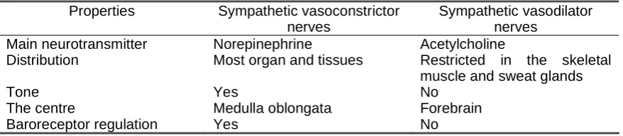
The sympathetic vasodilator nerves differ from the sympathetic vasoconstrictor nerves in several aspects as shown by Table 1. This system is a part of alerting response to mental stress, fear, danger, or simply anticipation of exercise2.
Table 1. Differential properties of the sympathetic vasoconstrictor and vasodilator nerves2
Properties Sympathetic vasoconstrictor nerves
Sympathetic vasodilator nerves
Main neurotransmitter Distribution
Tone The centre
Baroreceptor regulation
Norepinephrine
Most organ and tissues
Yes
Medulla oblongata Yes
Acetylcholine
Restricted in the skeletal muscle and sweat glands No
Forebrain No
5. Effectors
The working organs of the cardiovascular system are the heart and blood vessels. The heart is a pump that generates motive force to move the blood through the vessels, which act as a tube, but not a rigid one6.
Cardiovascular reflexes
1. Cardiovascular reflex to stimulation
of high pressure baroreceptors (baroreflex)
At the normal level of blood pressure, arterial baroreceptors (ARBs) burst at a slow rate. It tonically inhibits resting sympathetic nerve activity. A rise in arterial blood pressure causes vascular distension that stretches the carotid sinus and aortic arch, stimulating ARBs at a higher rate3. Cellular events can be described as follows: It is the role of mechanoreceptors ion channels on the nerve endings that mediate mechanoelectrical transduction in
Vol. 7 No. 1: 43-50, Januari 2007 decreases cardiac output and total
peripheral resistance, respectively. Therefore, these baroreflex will restore high arterial blood pressure back to the normal levels (depressor reflex) 4. When there is a drop in arterial blood pressure, the carotid sinus and aortic arch are less stretched and ARBs will be excited at a slower rate3. NTS will receive fewer ascending impulses2. In turn, it will excite CAC and inhibit CIC. As CAC is stimulated, the spinal centre of sympathetic nerves is more excited and thus sympathetic tone to the heart and blood vessels are increased. As CIC is inhibited, vagal tone to the heart is decreased and heart rate will rise. The overall results are tachycardia and vasoconstriction, which causes an increase in cardiac output and total peripheral resistance, and finally a rise in blood pressure4.
ARBs provide moment-to-moment negative feedback of blood pressure regulation7. The carotid baroreceptors are sensitive to pulse pressure (the difference between systolic and diastolic blood
pressure) as well as to mean arterial blood pressure. The greater the oscillation in blood pressure (meaning the greater the variance in pulse pressure), the greater ARBs would be stimulated, thus, the greater would be the depressor reflex. This signalling of pulse pressure is important during orthostasis or postural changes and behavioural stress2. ARBs also tonically inhibit the release of vasopressin and angiotensin II (AngII). When blood pressure drops, the decrease in the impulses of ARBs evokes increase in the sympathetic activity, which in turn will stimulate the juxtaglomerular cells in the kidneys to release renin. As a consequence, the renin-angiotensin-aldosteron system (RAAS) is activated. AngII will enhance the pressor reflex. In the kidneys, AngII and aldosteron cause salt retention. The decrease in the ARBs traffic also stimulates the posterior pituitary gland to release arginine vasopressin to the blood to enhance peripheral vasoconstriction and water retention (anti diuresis) as well2. It can be summarized in the below table.
Table 2. Hormonal changes during baroreflexes2
Hormones ARBs are stimulated ARBs are inhibited
RAAS Decrease Increase
Vasopressin Decrease Increase
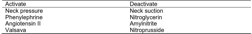
The decrease or increase of ABR activity to stimuli is known as baroreceptor sensitivity (BRS). BRS is defined as changes in the R-R interval (reciprocal of the heart rate) on the electrocardiogram (ECG) plotted as a function of the change in blood pressure during the preceding cardiac cycle. Change in BRS can be induced by any physiological or pharmacological maneuver. The assessment can be done qualitatively and quantitatively. Qualitatively, BRS can be measured at bedside by the change in heart rate from baseline during the Valsava maneuver for 15 seconds. The heart rate usually increases 10 – 30 bpm by the end of the maneuver. Quantitatively, assessment of BRS requires continuous monitoring of the ECG and arterial pressure. It can be
2. Cardiovascular reflex to stimulation
of low pressure baroreceptors (cardiopulmonary baroreceptors) Two types of veno-atrial stretch receptors, a) type A, which discharges during atrial systole, b) type B, which discharges during atrial filling, detect changes in the volume of the blood through changes in the central venous pressure Any increase in blood volume will activate type B receptors. The impulses are then transmitted to NTS in the medulla through the afferent fibres of the vagus nerves, resulting in tachycardia, natriuresis, and diuresis. The atrial natriuretic peptide, hormone released by atrial muscle, acts as the intermediary link of the natriuresis and diuresis effect. Atrial systole stimulates type A receptors. Similar to type B receptors, the impulses from type A receptors are then transmitted to NTS in the medulla oblongata through vagus nerves. It is followed by inhibition to cardiac and peripheral sympathetic outflow but excitatory to cardiac vagal outflow, resulting in bradycardia and vasodilatation2. The clinical syndrome of vasovagal syncope may occur by this mechanism10. The stimulation of type A veno-atrial stretch receptor is associated with the inhibition of the release of angiotensin II, catecholamine, and vasopressin2. The mechanoreceptors in the walls of the left ventricle are stimulated during ventricle systole since these receptors are sensitive to changes in the intra ventricular pressure. The reflex responses of their stimulation are bradycardia and vasodilatation2. The stretch receptors in the pulmonary artery are excited by a rise in the pressure of the pulmonary artery. The vessels of the systemic circulation dilate in response to the excitation of these mechanoreceptors and lead to a drop in blood pressure1. The signals from those
Table 3. Physiological and pharmacological maneuvers that modulate arterial baroreflex
Activate Deactivate Neck pressure
Phenylephrine Angiotensin II Valsava
Neck suction Nitroglycerin Amylnitrite Nitroprusside
cardiopulmonary baroreceptors modify input from arterial baroreceptors. They are sensitive to small changes in cardiac stretch and affect sympathetic discharge during low grade physiological change not detected by arterial baroreceptors10.
3. Cardiovascular reflex of stimulation
of peripheral chemoreceptors
Nerve endings in the carotid and aortic bodies are stimulated by hypoxia, hypercapnia, hyperkalemia, and acidosis. The impulses are transmitted to the cardiovascular centre in the medulla through the afferent fibres of the IXth and Xth cranial nerves, respectively. It actually causes bradycardia reflex and vasoconstriction of the resistance vessels except in the skin. However, the inspiratory centre in the medulla is stimulated also, which in turn causes an increase in tidal volume and excites the stretch receptor within the lungs. The final result is tachycardia and vasodilatation in most vascular beds2. Stimulation of metaboloreceptors in the skeletal muscle by the chemical released during exercise, notably K+ ions,
H----2---PO4 ions, and H+ ions, will result in tachycardia, increased myocardial contractility and peripheral vasoconstriction, resulting in rise of blood pressure2, known as the exercise pressor reflex11.
Vol. 7 No. 1: 43-50, Januari 2007 (asphyxia). Teleologically, this interaction is
important to survival fitness. However, inputs from chemoreceptors alter baroreflex function in more complicated ways. Both hypocapnia and hypercapnia will result in tachycardia12.
4. Cardiovascular reflex of stimulation
of exteroceptors
Stimulation of the exteroceptors causes reflex constriction of the vessels and sometimes also acceleration of the cardiac activity1. Cold applied to the skin is followed by constriction of the blood vessels of the skin, especially the arterio-venous anastomoses. Sudden load noise or unpleasant sight also could increase blood pressure and elicit tachycardia2. Somatic pain is associated with the increase in sympathetic activity (somato-sympathetic reflex) 3. However, severe visceral pain is associated with bradycardia, hypotension and even painting2.
Conclusion
Cardiovascular reflexes are part of homeostasis mechanism. Arterial baroreflex is response to changes in blood pressure. Cardiopulmonary baroreflex is response to small changes in blood volume. Cardiovascular reflex of stimulation of peripheral chemoreceptors is response to changes in blood pH and gas partial pressure. Cardiovascular reflex associated to stimulation of exteroceptors is conditioned reflex, such as seen in fight and flight mechanism. Interactions among these reflexes are seen clinically. All these reflexes are to maintain blood flow to part of body, mainly vital and demanded active organs.
References
1. Bykov KM, Konradi G. Text book of physiology. 2nd ed. Moscow: Peace Publishers; 1953.
2. Levick JR. An introduction to cardiovascular physiology. 3rd ed. London: Arnold; 2000.
3. Ganong WF. Review of medical physiology. 16th ed. Norwalk: Appleton & Lange; 1993.
4. Chauduri SK. Concise medical physiology. 2nd ed. Calcutta: New Central Book Agency; 1993.
5. Guyton AC, Hall JE. Text book of medical physiology. 9th ed. Philadelphia: W.B. Saunders Company; 1996.
6. Bijlani RL. Understanding medical physiology: A text book for medical student. 2nd ed. New Delhi: Jaypee Brothers Medical Publishers; 1997. 7. Chapleau MW. Arterial baroreceptors. In:
Izzo JL, Black HK, editors. Hypertension primer: The essential of high blood pressure. 2nd ed. Maryland: Lippincott Williams & Wilkins; 1999.
8. Taylor A. Evaluation of aortocarotid baroreflexes. In: Izzo JL, Black HK, editors. Hypertension primer: The essential of high blood pressure. 2nd ed. Maryland: Lippincott Williams & Wilkins; 1999.
9. Yambe T, Yoshizawa M, Sugita N, Tanaka A, Maruayam M, Konno S. Invention of the new diagnosis tool for the quantitative evaluation of the Baroreflex sensitivity. The 21st Scientific Meeting of the International Society of Hypertension: Abstract CD-ROM; 2006. 10. Mohanty PK. Cardiopulmonary baroreceptors. In: Izzo JL, Black HK, editors. Hypertension primer: The essential of high blood pressure. 2nd ed. Maryland: Lippincott Williams & Wilkins; 1999.
11. Kluess HA, Wood RH. Heart rate variability and the exercise pressor reflex during dynamic handgrip exercise and postexercise occlusion. Am J Med Sci 2005; 329(3):117-123.