Proximate Compositions and Biological Activities of Caulerpa lentillifera
Risa Nofiani*1, Sigit Hertanto1, Titin Anita Zaharah1, and Sutarman Gafur2
1
Department of Chemistry, Faculty of Mathematics and Natural Sciences, Universitas Tanjungpura, Pontianak, Indonesia
2
Department of Soil Science, Faculty of Agriculture, Universitas Tanjungpura, Pontianak, Indonesia
*
email: [email protected], [email protected]
Received September 14, 2018; Accepted November 25, 2018; Available online December 8, 2018
ABSTRACT
Caulerpa lentillifera is an edible and functional seaweed due to its high nutritional compositions and its biological activities. In this study, C. lentillifera was evaluated for its proximate compositions (moisture, ash, protein, lipid and fiber contents) and its biological activities (antimicrobial, anti-oxidant, and toxicity). Moisture content, crude lipid, crude protein, and crude fiber were determined using oven method, soxhlet extraction, semi-micro Kjeldhal, and hydrolysis, respectively. Fresh C. lentillifera of Natuna Island, Indonesia, showed its higher level content of ash, crude lipid, and crude fiber compared to that of fresh C.
lentillifera of Penghu, Taiwan. For its biological activity assays, the extracts were prepared from fresh and dry C. lentillifera (FC and DC). Both of the extracts showed the broad spectrum of weak antimicrobial using well-diffusion agar tests and antioxidant activities using a modified linoleic acid emulsion system. The toxicity for both extracts was determined using brine shrimp lethality test. DC extract showed its very low toxicity level and there was no toxicity for FC. Hemolytic activity was determined using red blood assay. Both extracts showed their low hemolytic activities (about 5-13%) for the concentration of 100 and 150
μg/mL, but the activity increased sharply (about 96%) on the concentration of 200 μg/mL. It was concluded that C. lentillifera has a potency as a functional food due to containing secondary metabolites with various biological activities.
Keywords: Caulerpa lentillifera, antimicrobial and antioxidant activity, toxicity, hemolysis, proximate composition
INTRODUCTION
Seaweeds have high nutritional constituents consisting of essential minerals, fatty acids, dietary fibers, amino acids and vitamins (Bhuiyan, Qureshi, Kamal, Aftab, & Siddique, 2016). Studies about chemical composition and nutritive value of edible seaweeds have increased due to their potential for food uses. For example, C. lentillifera from Malaysia and Thailand were reported having the nutritional composition such as: proximate composition, amino acids, vitamin C and E (Matanjun et al, 2009; Ratana-arporn and Chirapart, 2006). It was also reported that dry C. lentillifera (DC) which can be used as sources of food protein due to its high protein levels and balanced amino acid profiles (Ratana-arporn & Chirapart, 2006)
C. lentillifera is a green edible seaweed, classified into a Chlorophyta marine macro-algae (Mohamed, Hashim, & Rahman, 2012). It is widespread in Asian countries such as: Indonesia, Japan, Thailand, Malaysia, China, Philiphine, Korea and some other countries in Southeast Asia (Nguyen, Ueng, & Tsai, 2011).
In Natuna Island, Indonesia, C. lentillifera is naturally grown and known as Latuh and it is used to be consumed as a fresh salad, although it is lately rarely consumed by the community of Natuna.
Ismail, & Tawang, 2016). Extract of C. lentillifera has the potency to be used as an anti-diabetic agent.
A seaweed grown in different location, habitat, or environmental conditions, and harvested in different maturity levels had caused the different in its chemical compositions, consistency, color, quality, and its bioactive compounds (Ito and Hori 2009; Kılınç et al. 2013). The proximate composition of C. lentillifera of Malaysia and Thailand has been reported but C. lentillifera of Indonesia has not yet been studied and reported.
In this study, C. lentillifera from Natuna Island of Indonesia was evaluated for its proximate compositions as C. lentillifera fresh because it is eaten fresh in the community of Natuna. For its biological activities particularly for its antimicrobial, anti-oxidant, and its toxicity activities was also analyzed as fresh and dry C. lentillifera which probably benefit for health or is called as a functional food.
EXPERIMENTAL SECTION
Sampling
C. lentillifera was collected randomly from the coastal area of Kabupaten Natuna, Kepulauan Riau. Morphology of C. lentillifera was identified in the Laboratory of Biology, Faculty of Mathematics and Natural Sciences, Universitas Tanjungpura, Indonesia.
Preparation of C. lentillifera
Fresh C. lentillifera (FC) was rinsed using distilled water and used directly for its proximate analysis and biological activity assays. Dry C. lentillifera (DC) were prepared by drying of C. lentillifera directly under the sunshine. The FC was used for proximate analysis, while the FC and DC were used for its antioxidant, hemolysis, cytotoxicity and antimicrobial assays.
Proximate Analysis
Moisture content was determined using oven method based on Method of 925.10 from AOAC. Ash content was determined based on Method numberof 08-01 from AOAC. Crude lipid was determined using Soxhlet extraction with diethyl ether referred to Method of 30-25, AOAC. Crude fiber was determined using successive hydrolysis with 100°C 1.25% of H2SO4 and 3.25% of NaOH for 30 min each
based on SNI 01-2891-1992. Crude protein was determined using semi-micro Kjeldhal
methods referred to SNI (Indonesian National Standard 01-2891-1992).
Extraction of C. lentillifera
The FC (139.6 g) was ground using a a mortar and macerated using 80% of ethyl acetate. The maceration was carried out many times until colorless filtrate. The filtrate was evaporated using a rotary evaporator to get crude extract FC.The DC powder (21.6 g) was macerated using of methanol 80% until screened antimicrobial activities using well-diffusion methods (Okeke, Iroegbu, Eze, Okoli, & Esimone, 2001). Tested microorganisms were cultured by inoculation onto 10 mL of nutrient broth (NB) medium and incubated on a rotary shaker incubator at 130 rpm at 30 °C for 14-16 h. The culture was added on nutrient agar (NA), mixed and poured into the plate. After gel solid, the medium was punched using a sterilized pipette with diameter 0.4 mm. The extracts (FC and DC) with various concentrations (50, 100, 200, 400, and 500 μg/well) were poured into the well. After solvent from the extract was evaporated, the plate was incubated for 14-16 hours.
The extract having antimicrobial activities was signed with the formation of inhibition zone around the well then the diameter of inhibition zone (measured from the edge of the colony to the edge of the clear zone) was recorded. Furthermore, the extract having antimicrobial activities was tested for its bacteriostatic and bactericidal activities by the measuring its inhibition zone. Minimum inhibition concentrations (MICs) of each extract was counted based on the method of Bonev et al. (Bonev, Hooper, & Parisot, 2008).
Antioxidant Assay
The antioxidant assay was carried out using a slight modified linoleic acid emulsion system (Loganayaki, Siddhuraju, & Manian, 2013). A 10 μL of linoleic acid was mixed with 1 mL of ethanol in Eppendorf tube. The mixture was added with 1000 μg extract of C. lentillifera and incubated in the dark room at 25 °C.
% of AA = 1-measured at 490 nm. A blank absorbance was measured at t = 0 h and t = 24 h. Vitamin C (1000 μg) was used as a positive control. The antioxidant activity (AA) was calculated as a persentage of inhibition relative to the control using the Equation 1.
Erythrocyte suspensions were prepared according to Situ & Bobek (2000) and NaCl, 10 mM of sodium phosphate buffer, pH 7.4). The pellet was resuspended in PBS solution and adjusted to a hematocrit of 1%.
Hemolytic Assay incubated at 37 °C. After 30 min, the mixture was centrifuged at 1500 x g, 4 °C. Cell lysis was monitored by measuring the release of hemoglobin at 540 nm using a Microplate reader. As a positive and negative control was used 1% of sodium dodecyl sulfate (SDS) in PBS and PBS alone, respectively. Percentage of hemolysis was calculated as follows: [(A540
of the sample treated with the extract-A540
sample treated with buffer)]/[(A540 of the
sample treated with SDS -A540 sample treated
with buffer)] x 100%.
Preliminary Toxicity Test
Preliminary toxicity test was carried out using Brine Shrimp Lethality Test (BSLT) (Meyer et al., 1982). Ten shrimps Artemia salina Leach, of 2 days old, were added with the extracts of various concentrations (0 (for control), 10, 100, 200, 400, 600, 800 dan 1.000 μg/mL) and with artificial sea water (3 mg in 5 ml artificial sea water) to make a 9 mL solution. After 24 hours at room temperature, the nauplii survivors can be counted macroscopically and the percent deaths at each dose and control were determined. The significance of the difference between means was determined by LSD (P<0.05) using SPSS version 23 (Kokoska, 2015). Values expressed are means of three replicate determinations ± standard deviation.
RESULTS AND DISCUSSION
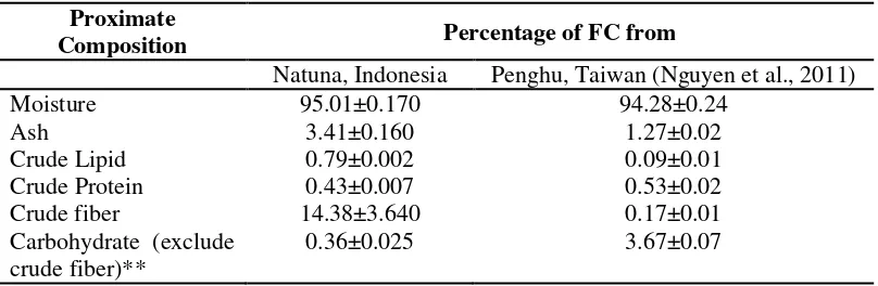
Proximate Composition of C. lentillifera Proximate composition of FC from Natuna District of Indonesia was different than that of FC from Penghu of Taiwan except for the moisture content (Table 1).
Table 1. Proximate Compositions of C. lentillifera from Natuna District (% FC)
Proximate
Composition Percentage of FC from
Natuna, Indonesia Penghu, Taiwan (Nguyen et al., 2011)
Moisture 95.01±0.170 94.28±0.24
Ash 3.41±0.160 1.27±0.02
Crude Lipid 0.79±0.002 0.09±0.01
Crude Protein 0.43±0.007 0.53±0.02
Crude fiber 14.38±3.640 0.17±0.01
Carbohydrate (exclude crude fiber)**
0.36±0.025 3.67±0.07
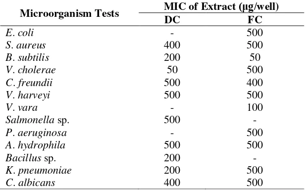
Table 2. Antimicrobial Activities of the extract DC and FC against Tested Microorganism
MicroorganismTests MIC of Extract (μg/well)
DC FC
E. coli - 500
S. aureus 400 500
B. subtilis 200 50
V. cholerae 50 500
C. freundii 500 400
V. harveyi 500 500
V. vara - 100
Salmonella sp. 500 -
P. aeruginosa - 500
A. hydrophila 500 500
Bacillus sp. 200 -
K. pneumoniae 200 500
C. albicans 400 500
Note: -: no antimicrobial activities
FC from Natuna had generally higher levels of ash, crude lipid, and crude fiber compared with that of FC from Penghu of Taiwan. DC of Malaysia, Taiwan, and Thailand showed a generally different of their proximate composition (Nguyen et al., 2011; Ratana-arporn and Chirapart, 2006; Matanjun et al., 2009). It might be due to the different in their habitat conditions, maturity levels, and their environmental conditions (Ito & Hori, 2009).
Antimicrobial Activity
In general, extract C. lentillifera FC and DC showed the broad spectrum of weak antimicrobial activities against tested microorganisms (E. coli, S. aureus, B. subtilis, V. cholerae, C. freundii, V. harveyi, P. aeruginosa, A. hydrophila, K. pneumoniae and C. albicans) based on their high minimum inhibition concentration (MIC) value (Table 2). The broad spectrum of antimicrobial activities was also shown by the other extract of Caulerpa such as: C. ashmeadii, C. paspaloides and C. prolifera (Freile-pelegrin & Morales, 2004). C. cupressoides, C. Mexicana, and C. racemose showed their
antimicrobial activities towards Bacillus subtilis (Freile-pelegrin & Morales, 2004). Caulerpin and caulerpenyne were probably contributed to antimicrobial activities of C. lentillifera which is commonly to occur in Caulerpa (Paul et al., 1987).
Hemolytic Assay
The hemolytic assay can be used for toxicity studies of compounds (Situ and Bobek 2000; Djouossi et al. 2015). The advantages using of this assay is it is sensitive, cheap, quick, and easy to monitor the lysis. Both extracts of FC and DC showed its low hemolytic activities (around 5-13%) for concentrations of 100 and 150 μg/mL, but the activity increases sharply (about 96%) for both concentration of 200 μg/mL (Table 3). The hemolytic activity will probably damage the red cell of the membrane and induces the hemolytic anemia (Zohra and Fawzia 2014). This activity probably results from the astringent phenolic content of C. lentillifera (Nguyen et al. 2011; Singh & Kaur 2008).
According to this result, C. lentillifera is not recommended to be consumed in excessive amount.
Table 3. Hemolytic activitiy of the extract C. lentillifera
Sample Pencentage of Hemolysis for Concentration (μg/mL)
100 150 200
Extract FC 13.32±0.23a 13.48±0.27a 96.71±1.40b
Extract DC 5.11±0.56a 9.20±1.43b 96.22±1.77c
Values are presented as mean±SD (n=3). Values with different superscript in each column
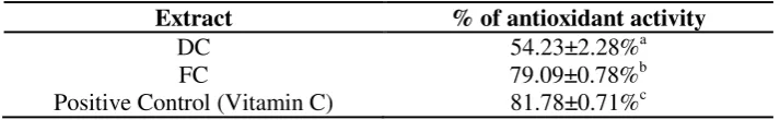
Table 4. Antioxidant Activity of C. lentillifera Extracts Using Linoleic Acid Emulsion System
Extract % of antioxidant activity
DC 54.23±2.28%a
FC 79.09±0.78%b
Positive Control (Vitamin C) 81.78±0.71%c
Values are presented as mean±SD (n=3). Values with different superscript in each row are significantly different from one another (p < 0.05)
Brine Shrimp Lethality Test (BSLT)
BSLT is a cheap and simple bioassay system which is used to detect a bioactive compound such as a cytotoxic drug (Meyer et al., 1982). BSLT result of both extracts of C. lentifera showed that was only extracted of DC caused death on larvae (nauplii) of A. salina but no extract of FC. The extract of DC gave LC50 value 258,360 μg/mL which was very
low toxicity due to an LC50 value greater than
100 μg/mL (Mbwambo, Moshi, Masimba, Kapingu, & Nondo, 2007). As a comparison, standard cytotoxic of drug (cyclophosphamide) and of gallic acid have LC50 of 531.0 and 323.6
μg/mL, respectively (Sonibare, 2017). According to Meyer et al., the LC50 value of
extracts ≤ 1000 μg/mL was predicted containing bioactive compounds (Meyer et al., 1982). Caulerpenyne, a cytotoxic sesquiter-penoid, is likely responsible for this activity due to its most abundant found in the genus of Caulerpa (Dumay et al., 2002).
Antioxidant Activity
Antioxidant activity of an extract of C. lentillifera from Taiwan have been analyzed using various assays which are DPPH (1,1-diphenyl-2-picryl hydrazyl) radical scavenging, ferric reducing, hydrogen peroxide scavenging and ferrous ion chelating (FIC) activity. The extract of C. lentillifera showed its low antioxidant activity compared with that of a positive control for assays using DPPH radical scavenging, ferric reducing, and hydrogen peroxide scavenging (Nguyen et al., 2011). The best antioxidant activity of the extract was resulted in using FIC assay.
In this study, the extract of C. lentillifera was also tested for its antioxidant activity using another method namely linoleic acid emulsion system. This system measures its inhibition of lipid peroxidation by certain compounds having antioxidant activity (Loganayaki et al., 2013). Linoleic acid is an unsaturated fatty acid with 2 double bonds which is oxidized easily to be peroxides. The peroxides are able to oxidize from Fe2+ ions to
Fe3+ ions. Fe3+ ions then react with ion thiocyanate to produce ferri thiocyanate complex (Fe(SCN)6) with red color. The red
color intensity showed the active peroxide formed and was measured at an absorbance of 490 nm wavelength.
Both extracts showed antioxidant activity, even though the antioxidant activity value of both was significantly lower (p<0.05) than that of in the positive control, vitamin C (Table 4). According to Matanjun et. al., C. lentillifera also contains a low level of vitamin C and vitamin E which probably contributes to antioxidant activity in this assay (Matanjun et al. 2009; Nguyen et al. 2011).
CONCLUSION
The proximate composition of C. lentillifera of Indonesia was different from that of Taiwan. Ash, lipid, and fiber contents of C. lentillifera of Indonesia were higher than that of C. lentillifera of Taiwan. Furthermore, C. lentillifera of Indonesia also contained secondary metabolites having various biological activities, even though it is not recommended to be consumed in excessive amount. According to this study, C. lentillifera is highly recommended to be a functional food and to be a potent secondary metabolite source with various biological activities.
REFERENCES
Abbot, W. (1925). A Method of Computing The Effectiveness of an Insecticide. Journal of Economic Entomology, 18(2), 265–267.
Bhuiyan, M. K. A., Qureshi, S., Kamal, A. H. M., Aftab, U. S., & Siddique, M. A. M. (2016). Proximate Chemical Composition of Sea Grapes Caulerpa racemosa (J. Agardh, 1873) Collected from a Sub-Tropical Coast. Virology and Mycology, 5(2). https://doi.org/10.4172/2161-0517.1000158
Principles of Assessing Bacterial Susceptibility to Antibiotics using the Agar Diffusion Method. Journal of pyretic Action of Caulerpa lentillifera, Hibiscus rosasinensis, and Piper sarmentosum Aqueous Extract in Mice. Asian Journal of Pharmaceutical and Clinical Research, 9(1), 9–11.
Djouossi, M. G., Tamokou, J., Ngnokam, D., Kuiate, J., Tapondjou, L. A., Harakat, D., & Voutquenne-nazabadioko, L. (2015). Antimicrobial and Antioxidant Flavonoids from the Leaves of Oncoba spinosa Forsk. (Salicaceae). BMC Complementary and Alternative
Medicine, 15, 4–11.
https://doi.org/10.1186/s12906-015-0660-1
Doty, S. M. (1966). Caulerpicin, a Toxic Constituent of Caulerpa. Nature, 211, 990.
Doty, S. M., & Aguilar-santos, G. (1970). Transfer of Toxic Alga Substances in Marine Food Chains. Pacific Science, 24, 351–355.
Dumay, O., Erard, G., Pergent-martini, C., & Amade, P. (2002). Variations in Caulerpenyne Contents in Caulerpa taxifolia and Caulerpa racemosa. Journal of Chemical Ecology, 28(2), 343–352. Freile-pelegrin, Y., & Morales, J. L. (2004).
Antibacterial Activity in Marine Algae from the Coast of Yucatan, Mexico. Botanica Marina, 47, 140–146. https://doi.org/10.1515/BOT.2004.014 Ito, K., & Hori, K. (2009). Seaweed : Chemical
Composition and Potential Food Uses. Food Reviews International, 5(1), 101– 144.
Kılınç, B., Cirik, S., & Turan, G. (2013). Seaweeds for Food and Industrial Applications. In Food Industry (pp. 735– 748). INTECH.
Kokoska, S. (2015). Introductory Statistics: A Problem-Solving Approach (Second). New York: W.H Freeman and Company. Loganayaki, N., Siddhuraju, P., & Manian, S.
(2013). Antioxidant Activity and Free Radical Scavenging Capacity of Phenolic Extracts from Helicteres isora L. and
Ceiba pentandra L. Journal of Food Science and Technology, 50(4), 687–695. https://doi.org/10.1007/s13197-011-0389-x
Matanjun, P., Mohamed, S., Mustapha, N. M., & Muhammad Kharidah. (2009). Nutrient Content of Tropical Edible Seaweeds, Eucheuma cottonii, Caulerpa lentillifera and Sargassum polycystum. Journal of Applied Phycology, 21, 75–80. https://doi.org/10.1007/s10811-008-9326-4
Mbwambo, Z. H., Moshi, M. J., Masimba, P. J., Kapingu, M. C., & Nondo, R. S. O. (2007). Antimicrobial Activity and Brine Shrimp Toxicity of Extracts of Terminalia brownii roots and stem. BMC Complementary and Alternative
Medicine, 7(9), 5–9.
https://doi.org/10.1186/1472-6882-7-9 Meyer, B. N., Ferrigni, N. A., Putnam, J. E.,
Jacobsen, L. B., Nichols, D. E., & Mclaughlin, J. L. (1982). Brine Shrimp : A Convenient General Bioassay for Active Plant Constituents. Journal of Medicinal Plant Research, 45, 31–34. Mohamed, S., Hashim, S. N., & Rahman, A.
(2012). Seaweeds : A Sustainable Functional Food for Complementary and Alternative Therapy. Trends in Food Science & Technology, 23(2), 83–96. https://doi.org/10.1016/j.tifs.2011.09.001 Nguyen, T. Van, Ueng, J.-P., & Tsai, G.-J.
(2011). Proximate Composition, Total Phenolic Content, and Antioxidant Activity of Sea grape (Caulerpa lentillifera). Journal of Food Science, 76
(November), C950-c958. owerrience for Antibacterial Activity. Journal of Ethnopharmacology, 78, 119– 127. https://doi.org/10.1016/S0378-8741(01)00307-5
Paul, J. V., Littler, M. M., Littler, S. D., & Fenical, W. (1987). Evidence for Chemical Defense in Tropical Green
Alga Caulerpa ashmeadii
Ratana-arporn, P., & Chirapart, A. (2006). Nutritional Evaluation of Tropical Green Seaweeds Caulerpa lentillifera and Ulva reticulata. Kasetsart J. (Nat. Sci), 40(September), 75–83.
Saengkhae, C., Arunnopparat, W., & Sungkhajorn, P. (2007). Antioxidative Activity of the Leaf of Nelumbo nucifera Gaertn. on Oxidative Stress-Induced Erythrocyte Hemolysis in Hypertensive and Normotensive Rats. Thai Journal of Physiological Sciences, 20(2), 70–78. Singh, R. P., & Kaur, G. (2008). Hemolytic
Activity of Aqueous Extract of Livistona chinensis Fruits. Food and Chemical Toxicology, 46, 553–556. https://doi.org/10.1016/j.fct.2007.08.037 Situ, H., & Bobek, A. L. (2000). In Vitro
Assessment of Antifungal Therapeutic
Potential of Salivary Histatin-5, Two Variants of Histatin-5, and Salivary Mucin ( MUC7 ) Domain 1. Antimicrobial Agents and Chemotheraphy, 44(6), 1485–1493. Sonibare, A. A. A. M. A. (2017). In Vitro
Antioxidant Activity, Brine Shrimp Lethality and Assessment of Bioactive Constituents of Three wild Dioscorea Species. Journal of Food Measurement and Characterization, 11(2), 685–695. https://doi.org/10.1007/s11694-016-9438-5