Volume 21| No. 2| June | 2016
Corresponding Author: Rini Devijanti Ridwan
Department of Oral Biology, Faculty of Dentistry Universitas Airlangga
Jl. Mayjen Prof. Dr. Moestopo 47 Surabaya 60132 – Indonesia phone : +6281331049714
e-mail : [email protected]
Rini Devijanti Ridwan
1, Tuti Kusumaningsih
1, Sidarningsih
1, Soetjipto
21Department of Oral Biology, Dental Medicine Faculty Airlangga University 2Department of Biochemistry, Faculty of Medicine Airlangga University Surabaya-Indonesia
ABSTRACT
Adhesion is a powerful survival mechanism as well as a virulence mechanism for bacterial pathogens. Bacterial adhesin is a media for bacteria to invade the host. Bacterial adhesin,is a medium for bacteria to invade the host. Baterial adhe-sion, moreover, is depend on the ligand interaction as a signaling mediator that will influence the invasion process and increase pro and anti-inflammatory due tob the influence of the receptors of innate immune response. Aggregatibacter actimycetemcomitans (A.actinomycetemcomitan) have many virulence factors that may result in tissue and alveolar bone damage. One of the virulence factors is adhesin that can be isolated from the fimbriae. This research purposed to analyze the ability of adhesin protein from A.actinomycetemcomitan that cause the destruction of alveolar bone. Thus, the number of osteoblasts and osteoclasts as well as osteocalcin expression can be used as a marker of damage on the alveolar bone of Wistar rats. The research was conducted through several processes. First, the adhesin of A.actinomycetemcomitan with a molecular weight (MW) of 24 kDa is induced into Wistar rats. Next, to determine the number of osteoblasts and osteoclasts performed, hematoxylin eosin staining is conducted. Meanwhile, to determine osteocalcin expression performed, immunohistochemical techniques is used. This research shows the decreasing of the number of osteoblasts and increasing of the number of osteoclasts in the treatment groups induced by adhesin proteins, A. actinomycetemcomitans + adhesin protein, and A.actinomycetemcomitan compared those in the control group. It also shows the increasing of osteocalcin expressions on the alveolar bone of Wistar rats in the groups induced by adhesin proteins, A. actinomycetemcomitans + adhesin protein, and A. actinomycetemcomitans than those in the control group. It can be concluded that the adhesin protein of A. actinomycetemcomitans plays an important role in the destruction of alveolar bone through the reduction of the number of osteoblasts, the increasing of the number of osteoclasts and oste-ocalcin expression in aggressive periodontitis.
Keywords:Adhesin, A. actinomycetemcomitans, osteoblast, osteoclast, osteocalcin expression
Aggressive periodontitis is a disease found on tis-sues supporting teeth, and characterized by rapid deterio-ration in periodontal ligament and alveolar bone. Aggres-sive periodontitis is usually found in young patients, who are under 30 years old. In the aggressive periodontitis, moreover, the loss of tissue attachment and the recession of gingival can occur four times (4x) faster than in chron-ic periodontitis (Newman et al., 2006; Velden et al., 2006). Until now, the occurence of aggressive periodonti-tis in young age has been a problem that cannot be ex-plained comprehensively in dentistry.
The pathogenesis of periodontitis is actually affected by the interaction of the host and bacterial factors domi-nated by Aggregatibacter actinomycetemcomitans (A. actinomycetemcomitans). In other words, the presence of these bacteria in dental plaque can trigger the aggression
of periodontal tissue destruction that may also be exacer-bated by genetic and environment factors ( Korman 2000). The direct contact between infectious agents and host cells is actually started with the process of adhesion (attachment). The process of adhesion is one of the viru-lence properties of pathogenic bacteria, which are crucial for the colonization, the invasion and the onset of infec-tious diseases ( Doig et al., 1988). It is because A. actino-mycetemcomitans have fimbriae which functions as the adhesion and the invasion. The fimbriae, thus, can be con-sidered as the virulence factors in infection process in oral cavity. When periodontitis actively becomes progressive, as a result, the level of MMP-8, in gingival crevicular fluid (GCF) significantly increases, causing damage to periodontal tissues and alveolar bone. This study pur-posed to analyze the ability of adhesin protein from A.actinomycetemcomitan local isolate that cause the destruction of alveolar bone.
Culture for A. actinomycetemcomitans
The culture for A. actinomycetemcomitans should be prepared in Luria Berthani medium as much as 200 mL to be used in each group (20 mice). Thus, it must be
pre-METHODS
STUDY OF ADHESIN FROM
Aggregatibacter actinomycetemcomitans
LOCAL ISOLATE ON ALVEOLAR BONE DESTRUCTION IN
AGGRESSIVE PERIODONTITIS DISEASE
pared at least as much as 5 ml at a density of 108 A. acti-nomycetemcomitans and given in at least 7 days.
Adhesin of A. actinomycetemcomitans
The adhesin of A. actinomycetemcomitans should be prepared with a molecular weight (MW) of 24 kDa. It means that the induction for each group is about 200 µl with 200 mg / ml protein and it is given for at least 7 days.
Inducing adhesin protein into Wistar rats
First, Wistar rats we are divided into four groups, one control group and three treatment groups. Each of the groups consisteds of ten rats. Next, the first group as a negative control was induced by 0.9% NaCl, while the second group was induced by adhesin. Meanwhile the third group was induced by adhesin and the whole cell of A.actinomycetemcomitan. The last group as the positive control was induced by the whole cell of A. actinomy-cetemcomitans. The induction of adhesin was done with 200 mL with 200 mg/ml protein at the density of 108 A. actinomycetemcomitans, and was given at least 7 days to acquire real aggressive periodontitis symptoms (Zhou et al., 2005). The induction was carried out in the pocket of the right first molar of the Wistar rats as explained in Dumitrescu method (Dumitrescu 2006).
Immunohistochemical technique
The use of immunohistochemical methods in exam-ining osteocalcin expression was conducted through the following stages:
1. Making histopathology preparations based on
Hu-mason’s method (1972) as quoted from Sudiana
(Sudiana 2015).
2. Calculating the results of immunohistochemical stain-ing as quoted from Soini et al., and Pizem and Cor (Soini et al.,1997 ; Pizem and Cor 2003).
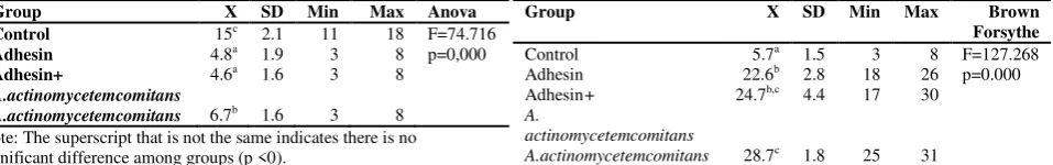
Based on the results of calculating the number of os-teoblasts on the alveolar bone, it is known that the distri-bution of the data is normal and homogeneous with a sig-nificance value > 0.05 (0.68). From the results of ANO-VA test, it is also known that the data have p-value of 0.000 (<0.05), indicating that there is a significant differ-ence between the number of osteoblasts in the treatment group and that in the control group. LSD test results, moreover, shows that there are significant differences between the number of osteoblasts in the control group and that in the groups induced by adhesin, adhesin + A. actinomycetemcomitans, and A. actinomycetemcomitans.
Figure 1. Alveolar bone tissue (Magnification 400X). A. Control Group; B. Group induced by A.actinomycetemcomitans; C. Group induced by adhesin protein; D. Group induced by A.actinomycetemcomitan + adhesin protein. Black arrow points to osteoclasts, while yellow arrow points to osteoblasts.
Table 1. Means and standard deviations of the number of osteoblasts in alveolar bone
Note: The superscript that is not the same indicates there is no significant difference among groups (p <0).
Table 2. Means and Standard Deviations of the Number of Osteoclasts on Alveolar Bone significant difference among groups (p <0.05).
Similar result is also shown among treatment group, except between that in the group induced by adhesin and that in the one induced by adhesin + A. actinomycetem-comitans. The results also show that there are significant differences between the number of osteoblasts in the con-trol group and that in the treatment groups induced by adhesin, adhesin + A. actinomycetemcomitans, and A.actinomycetemcomitan as well as among the treatment groups, except between that in the group induced by ad-hesin and that in the one induced by adad-hesin + A. actino-mycetemcomitans.
Furthermore, the results of calculating the number of osteoclasts on the alveolar bone show, that the distribu-tion of the data is normal, but not homogeneous with a significance value <0.05 (0.02). Based on the results of Brown Forsy test, it is known that p-value is about 0.000 (<0.05), indicating that there are significant differences between the number of osteoclasts in the treatment groups and that in the control group. Meanwhile, the results of Games Howell test, show that there are significant differ-ences between the number of osteoclasts in the control group and that in the treatment groups induced by adhe-sin, adhesin + A. actinomycetemcomitans, and A.
actino-RESULTS
mycetemcomitans as well as among the treatment groups, except between that in the group induced by adhesin and that in the group induced by adhesin + A. actinomycetem-comitans. Similar results are also between that in the group induced by adhesin + A. actinomycetemcomitans and that in the group induced by A. actinomycetemcomi-tans. Thus it may be concluded that there are significant differences between the number of osteoclasts in the con-trol group and that in the group induced by adhesin,
adhe-sin + A. actinomycetemcomitans, and A. actinomycetem-comitans, as well as among the treatment groups, except between that in the group induced by adhesin and that in the group induced by adhesin + A. actinomycetemcomi-tans, and also between that in the group induced by adhe-sin + A. actinomycetemcomitans and that in the group induced by A. actinomycetemcomitans. The Means and Standard Deviations of the Number of Osteoclasts on Alveolar Bone can seen in table 2 and figure 2.
Figure 2. The Average Number of Osteoblasts and Osteoclasts on Alveolar Bone
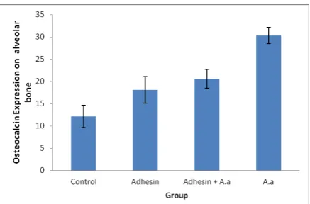
Figure 3. The Average of Osteocalcin Expression on Alveolar Bone
Figure 4. Osteocalcin expression on alveolar bone of wistar rats (black arrow) (magnification 400X). A. control, B. adhesin, C. adhesin + A. actinomycetemcomitans, D. A.actinomycetemcomitans
The data of positive activation of osteocalcin expres-sions are obtained from the observation of osteoblasts on alveolar bone by using immunohistochemical method in the control group and the treatment groups induced by adhesin, adhesin + A. actinomycetemcomitans, and A. actinomycetemcomitans. The result of the observation can be seen in Figure 3.
Osteocalcin expression can be observed through the number of osteoblasts which function as the producing cells of osteocalcin. Based on the results of
Kolmogorov-Smirnov’s test, it is known that the osteocalcin expre s-sions on the alveolar bone in the rats are normally distrib-uted (p> 0.05). Moreover from the results of Levene's test, it is shown that the relation among the groups has a variety of homogeneity (p> 0.05 = 0.48). Because the data were normally distributed and homogeneous, thus, to de-termine the difference of the osteocalcin expressions among the groups, ANOVA analysis is used. Based on the LSD test, the result shows that the value of p is <0.05, indicating that the expressions of osteocalcin in all four treatment groups have significant differences. The significant differences of osteocalcin expressions occur between those in the control group and those in the treat-ment groups induced by adhesin, adhesin + A.
actinomy-cetemcomitans, and A. actinomycetemcomitans, as well as among those in treatment groups. However, there is no significant difference between osteocalcin expression in the group induced by adhesin and those induced by adhe-sin + A. actinomycetemcomitans. Finally, the means and standard deviations of osteocalcin expression on alveolar bone are shown in the following the figure 3.
Based on the results above, it is shown that the num-ber of osteoblasts found on alveolar bone in the control group is significantly different from that in the treatment groups induced by adhesion, adhesin + A. actinomycetem-comitans, and A. actinomycetemcomitans. It indicates that there is no inflammation (aggressive periodontitis) in the control group. On the other hand there is inflammation in the treatment groups induced by adhesin, A. actinomy-cetemcomitans + adhesion, and A. actinomycetemcomi-tans.
Osteoblasts actually play an important role in miner-alization process through the deposition of hydroxyap-atite. It is because osteoblasts regulate calcium and phos-phate concentrations that are useful in the formation of
DISCUSSION
hydroxyapatite. Osteoblasts also show alkaline phospha-tase in high quantities in plasma membrane. This alkaline phosphatase is essential in the process of bone mineraliza-tion. Osteoblasts, moreover, also express a variety of cy-tokines, namely Colony Stimulating Factor 1 (CSF 1), RANKL, and Osteoprotegerin (OPG) (Manolagas 2000 ; Morawati 2009). The highest number of osteoclasts found on the alveolar bone is in the treatment group induced by A. actinomycetemcomitans. This condition is significantly different from the number found in the control group and in the treatment groups induced by adhesin and adhesin + A. actinomycetemcomitans.
That condition discussed previously indicates that the induction of A. actinomycetemcomitans has caused adhesion, colonization, and invasion to the host. At the time of the invasion to the host, A. actinomycetemcomi-tans secretes virulence factors, one of them is LPS. LPS is a major factor of the bacteria that has an ability to per-form bone resorption by conducting osteoclasts stimula-tion, in which LPS activates osteoblasts to secrete factors that can attract and or activate osteoclasts. LPS, according to a research conducted by Nair et al, also inhibits the synthesis of collagen and non-collagen protein (Nair et al., 1996). In addition, LPS, according to a research con-ducted by Nishihara, (1994), can cause bone resorption on murine calvarial by stimulating murine macrophages. Thus, it ca ben be said that A. actinomycetemcomitans can cause damage to alveolar bone and create antibody re-sponses in the three strains of mice, namely Hypertensi-tive Fawn Hooded (FHH), Dahln Salt SensiHypertensi-tive (DSS), and Brown Norway (BN) ( Nishihara et al., 1994; Schreiner et al., 2006)
Basically, periodontitis occurs through four stages, namely the accumulation and the presence of bacteria in gingival sulcus (colonization), the invasion of bacteria on the epithelium and gingival tissue, the stimulation of the host response, the activation of the acquired and innate immune response (inflammation), and finally the destruc-tion of the connecting tissue attached to tooth surface and bones causing irreversibel damage (Graves et al., 2010). The reduction of the number of osteoblasts caused by the induction of A. actinomycetemcomitans, A. actinomy-cetemcomitans + adhesion, and adhesin causes stimula-tion on RANKL. As a result, RANKL will be attached to RANK to stimulate TRAF 6, and then will activate osteo-clast progenitor causing differentiation. Next, the activa-tion of osteoclasts causes the increasing of osteoclasts. Finally, the increase of the osteoclasta. Finally, the in-crease of osteoclast causes alveolar bone damage.
From the research, it is known that osteocalcin ex-pressions on alveolar bone are increasing. The research also shows that there are significant differences between osteocalcin expression found in the group induced by A. actinomycetemcomitans and that in the groups induced by A. actinomycetemcomitans + adhesin and adhesin, as well as that in the control group. It indicates that the adhesin of A. actinomycetemcomitans act as adhesin causing adhe-sion to the host. After the adheadhe-sion of the adhesin of A. actinomycetemcomitans to the adhesin receptor on the host, A. actinomycetemcomitans secrete a variety of viru-lence factors triggering inflammatory process and
increas-ing osteocalcin expression on alveolar bone, which is one of indicators of bone damage.
The main function of adhesin in A. actinomycetem-comitans, actually, is as the adhesion factor to improve bacterial colonization, so bacteria can invade the host. In addition , it is also considered as the main component stimulating the host immune response. This is supported by the statement of Amano that serum IgG anti Flp is sig-nificantly higher in patients with periodontitis than that in healthy periodontal tissues. Flp on bacterial is identified as adhesin factor increasing the adhesion and colonization of bacteria in the host tissue. Adhesin has a primary func-tion as an antigenic component that can evoke an immune response of the host, so it will stimulate the proinflamma-tory cytokines, especially TNF-α and IL-1, stimulating RANKL. RANKL itself acts as a stimulant to increase osteoclast (Amano 2010). Thus, the increasing of osteo-clasts will cause the increasing of osteocalcin triggering the resorption of alveolar bone. This situation is supported by the statement of Bullon in 2007 that the increasing of osteocalcin is a marker in the absence of inhibition of bone formation. Finally, wreiter can conclude that the high level of osteocalcin in serum is associated with the rate of bone damage. It is strongly shown by a research on animals showing the role of osteocalcin in bone alveolar resorption (Bullon et al.,2007).
This research is supported by Airlangga University, Surabaya Indonesia (University Excellent Research fund-ed by DIPA BOPTN, 2013).
Amano A. 2010. Bacterial adhesin to host component in periodontitis. Periodontology
2000. 52: 12-37.
Bullon P, Chandler L, Egea JJS, Cano RP, Sahuquillo AM. 2007. Oste-ocalcin in serum, saliva and gingival crevicular fluid : their re-lation with periodontal treatment outcome in postmenopausal women.Med Oral Patol Oral Cir Bucal. 12:E 193-7
Doig P, Todd T, Sastry PA, Lee KK, Hodges RS, Paranchych W and Irvin RT. 1988. Role of Pili in adhesion of Pseudomonas aer-oginosa to human respiratory epithelial cell. Infect Immun.. pp. 128-9.
Dumistrescu AL. 2006. Histological comparison of periodontal inflam-matory changes in two models of experimental periodontitis the rat: a pilot study. TMJ 56(2):211-217.
Graves DT, Oates T and Garlet GP. 2011. Review of osteoimmunology and the host response in endodontic and periodontal lesion. Journal of Microbiology. 3. 5304.
Korman, KS. 2000. Genetics factors in the pathogenesis of periodontitis of Actinobacillus actinomycetemcomitans. Periodontol 20:135-167.
Manolagas SC. 2000. Birth and death of bone cells: basic regulatory mechanisms and implications for pathogenesis and treatment of osteoporosis. Endocrinol Rev. 21:115-37.
Morawati S. 2009. Kadar β-Cross-Links telopeptide pada wanita post-menopause dengan osteoporosis atau osteopeni. Fakultas Kedokteran USU Medan.Tesis.
Nair SP, Meghji S, Wilson M, Reddi K, White P, Henderson B. 1996. Bacterially induced bone destruction: Mechanisms and mis-conceptions. Infection and Immunity. 64: 2371-2380. Newman MG, Takei N, Klokkevold P, Carranza F. 2006. Carranza’s
Clinical Periodontology. 10th ed. WB Saunders Co. Philadelph-ia, New York, London. p: 168-181, 409-414, 675-688.
REFERENCES
Nishihara T, Ohsaka N, Ueda N, Saito N, Mundy GR. 1994. Mouse interleukin- 1 receptors antagonist induced by Actinobacillus actinomycetemcomitans lipopolysaccharide blocks the effects of interleukin-1 on bone resorption and osteoclast –like cell formation. Infect. Immun. 62:390-397.
Pizem, J, Cor A. 2003. Detection of Apoptosis Cells in Tumour Paraffin Section. Radiol. Oncol. 37(4): 225-232.
Schreiner H, Markowitz K, Miryalkar M, Moore D, Diehl S, Fine DH. 2010. Aggregatibacter actinomycetemcomitans induced bone loss and antibody response in three rat strain. Journal of Perio-dontology .1-16.
Soini Y, Paakko P. and Lehto V-P. 1997. Histopathological Evaluation of Apoptosis
Cancer, American Journal of Pathology.153(4): 1041-1048. Sudiana K. 2005.Teknologi Ilmu Jaringan dan Imunohistokimia.
Uni-versitas Airlangga. Sagung Seto, Jakarta.
Velden, V, Abbas, F, Armand S, Loos BG, Timmerman MF, Weijden V. 2006. Java project on periodontal diseases. The natural devel-opment of periodontitis : risk factor, risk predictors and risk de-terminants. J Clin. Periodontol. 33:540-549.