ASUHAN KEPERAWATAN DENGAN PENERAPAN TEKNIK DISTRAKSI RELAKSASI (NAFAS DALAM) UNTUK MENURUNKAN
KECEMASAN PADA IBU HAMIL DENGAN PEB DI RSUD KEBUMEN
Karya Tulis Ilmiah
Disusun Sebagai Salah Satu Prasyarat Untuk Menyelesaikan Program Diploma III Keperawatan
DWI SUDARYANI NIM: A01401880
STIKES MUHAMMADIYAH GOMBONG /PROGRAM STUDI DIII KEPERAWATAN
i
ASUHAN KEPERAWATAN DENGAN PENERAPAN TEKNIK DISTRAKSI RELAKSASI (NAFAS DALAM) UNTUK MENURUNKAN
KECEMASAN PADA IBU HAMIL DENGAN PEB DI RSUD KEBUMEN
Karya Tulis Ilmiah
Disusun Sebagai Salah Satu Prasyarat Untuk Menyelesaikan Program Diploma III Keperawatan
DWI SUDARYANI NIM: A01401880
STIKES MUHAMMADIYAH GOMBONG PROGRAM STUDI DIII KEPERAWATAN
v
KATA PENGANTAR
Alhamdulillah, Puji syukur ke hadirat Allah SWT atas rahmat dan hidayah yang diberikan-Nya sehingga Karya Tulis Ilmiah yang berjudul “ASUHAN KEPERAWATAN DENGAN PENERAPAN TEKNIK
DISTRAKSI RELAKSASI (NAFAS DALAM) UNTUK
MENURUNKAN KECEMASAN PADA IBU HAMIL DENGAN PEB DIRSUD KEBUMEN”dapat terselesaikan.Tak lupa Sholawat dan salam tetap tercurahkan kepada junjungan kita Nabi Agung Muhammad SAW yang senantiasa kami nantikan syafaatNya dan yang selalu menerangi dunia ini dengan cahaya Islam.
Karya Tulis Ilmiah ini disusun sebagai prasyarat untuk memenuhi tugas akhir Program Studi DIII Keperawatan. Tentu suksesnya hasil laporan ini berkat bimbingan dari semua pihak yang membantu kami selama pembuatan Karya Tulis Ilmiah ini.Dengan ini kami mengucapkan terima kasih kepada:
1. Allah SWT, yang telah memberikan kami karunia-Nya sehingga kami dapat menyelesaikan Karya Tulis Ilmiah ini.
2. Hj.Herniatun,M.Kep.Sp.Mat selaku ketua STIKES Muhammadiyah Gombong
3. Nurlaila,S.Kep.Ns.M.Kep selaku ketua Prodi DIII Keperawatan. 4. Eka Riyanti, M.Kep, Sp.Kep.Mat selaku penguji Karya Tulis Ilmiah. 5. Diah Astutiningrum,S.Kep.Ns.M.kep selaku pembimbing Karya Tulis
Ilmiah ini,.
6. Seluruh dosen dan karyawan Prodi DIII Keperawatan yang telah membantu dalam penyusunan Karya Tulis ilmiah ini.
7. Kedua orang tua yang telah memberikan do’a dan motivasi dalam menyusun Karya Tulis Ilmiah ini.
8. Teman-teman serta semua pihak yang telah membantu penyelesaian Karya Tulis Ilmiah ini,
Penyusun menyadari, bahwa dalam pembuatan Karya Tulis Ilmiah ini masih jauh dari kesempurnaan. Oleh karena itu, kami mengharapkan kritik dan saran yang bersifat membangun, semoga kedepannya bisa lebih baik lagi.
Semoga bimbingan dan kebaikan yang telah diberikan kepada kami akan mendapatkan ridho dari Allah SWT. Kami berharap semoga Karya Tulis Ilmiah ini dapat bermanfaat bagi semua pihak baik institusi, masyarakat maupun lembaga terkait dan penulis sendiri.
Gombong, Agustus 2017
vi
DAFTAR ISI
HALAMAN JUDUL ... i
HALAMAN ORISINALITAS ... ii
HALAMAN PERSETUJUAN ... iii
LEMBAR PENGESAHAN ... iv
KATA PENGANTAR ... v
DAFTAR ISI ... vi
ABSTRAK ... viii
BAB I PENDAHULUAN A. Latar Belakang ... 1
B. Rumusan Masalah ... 3
C. Tujuan Studi Kasus ... 3
D. Manfaat Studi Kasus ... 3
BAB II TINJAUAN PUSTAKA A. Teori Dasar Kehamilan ... 5
1. Definisi Kehamilan ... 5
2. Tanda – Tanda Kehamilan ... 5
3. Perubahan Fisiologis Pada Kehamilan Trimester III ... 8
B. Kecemasan ... 12
1. Teori Kecemasan ... 12
2. Tingkat Kecemasan ... 12
3. Penyebab Kecemasan ... 13
4. Tanda Gejala Kecemasan ... 14
5. Kecemasan Pada Ibu Hamil ... 14
C. Pre-Eklampsia ... 15
1. Definisi Pre-Eklampsia ... 15
2. Etiologi Pre-Eklampsia ... 16
3. Manifestasi Klinis ... 16
4. Patofisiologi ... 17
5. Penatalaksanaan dan Penanganan ... 17
D. Distraksi Relaksasi (Nafas Dalam) ... 18
E. Konsep Dasar Keperawatan PEB ... 20
1. Pengkajian ... 20
2. Diagnosa Keperawatan... 22
3. Perencanaan... 22
F. Kerangka Teori... 29
G. Kerangka Konsep ... 30
BAB III METODE STUDI KASUS A. Jenis/Desan/Rancangan Studi Kasus Karya Ilmiah ... 31
B. Subyek Studi Kasus ... 31
C. Fokus Studi Kasus ... 31
D. Definisis Operasisonal... 31
E. Instrumen Studi Kasus ... 32
F. Metode Pengumpulan Data ... 32
vii
H. Analisis Data dan Penyajian Data ... 33
I. Etika Studi Kasus ... 33
BAB IV HASIL DAN PEMBAHASAN A. Hasil Studi Kasus ... 35
1. Gambaran Umum Lingkungan Studi Kasus ... 35
2. Pemaparan Data Dan Hasil Studi Kasus ... 35
B. Pembahasan ... 39
1. Kecemasan Pada Pasien PEB ... 39
2. Pemberian Teknik Relaksasi Nafas Dalam ... 42
C. Keterbatasan Studi Kasus ... 43
BAB V KESIMPULAN DAN SARAN A. Kesimpulan ... 44
B. Saran ... 44 DAFTAR PUSTAKA
viii
Program Studi DIII Keperawatan
Sekolah Tinggi Ilmu Kesehatan Muhammadiyah Gombong Karya Tulis Ilmiah, 2017
Dwi Sudaryani1, Diah Astutiningrum2
ABSTRAK
ASUHAN KEPERAWATAN DENGAN PENERAPAN TEKNIK DISTRAKSI RELAKSASI (NAFAS DALAM) UNTUK MENURUNKAN KECEMASAN PADA IBU HAMIL
DENGAN PEB DI RSUD KEBUMEN
Latar Belakang: Sebuah penelitian menunjukkan bahwa gangguan suasana perasaan dan kecemasan pada ibu hamil merupakan faktor pre-eklampsia.
Tujuan: Mengetahui gambaran asuhan keperawatan dengan penerapan teknik distraksi relaksasi (nafas dalam) untuk menurunkan tingkat kecemasan pada ibu hamil dengan pre-eklampsia.
Metode: Penulis menggunakan jenis studi deskriptif dengan studi kasus. Dilaksanakan di RSUD Kebumen pada tanggal 7 – 9 Juli 2017. Subyek adalah seorang ibu hamil trimester III dengan maslaah kecemasan.
Hasil: Setelah dilakukan penerapan teknik distraksi relaksasi (nafas dalam) ibu merasa nyamn dan rileks, sehingga terjadi penurunan tekanan darah meskipun tidak signifikan.
Kesimpulan: Teknik relaksasi 9nafas dalam) daapt meurunkan tingkat kecemasan pada ibu hamil dengan pre-eklampsia.
Kata Kunci: kecemasan, relaksaai, peb 1. Mahasiswa
ix
DIII Program of Nursing Department
Muhammadiyah Health Science Institute of Gombong Scientific Paper, August 2017
Dwi Sudaryani1, Diah Astutiningrum2
ABSTRACT
THE APPLICATION OF DISTRACTION RELAXATION (DEEP BREATHING) IN NURSING CARE TO DECREASE ANXIETY OF PREGNANT MOTHER WITH
PREECLAMPSIA
IN Dr. SOEDIRMAN HOSPITAL OF KEBUMEN
Background: A studies have shown that mood disorder and anxiety of pregnant mother are factors of the incidence of preeclampsia.
Objective: To describe the application of distraction relaxation (deep breathing) in nursing care to decrease anxiety level of pregnant mother with preeclampsia.
Method: This study isananalytical descriptive with a case study approach. It was conducted on July 7 - 9, 2017 in dr. Soedirman hospital of Kebumen. The subject was a pregnant mother in trimester III having anxiety problem.
Result: After having application of relaxation (deep breathing), the mother feel comfortable and rilaxed. And at last there was a decrease in blood pressure although not significant.
Conclusion: The application of distraction relaxation (deep breathing) in nursing can decrease anxiety level of a pregnant mother with preeclampsia.
Keywords: anxiety, relaxation, pre-eclampsia
1
BAB I PENDAHULUAN
A. Latar Belakang
Kehamilan pada umumnya merupakan ujian berat bagi seorang
wanita dan menimbulkan ketakutan serta kecemasan pada dirinya. Kecemasan yang timbul biasanya akibat informasi yang salah mengenai kehamilan dan kelahiran, harapan yang ada dalam pemikirannya dan penolakan bayi dalam kandungannya. Beberapa penelitian menunjukkan bahwa gangguan suasana perasaan dan kecemasan pada ibu merupakan faktor terjadinya pre-eklampsia. Salah satu faktor yang dapat meningkatkan resiko hipertensi adalah tingkat kecemasan pada ibu hamil. Ibu hamil yang mengalami kecemasan akan meningkatakan resiko pre-eklampsia (Astria, 2009).
Diperkirakan lebih dari 500.000 kehamilan di Amerika setiap tahunnya mengalami gangguan psikologis yang muncul selama kehamilan. Beberapa peneliti menemukan bahwa depresi, kecemasan, dan psikopatologi yang lain selama kehamilan merupakan faktor risiko terjadinya kelahiran prematur, janin tumbuh lambat, dan skor APGAR rendah (Qiu et al., 2009).
. Penyakit yang masih menjadi faktor mordibitas dan mortalitas ibu dan janin di Indonesia adalah hipertensi pada kehamilan (Justitia, 2009). Gambran klinis yang utama dan harus terpenuhi pada pre-eklampsia
adalah terdapatnya huipertensi dan proteinuria, karena organ target yang utama terpengaruhi adalah ginjal (glomerular endoteliosis). Patogenesisnya sangat kompleks, dipengaruhi oleh genetik, imunologi, dan interaksi faktor longkungan (Adi dkk.2015).
2
berkisar antara 3,6% hingga 9,1%, preeklamsia 1,4% hingga 4,0% dan tanda awal preeklamsia sebanyak 0,3% hingga 0,7% (Roberts,2011).
Faktor resiko pre-eklampsia meliputi kehamilan pertama, riwayat komponen genetik pre-eklampsia pada keluarga, riwayat pre-eklampsia
sebelumnya, indeks masa tubuh sama dengan atau lebih dari 35 kg saat kunjungan antenatal pertama, kehamilan kembar, mola hidatidosa, penyakit ginjal, hipertensi kronik, diabetes mellitus, penyakit kolagen vaskuler, isoimunisasi rhesus, usia ekstrem (dibawah 20 tahun atau diatas 35 tahun), perubahan patrenitas (peran perlindungan pajanan antigen sebelumnya (Widiarti, 2013).
Pada ibu hamil dengan pre-eklampsia dibutuhkan cara untuk mengatasi kecemasan dalam menghadapi persalinan, hal ini melibatkan strategi coping untuk mengatasi keadaan dari situasi yang menekan, menantang atau mengancam. Disisi lain perawat berperan besar dalam penanggulangan hipertensi melalui pendekatan non farmakologi. Intervensi yang termasuk dalam pendekatan non farmakologis salah
satunya adalah dengan teknik ditraksi relaksasi (nafas dalam). Melakukan
distraksi relaksasi nafas dalam dapat meningkatkan ventilasi paru dan meningkatkan oksigenasi darah. Hal ini dikarenakan nafas dalam merupakan suatu usaha untuk inspirasi dan ekspirasi sehingga berpengaruh terhadap peregangan kardiopulmonal (Izzo, Sica, & Black, 2008).
Adanya deteksi dini pada asuhan Antenatal Care (ANC) juga merupakan cara penting untuk memonitor dan mendukung serta
mendeteksi kesehatan ibu hamil agar tidak terjadi komplikasi pada kehamilan (Saifuddin,2008).
3
B. Rumusan Masalah
Berdasarkan latar belakang di atas maka dapat ditarik perumusan masalah dalam studi kasus ini adalah “Bagaimana gambaran asuhan keperawatan penerapan teknik distraksi relaksasi (nafas dalam) untuk
menurunkan tingkat kecemasan pada ibu hamil dengan pre-eklmpsia berat?”.
C. Tujuan Studi Kasus 1. Tujuan Umum
Mampu memahami gambaran asuhan keperawatan penerapan distraksi relaksasi (nafas dalam) untuk menurunkan tingkat kecemasan pada ibu hamil dengan pre-eklmpsia berat.
2. Tujuan Khusus
Setelah melaksanakan asuhan keperawatan penerapan teknik relaksasi (nafas dalam) untuk menurunkan tingkat kecemasan pada ibu hamil dengan pre-eklmpsia berat diharapkan :
a. Melakukan asuhan keperawatan pada ibu hamil dengan Pre-Eklampsia Berat.
b. Melakukan analisa dari tindakan penerapan distraksi relaksasi (nafas dalam) pada ibu hamil dengan Pre-Eklampsia Berat.
c. Teknik distraksi relaksasi (nafas dalam) dapat menurunkan tingkat kecemasan pasien ibu hamil dengan Pre-Eklampsia Berat.
D. Manfaat Studi Kasus
Studi Kasus ini , diharapkan memberikan manfaat bagi:
1. Masyarakat, khususnya ibu hamil dalam menjaga kesehatan kehamilan, mencegah dan menurunkan terjadinya tingkat kecemasan pada ibu hamil dengan Pre-eklampsia.
2. Bagi Pengembangan Ilmu dan Teknologi Keperawatan:
4
3. Penulis:
Memperoleh pengalaman dalam mengaplikasikan hasil riset
keperawatan, khususnya studi kasus tentang pemeliharaan kesehatan kehamilan, mencegah dan menurunkan tingkat kecemasan pada ibu
DAFTAR PUSTAKA
Bahiyatun, 2009. Buku ajar asuhan kebidanan nifas normal. Jakarta: EGC Dewi, D., Setyoadi, dan N. M. Widastra. 2009. Pengaruh teknik relaksasi
nafas dalam terhadap penurunan persepsi nyeri pada lansia dengan artritis reumatoid. Jurnal Keperawatan, Volume 4, No.2 2009.
Fraser, D. Margaret A. Cooper(Ed), 2009. Myles buku ajar bidan. Edisi 14. Cetakan 1. Jakarta: EGC
Izzo, J.L., Sica, D., & Black, H.R. (2008). Hypertension primer : the essentials of high blood pressure basic science, population science, and clinical management, Edisi ke 4. Philadelphia. USA. Lippincott Williams & Wilkins.
Manuaba, I.G.B. (2009). Pengantar Kuliah Obstretri. Jakarta. EGC
Mitayani, (2009). Asuhan Keperawatan Maternitas.Jakarta: Salemba Medika.
Muslihatun, W,N (2010). Asuhan Neonatus Bay dan Balita. Yogyakarta: Fitramaya.
Notoadmodjo, Soekidjo. 2012. Metodologi Penelitian Kesehatan. Jakarta: Rineka Cipta.
Pribadi, Adi, Mose, Johanes.C & Anwar, Anita Deborah.2015.Kehamilan Resiko Tinggi Perkembangan, Implikasi Klinis & Kontroversi.Jakarta: CV Sagung Seto.
Prawirohardjo,S. (2009) Ilmu Kebidanan, Jakarta: PT Bina Pustaka. _____(2010) Ilmu Kebidanan, Jakarta: PT Bina Pustaka.
_____(2011) Ilmu Kebidanan, Jakarta: PT Bina Pustaka.
Rahayu, Lina D.P & Suryandari, Arthati Eka. (2014). Hubungan Kebiasaan Konsumsi Junk Food Dengan Kejadian Pre-Eklampsia Pada Ibu Hamil Di RSUD Prof. Dr.Margono Soekarjo. Jurnal Involusi Kebidanan, Vol.4 No.8 Juni 2014.Klaten
Rustam Mochtar, (2011). Sinopsis Obstetri Fisiologi/ Patologi
Diterjemahkan oleh Sofian,Amru.Jakarta : EGC
Setyoadi, Kushariyadi (2011). Terapi modalitas keperawatan pada klien psikogeriatrik. Salemba Medika. Jakarta.
Smeltzer, S.C., & Bare, B.G. (2013). Buku ajar keperawatan medikal bedah Brunner Suddarth. Alih bahasa Agung Waluyo. Jakarta: EGC.
Sugiyono (2013).Metode Penelitian Kuantitatif, Kualitatif Dan R&DCetakan Ke-19..Alfabeta, CV. Bandung
Sugiyono, 2010. Metode Penelitian Bisnis. Bandung: Alfabeta
Sukarni, (2014) Patologi Kehamilan, Persalinan, Nifas, Neonatus Resiko Tinggi, Yogyakarta: Nuh Medika.
Sulistyawati, A. (2009). Buku ajar asuhan kebidanan pada ibu nifas. Yogyakarta: ANDI
Situmorang, Tigor H et.all. (2016).Faktor - Faktor Yang Berhubungan Dengan KejadianPreeklampsia Pada Ibu Hamil Di Poli Kia Rsu Anutapura Palu.Jurnal Kesehatan Tadulako Vol. 2 No. 1, Januari 2016 : 1- 75.
Stuart, G.W, & Sundeen, S.J. (2007). Buku Saku Keperawatan Jiwa, Edisi 5. Jakarta; EGC
Widiarti, Dwi.(2013).Managing Cildbirth Emergencies in Community Settings.Jakarrta: EGC.
Young & Koopsen (2007). Spiritual, Kesehatan, dan, Penyembuhan. Bina Perintis. Medan. Universitas Sumatera Utara
Nama Responden : Ny.T
Alamat : Jintung, Ayah
No Hari/tanggal Waktu Hasil Keterangan
TD Edema Proteinuria DJJ
1 7/72017 17.00 200/100
mmHg ya +2
151
x/menit Udem derajat satu
2 17.30 210/120
mmHg
3 18.00 220/115
mmHg
4 18.30 220/120
mmHg
5 19.00 190/110
mmHg
145 x/menit
6 19.30 200/115
mmHg
7 20.00 210/100
mmHg
8 8/7/2017 13.00 188/120
mmHg ya
136 x/menit
Udem derajat satu berkurang
9 14.00 180/110
mmHg 10
15.00 174/112 mmHg
138 x/menit
11 16.00 180/100
mmHg
12 17.00 199/123
mmHg
132 x/menit
mmHg
14 19.00 166/112
mmHg
15 20.00 177/110
mmHg
145 x/menit
16 9/7/2017 07.00 180/120
mmHg ya
146
x/menit Udem berkurang
17 08.00 187/120
mmHg
18 09.00 166/100
mmHg
1 10.00 133/81
mmHg
9 11.00 140/89
mmHg
154 x/menit
20 12.00 178/113
mmHg
21 13.00 178/115
mmHg
22 14.00 170/110
mmHg
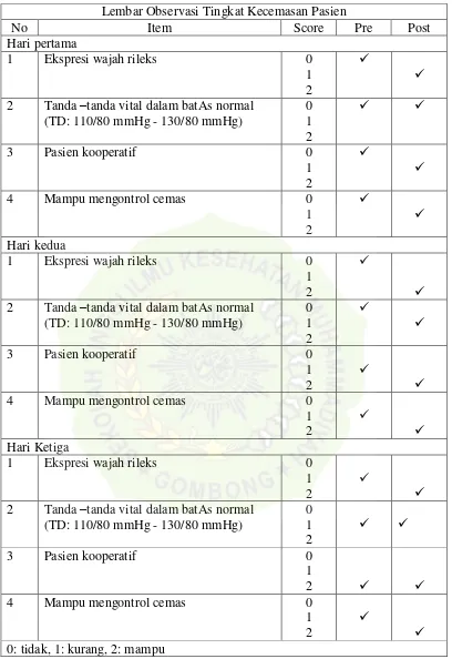
Gambar 4.1 : Tabel hasil observasi kecemasan pasien Lembar Observasi Tingkat Kecemasan Pasien
No Item Score Pre Post
2 Tanda –tanda vital dalam batAs normal (TD: 110/80 mmHg - 130/80 mmHg) 2 Tanda –tanda vital dalam batAs normal
(TD: 110/80 mmHg - 130/80 mmHg) 2 Tanda –tanda vital dalam batAs normal
INFORM K CONSENT
(PERSETUJUAN MENJADI PARTISIPAN)
Saya yang bertanda tangan di bawah ini menyatakan bahwa saya telah mendapat penjelasan secara rinci dan telah menegerti mengenai studi kasus yang akan dilakukan oleh Dwi Sudaryani dengan judul “ASUHAN KEPERAWATAN DENGAN PENERAPAN TEKNIK RELAKSASI (NAFAS DLAM) UNTUK MENURUNKAN KECEMASAN PADA IBU HAMIL DENGAN PEB DI RSUD KEBUMEN”.
Saya memutuskan setuju untuk ikut berpartisipasi pada penelitian secra sukarela tanpa paksaan. Bila selama penelitian saya menginginkan mengundurkan diri, maka saya dpat mengundurkan diri sewaktu – waktu tanpa sanksi.
Kebumen, 7 Juli 2017
Yang memberikan persetujuan
Saksi
... ... Kebumen, 7 Juli 2017
Mahasiswa
PENJELASAN UNTUK MENGIKUTI STUDI KASUS
1. Kami adalah Mahasiswa berasal dari STIKES Muhammadiyah Gombong, Program Studi DIII Keperawatan dengan ini meminta anda untuk berpartispiasi dengan sukarela dalam studi kasus yang berjudul “ASUHAN KEPERAWATAN DENGAN PENERAPAN TEKNIK
RELAKSASI (NAFAS DALAM) UNTUK MENURUNKAN
KECEMASAN PADA IBU HAMIL DENGAN PEB DI RSUD KEBUMEN”.
2. Tujuan dari studi kasus ini adalah memahami gambaran asuhan keperawatan dengan penerapan teknik relaksasi (nafas dalam) untuk menurunkan tingkat kecemasan pada ibu hamil dengan pre-eklampsia berat, yang dapat memberi manfaat berupa menjaga kesehtaan kehamilan serta mencegah dan menurunkan tingkat kecemasan pada ibu hamil dengan pre-eklampsia berat, studi kaus ini akan berlangsusng selama 3-4 hari
3. Prosedur pengambilan bahan data dengan cara wawancara terpimpin menggunakan pedoman wawancara yang akan berlangsung 15-20 menit. Cara ini mungkin menyebabkan ketidaknyamanan tetapi anda tidak perlu khawatir karena studi kasus ini untuk kepentingan pengembangan asuhan atau pelayanan keperawatan.
4. Keuntungan yang anda peroleh dalam keikutsertakan pada penelitian ini adalah anda turut terlibat aktif mengikuti perkembangan asuhan atau tindakan yang diberikan.
5. Nama jati diri anda seluruh informasi yang saudara sampaikan akan tetap dirahasiakan.
6. Jika saudara membutuhkan informasi sehubungan dengan penelitian ini, silahkan menghubungi peneliti pada nomor Hp: 082298063606.
Mahasiswa,
OBSTETRICS
Etiology and management of
postpartum
hypertension-preeclampsia
Baha M. Sibai, MD
Hypertensive disorders of pregnancy
are a major cause of maternal
mortality and morbidity, especially
in developing countries.1 Hypertension
may be present before or during
pregnancy or postpartum.2 Postpartum
hypertension can be related to
persistence of gestational hypertension
(GH), preeclampsia, or preexisting
chronic hypertension, or it could develop
de novo secondary to other
causes.3
During the past decades, there has
been extensive research regarding the incidence,
risk factors, pathogenesis, prediction,
prevention, and management of
GH-preeclampsia.4 However, patients
who were readmitted with postpartum
hypertension-preeclampsia were not
considered in reported studies.2,4 In addition,
the available data in the medical
literature have primarily focused on antenatal
and peripartum management of
such patients,4,5 even though some patients
can develop de novo eclampsia
and hemolysis, elevated liver enzymes,
and low platelet (HELLP) syndrome in
the late postpartum period.6-9 Thus,
there are few data regarding the evaluation,
management, and complications in
women who are rehospitalized with diagnosis
of postpartum hypertension.
3,10,11 Therefore, this report will focus
on the prevalence, etiology, and
who have de novo or persistent
is difficult to ascertain. In
clinical practice, most women will not
have their blood pressure (BP) checked
until the 6 weeks’ postpartum visit in physician’s offices or in postpartum clinics.
As a result, women with mild hypertension
who are asymptomatic are usually
not reported. In addition, postpartum
women who have hypertension in association
with symptoms such as headaches or
blurred vision are often seen and managed
in the emergency department and will not
be coded as hypertensive unless they are
hospitalized.
Research studies dealing with postpartum
hypertension are usually limited
by analysis of data from a single
center, focused on inpatients in the immediate
postpartum period (2-6 days),
or describing patients who were readmitted
because of preeclampsia-eclampsia,
severe hypertension, or complications
related to hypertension.12-17
Despite the limitations, the reported
prevalence of de novo postpartum hypertension
or preeclampsia ranges
from 0.3–27.5%.
Etiology and differential diagnosis
The etiology and different diagnosis of
postpartum hypertension is extensive
(Table), but it can be focused based on
clinical and laboratory findings as well as
(new onset or preexisting
prior to delivery) is the most common
cause, however, other life-threatening
conditions such as
pheochromocytoma
and cerebrovascular accidents should
also be considered.
New-onset postpartum
hypertension-preeclampsia
Normal pregnancy is characterized by
increased plasma volume in association
with sodium and water retention in the
interstitial tissue. This is further exaggerated
in women with multifetal gestation.
In addition, many women receive intravenously
a large volume of fluids during
labor, delivery, and postpartum. Large
volumes of fluids are also given because
of regional analgesia-anesthesia or during
cesarean section. In some women,
acute or delayed mobilization of large
From the Department of Obstetrics and
Gynecology, Division of Maternal-Fetal
Medicine, University of Cincinnati, Cincinnati,
Ohio.
Received June 14, 2011; revised Aug. 17,
2011; accepted Sept. 6, 2011.
The author reports no conflict of
Postpartum hypertension can be related to persistence of gestational hypertension, preeclampsia,
or preexisting chronic hypertension, or it could develop de novo postpartum
diagnosis, and management of postpartum hypertension-preeclampsia. The differential
diagnosis is extensive, and varies
cerebrovascular accidents. Therefore, medical providers caring for postpartum women
should be educated about continued monitoring of signs and symptoms and prompt
management of these women in a timely fashion. Evaluation and management should be
performed in a stepwise fashion and may require a multidisciplinary approach that considers
predelivery risk factors, time of onset, associated signs/symptoms, and results of
selective laboratory and imaging findings. The objective of this review is to increase
awareness and to provide a stepwise approach toward the diagnosis and management of
women with persistent and/or new-onset hypertension-preeclampsia postpartum period.
Key words: etiology, management, postpartum hypertension-preeclampsia
Clinical Opinion www.AJOG.org
470 American Journal of
Obstetrics & Gynecology JUNE 2012
volume of fluid into the intravascular
space, particularly in association with
suboptimal renal function, can lead to a
state of volume overload resulting in
hypertension.11,13
Some medications that cause vasoconstriction
are often used for pain relief,
in women having perineal lacerations,
episiotomy, or cesarean delivery. Such
women usually require large doses of
nonsteroidal antiinflammatory drugs
such as ibuprofen or indomethacin that
are associated with vasoconstriction and
which can result in severe hypertension.
18 In addition, some women receive
frequent injections of ergot alkaloids (ergometrine
or methylergonovine) for
treatment of uterine atony. The action of
these medications is mediated through
alpha adrenergic receptors, which can
lead to peripheral vasoconstriction with
resultant hypertension or aggravation of
hypertension, cerebral vasoconstriction,
and stroke.19 These medications are also
associated with nausea, vomiting, and
headaches, symptoms that are similar to
those in severe GH-preeclampsia.
Persistence/exacerbation of
hypertension-proteinuria in
women
with preexisting GH-preeclampsia
Maternal hypertension and proteinuria
will usually resolve during the first week
postpartum in most women with GH or
preeclampsia, however, there are conflicting
data regarding the time it takes
for resolution in such women.20-25 The
differences among various studies are
due to the population studied, severity of
disease process (mild, severe, with superimposed
preeclampsia, HELLP syndrome),
duration of follow-up, management
(aggressive vs expectant), and
criteria used for hypertension or proteinuria.
21-25 In women with preeclampsia,
there is a decrease in BP within 48
hours, but BP increases again between
cerebral manifestations and/or deterioration
in maternal laboratory findings will
manifest for the first time postpartum
leading to the development of eclampsia
and/or HELLP syndrome.6,8,26-31
Persistence/exacerbation of
hypertension in chronic
hypertension
Women with chronic hypertension during
pregnancy are at increased risk for
exacerbation of hypertension and/or superimposed
preeclampsia.32 The risk depends
on severity of hypertension, presence
of associated medical conditions
(obesity, type 2 diabetes, renal disease),
or whether antihypertensive medications
were used during pregnancy.32,33
Hypertension or exacerbation of hypertension
postpartum may be due to either
undiagnosed essential chronic hypertension
(women with limited medical care
prior to or early in pregnancy), or due to
exacerbation of hypertension after delivery
in those with superimposed preeclampsia.
Two studies in patients with superimposed
preeclampsia suggest that systolic
and diastolic BP increases at 3-6 days
postpartum in such women.17,33
TABLE
Etiology/differential diagnosis of postpartum hypertension
Etiology Key findings to consider
New-onset hypertension-preeclampsia Onset 3-6 d postpartum without headaches fluids, regional analgesia, delayed mobilization
... ... ... ...
Medications/drugs Nonsteroidal analgesics, ergot derivatives
...
Ibuprofen, indomethacin Peripheral and cerebral vasoconstriction, headaches
Phenylpropanolamine, ephedrine Peripheral and cerebral vasoconstriction, headaches
Ergotamine, ergonovine Vasoconstriction, headaches, nausea, vomiting, seizures
Persistence of GH-preeclampsia Preexisting condition antepartum/in labor
Late-onset eclampsia Headaches, visual changes, seizures, absent neurologic deficits
HELLP syndrome Nausea/vomiting, epigastric pain, low platelets, increased liver enzymes
Preexisting/undiagnosed
hypertension Hypertension prior to pregnancy, or _20 wk
...
Preexisting renal disease Proteinuria or hematuria _20 wk
...
Hyperthyroidism Palpitations tachycardia, sweating, dry skin, heart failure
Primary hyperaldosteronism Refractory hypertension, hypokalemia, metabolic alkalosis
...
Pheochromocytoma Paroxysmal hypertension, headaches, chest pain, hyperglycemia
Renal artery stenosis Hypertension that is refractory to treatment
...
Cerebral vasoconstriction syndrome Sudden thunderclap headaches, visual changes, neurologic deficits
...
Cerebral venous thrombosis/stroke Onset 3-7 d, gradual or acute headaches, seizures, neurologic deficits
... ... ...
TTP/hemolytic uremic syndrome
Hemolysis, severe
thrombocytopenia, neurologic symptoms, normal liver enzymes
...
GH, gestational hypertension;
HELLP, hemolysis, elevated liver enzymes, and low platelet; TTP, thrombotic thrombocytopenic purpura.
Sibai. Postpartum hypertension-preeclampsia. Am J Obstet Gynecol 2012.
www.AJOG.org Obstetrics Clinical Opinion
JUNE 2012 American Journal of Obstetrics & Gynecology 471
Postpartum hypertension or preeclampsia
can also be secondary to_1 of
the underlying medical disorders listed
in the Table.11,17,34-47
Maternal complications
Maternal complications depend on _1
of the following: severity and etiology of
the hypertension, maternal status at presentation
(presence of organ dysfunction),
and the quality of management
used. Potential life-threatening complications
include cerebral infarction
or hemorrhage, congestive heart failure
or pulmonary edema, renal failure, or
death. Maternal outcome is usually
pheochromocytoma,38,39 stroke, thrombotic
thrombocytopenic purpura/hemolytic
uremic syndrome,44,45 and with
delayed diagnosis and inadequate control
of persistent severe hypertension.
Evaluation and management of
Evaluation of patients with postpartum
hypertension should be performed in a
stepwise fashion and may require a multidisciplinary
approach. Consequently,
management requires a well-formulated
plan that takes the following factors into
consideration: predelivery risk factors,
time of onset in relation to delivery, presence
of signs/symptoms, results of laboratory/
imaging findings, and response to
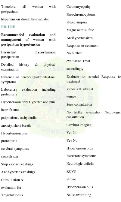
initial therapy (Figure).
The most common cause for persistent
hypertension beyond 48 hours after
delivery is GH, preeclampsia, or essential
chronic hypertension (either preexisting
prior to delivery or developing de novo).
Initial management will depend on their
history, clinical findings, presence or absence
of associated symptoms, results of
laboratory findings (urine protein, platelet
count, liver enzymes, serum creatinine,
and electrolytes), and response to
treatment of hypertension.
There are several medications that are
frequently prescribed in the postpartum
period such as ibuprofen, ergonovine,
and anticongestants. Use of large or frequent
doses of these agents can aggravate
preexisting hypertension or results in
new-onset hypertension.18,19 The use of
these drugs is also associated with cerebral
symptoms, nausea, and vomiting.
Many physicians and consultants are not
Therefore, all women with postpartum
hypertension should be evaluated
FIGURE
Recommended evaluation and
management of women with
postpartum hypertension
Persistent hypertension
postpartum
Detailed history & physical examination
Presence of cerebral/gastrointestinal symptoms
Laboratory evaluation including proteinuria
Hypertension only Hypertension plus
heart failure
palpitations, tachycardia
anxiety, short breath
Hypertension plus
proteinuria
cerebral symptoms
convulsions
Stop vasoactive drugs
Antihypertensive drugs
Response to treatment
No further
evaluation Treat
accordingly
Evaluate for arterial Response to treatment
stenosis & adrenal
tumors
Seek consultation
Epigastric pain
Elevated liver enzymes
Low platelets
HELLP syndrome
Magnesium sulfate
Antihypertensives
Supportive care
Response to treatment
No further
AFLP, acute fatty liver of pregnancy;
APAS, antiphospholipid antibody syndrome; HELLP, hemolysis, elevated liver enzymes, and low platelet; HUS, hemolytic uremic syndrome; RCVS, reversible cerebral vasoconstriction syndrome; TTP, thrombotic thrombocytopenic purpura.
Sibai. Postpartum hypertension-preeclampsia. Am J Obstet Gynecol 2012.
Clinical Opinion Obstetrics
www.AJOG.org
472 American Journal of
Obstetrics & Gynecology JUNE 2012
in regards to receiving these medications,
and discontinued if they are being used.
Subsequent management includes control
of hypertension and close observation until
resolution of hypertension and associated
symptoms.18,19
If the patient has hypertension only
with absent symptoms, no proteinuria,
and normal laboratory findings, the next
step is to control BP. Antihypertensive
medications are recommended if systolic
BP remains persistently _150 mm Hg
Hg.3,10,17 Bolus intravenous injections of
either labetalol or hydralazine are used
initially if there is persistent elevations in
BP to levels _160 mm Hg systolic or
_110 mm Hg diastolic; this is subsequently
followed by oral medication to
keep systolic BP _150 mm Hg and diastolic
BP_100mmHg. There are several
antihypertensive drugs to treat postpartum
hypertension.10,11,48 In GH-preeclampsia,
I recommend short-acting oral
nifedipine (10-20 mg every 4-6 hours) or
long-acting nifedipine XL (10-30mgevery
12 hours). Alternatively, one can use oral
labetalol 200-400 mg every 8-12 hours. As
comparedto labetalol, oral nifedipine is associated
with improved renal blood flow
with resultant diuresis, which makes it the
drug of choice in postpartum women with
volume overload.48 In some, it is necessary
to switch to a new agent such as angiotensin-
converting enzyme inhibitor, which is
the drug of choice in those with pregestational
diabetes mellitus or cardiomyopathy.
In addition, thiazide or loop diuretics
may be needed in women with
to add potassium supplementation.
Antihypertensive agents such as methyldopa,
hydrochlorothiazide, furosemide,
captopril, propranolol, and enalapril are
compatible with breast-feeding.11,49 If the
symptoms, the patient is then discharged
home with instructions for daily
BP measurements (self or by a
medications are then discontinued if the
doses of antihypertensive medications
require evaluation for the presence
of either renal artery stenosis or primary
hyperaldosteronism. In most women
with hyperaldosteronism, the elevated
progesterone levels act like spironolactone
reversing the hypokalemia and the
hypertension as well, with rapid exacerbations
of hypertension and falling potassium
levels in the postpartum period.
The diagnosis should be suspected in the
presence of hypokalemia (serum potassium
levels _3.0 mEq/L) in association
with metabolic acidosis, and then confirmed
by either computed tomography
(CT) or magnetic resonance imaging
(MRI) of the abdomen revealing presence
of adrenal tumor.17,38 Evaluation
and management should be made in
consultation with a nephrologist.17
Women presenting with hypertension
in association with shortness of breath,
should be evaluated for possible pulmonary
edema and/or postpartum cardiomyopathy,
hyperthyroidism, or
pheochromocytoma.
36,37,39,50,51 Indeed, 23-46% of
women with peripartum cardiomyopathy
will have associated
hypertension.50,51
Such women should receive a chest x-ray
and echocardiography and then be managed
in association with a cardiologist according
to the demonstrated etiology.50,51
Patients with Graves’ disease during
pregnancy can develop exacerbation of
the hyperthyroidism in the postpartum
period.36,37 In addition, new-onset hyperthyroidism
postpartum can be due to
the hyperthyroid phase of postpartum
thyroiditis (first 1-2 months postpartum).
37 The hypertension in hyperthyroidism
is mainly systolic, and is associated
with wide pulse pressure, tachycardia,
palpitations, and heat intolerance.36,37
Womenwith these findings should receive
thyroid function tests (thyroid stimulating
hormone and free thyroid 4 levels) and
then be managed in consultation with an
endocrinologist.Womenwith Graves’ disease
are treated with prophythiouracil
(100-300mgdaily) or methimazole (10-20
mg daily), and then followed with measurements
of thyroid stimulating hormone
and free thyroid 4 levels. Both of
these medications are compatible with
breast-feeding.36 Women with hyperthyroid
not require antithyroid drugs since the condition
resolves spontaneously.
Pheochromocytoma is a rare adrenal
or extraadrenal tumor that produces catecholamines
resulting in paroxysmal hypertension,
headaches, palpitations and
excessive sweating, chest pain, dizziness,
and postural hypotension.38,39 Maternal
of 24-hour urine epinephrine,
norepinephrine, and their metabolites
(metanephrine and normetanephrine)
and is then confirmed byCTscan orMRI
of the abdomen. Management of pheochromocytoma
should be made in consultation
with a nephrologist and a surgeon
and will include initially medical
therapy with alpha blockers followed
by surgical removal of the adrenal
tumor.38,39
Women with postpartum hypertension
in association with new-onset persistent
headaches and/or visual changes
or new-onset proteinuria should be considered
to have severe preeclampsia. If
there is hypertension with seizure, she
should be initially treated as having eclampsia.
It is important to emphasize
that many of these women will be first
seen and evaluated in the emergency department,
and many emergency room
physicians may not be aware that preeclampsia-
eclampsia can present postpartum.
8,16 In patients presenting with
these findings, magnesium sulfate therapy
prophylaxis and/or treatment. In
addition, intravenous
antihypertensive
medications are recommended to lower
BP to the desired goal while considering
an alternative cause for the cerebral
symptoms. Magnesium sulfate is given
24 hours.2 If the patient continues to
have cerebral symptoms and/or if she develops
seizures or neurologic deficits despite
magnesium sulfate and adequate
www.AJOG.org Obstetrics Clinical Opinion
JUNE 2012 American Journal of Obstetrics & Gynecology 473
BP control, then she should receive neurodiagnostic
evaluation and be managed
in consultation with a neurologist.
Women presenting with hypertension
in association with refractory and/or
thunderclap headaches, visual disturbances,
or neurologic deficits should be
evaluated for possible cerebrovascular
complications such as reversible cerebral
vasoconstriction syndrome (RCVS) or
stroke. These women will require selective
diagnostic neuroimaging and consultation
with neurology and/or neurosurgery. Such
an evaluation may include CT scan for
hemorrhage, MRI for detection of vasogenic
edema and/or ischemia or infarction,
cerebral angiography for diagnosis of
RCVS, and cerebral venography for detection
of cerebral venous thrombosis (CVT).
etiology.19,41,42,44,45
RCVS is a poorly understood form of
angiopathy that develops between postpartum
days 3-14.40,41 The presenting
symptoms are a thunderclap headache
(89%) in association with other neurologic
manifestations such as seizures and
visual disturbances. Hypertension is
present in 60% of cases, and multifocal
neurologic deficits may be present.40
MRI findings in this syndrome overlap
with those of eclampsia (posterior reversible
encephalopathy syndrome),
however, cerebral MRI or traditional angiography
reveal the presence of segmental
vasoconstriction.41 These latter
findings are consistently absent in eclampsia.
40-42 Prognosis is favorable in
most cases, however, if the vasoconstriction
is severe and persistent or recurrent,
it can lead to cerebral hemorrhage or infarction
with permanent neurologic deficits.
40-42 In some patients, serum creatinine
may be extremely low suggesting
massive volume overload as a cause and
thus rapid diuresis with diuretics is beneficial
in such cases.52 Additional therapy
may include the use of a calcium
Reported risk factors for postpartum
stroke include hypertension, advanced
maternal age, and dehydration.43-45 Potential
causes of stroke include CVT, aneurysmal
subarachnoid hemorrhage, intraparenchymal
encephalopathy.43-45 Cerebral hemorrhage
and CVT secondary to major dural
sinus thrombosis can lead to increased intracranial
pressure with compensatory peripheral
vascular hypertension. In addition,
the signs and symptoms of stroke
(headache, visual changes, seizures, nausea,
eclampsia, and HELLP syndrome.
45 The definitive diagnosis is made
by cerebralMRIand/or angiography (both
arterial and venous).43-45
Women with hypertension with persistent
nausea, vomiting, or epigastric
pain should be evaluated for HELLP syndrome
since up to 30% who develop the
syndrome do so postpartum.6 The time
of onset of the clinical and laboratory
findings ranges from 1-7 days postpartum.
6 Management of these women is
similar to that before delivery, which includes
the use of magnesium sulfate, antihypertensives,
and close monitoring of
vital signs and laboratory values.6 In general,
patients with HELLP syndrome will
demonstrate an improvement in clinical
and laboratory findings within 48 hours
after treatment. If there is either no improvement
or a deterioration in these
findings, then it is important to obtain
consultation with appropriate specialists
for evaluation and subsequent management.
include thrombotic thrombocytopenic
purpura, hemolytic uremic syndrome,
acute fatty liver of pregnancy, and exacerbation
of lupus nephritis.6,46,47
In summary, there are several causes
for postpartum hypertension; some may
be benign (mild GH or mild chronic hypertension)
whereas others can be life
threatening such as severe preeclampsia
or stroke. Therefore, a high index of suspicion
for secondary dangerous causes
of hypertension should be considered
when evaluating such women. By directing
efforts and educating health care
providers about the continued monitoring,
reporting, and prompt evaluation of
symptoms in the postpartum period, it is
expected that some of the maternal complications
will be avoided. Evaluation
and management of women with postpartum
hypertension should be guided
by obtaining a detailed history, careful
physical examination, selective laboratory
and imaging studies, and response
to initial treatment. f
REFERENCES
1. Goldenberg RL, McClue EM, MacGuire ER,
Kamath BD, Jobe AH. Lessons for low-income
regions following the reduction in hypertensionrelated
maternal mortality in high-income countries.
Int J Gynecol Obstet 2011;113:91-5.
2. Sibai BM. Diagnosis and management of
gestational hypertension and preeclampsia.
Obstet Gynecol 2003;102:181-92.
3. Tan L, de Swiet M. The management of postpartum
4. Report of the national high blood pressure
education program working group on high
blood pressure in pregnancy. Am J Obstet Gynecol
2000;183:S1-22.
5. American College of Obstetricians and Gynecologists.
ACOG practice bulletin no. 33: diagnosis
and management of preeclampsia and
eclampsia. Washington, DC: The College; 2002.
6. Sibai BM. Diagnosis and management of
HELLP syndrome. Obstet Gynecol 2004;105:
402-10.
7. Sibai BM. Diagnosis, prevention and management
of eclampsia. Obstet Gynecol 2005;
105:402-10.
8. Chames MC, Livingston JC, Ivester TS, Barton
JR, Sibai BM. Late postpartum
of atypical preeclampsia-eclampsia. Am J
Obstet Gynecol 2009;200:481.e1-7.
10. Magee L, Sadeghi S. Prevention and treatment
of postpartum hypertension. Cochrane
Database Syst Rev
2005;1:CD004351.
11. Ghuman N, Rhiener J, Tendler BE, White
WB. Hypertension in the postpartum woman:
clinical update for the hypertension specialist.
J Clin Hypertens (Greenwich) 2009;11:726-33.
12. Piver MS, Corson SL, Bolognese RJ. Hypertension
6 weeks’ postpartum in apparently
Swiet M. Blood pressure in the puerperium. Clin
Sci (Colch) 1986;71:589-94.
14. Attrebury JL, Groome LJ, Hoff C. Blood
pressure changes in normotensive women readmitted
to the postpartum period with severe
preeclampsia/eclampsia. J Matern Fetal Med
1996;5:201-5.
15. Clark SL, Belfort MA, Dildy GA, et al. Emergency
department use during the postpartum
period: implications for current management of
Clinical Opinion Obstetrics
www.AJOG.org
474 American Journal of
Obstetrics & Gynecology JUNE 2012
eclampsia: an experience of 151 cases. Am J
Obstet Gynecol 2004;190:1464-6.
17. Podymow T, Aujgust P. Postpartum course
of gestational hypertension and preeclampsia.
Hypertens Pregnancy 2010;29:294-300.
18. Makris A, Thornton C, Hennessy A. Postpartum
hypertension and nonsteroidal analgesia.
Am J Obstet Gynecol 2004;190:577-8.
19. Singhal AB, Bernstein RA. Postpartum angiopathy
and other cerebral vasoconstriction
syndromes. Neurocrit Care 2005;3:91-7.
20. Walters BN, Walters T. Hypertension in the
puerperium. Lancet 1987;2:330.
21. Ferrazzani S, DeCardis S, Pomini F, et al.
The duration of hypertension in the puerperium
impairment and week of delivery. Am J Obstet
Gynecol 1994;171:506-12.
22. Stepan H, Nordmeyer A, Faber R. Proteinuria
in hypertensive pregnancy diseases is associated
importance of urinary protein excretion during
conservative management of severe preeclampsia.
Am J Obstet Gynecol 1996;175:
1313-6.
24. Berks D, Steegers EAP, Molas M, Visser W.
Resolution of hypertension and proteinuria after
preeclampsia. Obstet Gynecol 2009;114:
experience with eclampsia in Singapore. Singapore
Med J 2003;44:88-93.
27. Knight M, on behalf of UKOSS. Eclampsia
in the United Kingdom 2005. BJOG 2007;114:
1072-8.
28. Conde-Agudelo A, Kafury-Goeta AC. Epidemiology
of eclampsia in Columbia. Int J
Gynaecol Obstet 1998;61:1-8.
29. Katz V, Farmer R, Kuller JA. Preeclampsia
into eclampsia: toward a new paradigm. Am J
Obstet Gynecol 2000;182:1389-96.
30. Mattar F, Sibai BM. Eclampsia VIII; risk factors
for maternal morbidity. Am J Obstet Gynecol
31. Martin JN Jr, Rose CH, Briery CM. Understanding
and managing HELLP syndrome: the
integral role of aggressive glucocorticoids for
Obstet Gynecol 2002;100:369-77.
33. Peterson E, Craigo S, House M. Risk factors
for postpartum antihypertensive medication
requirement in severe preeclampsia. Hypertens
in a tertiary center. Clin Rev Allergy Immunol
2010;38:77-81.
35. Smyth A, Oliveira GHM, Lahr BD, et al. A
systemic review and meta-analysis of pregnancy
outcomes in patients with systemic lupus
erythematosus and lupus nephritis. Clin J Am
Soc Nephrol 2010;11:1060-8.
36. Patil-Sioda K, Mestman JH. Graves hyperthyroidism
and pregnancy: a clinical update.
Endocr Pract 2010;16:118-29.
37. Girling J, Cotzias C. Thyroid and other endocrine
disorders in pregnancy. Obstet Gynaecol
Reprod Med 2007;17:349-55.
38. Nezu M, Miwa Y, Noshire T, Inoue M. Primary
aldosteronism as a cause of severe
hypertension from
pheochromocytoma: a
rare and challenging entity. Am J Hypertens
40. Sattar A, Manousakis G, Jensen MB. Systematic
review of reversible cerebral vasoconstriction
syndrome. Expert Rev Cardiovasc Ther
postpartum angiopathy. Neurocrit Care 2009;
Gynecol Obstet 2011;283:663-8.
43. Lanska DJ, Kryscio RJ. Risk factors for peripartum
and postpartum stroke and intracranial
venous thrombosis. Stroke 2000;31:1274-82.
44. Treadwell SD, Thanui B, Robinson TG.
Stroke in pregnancy and the puerperium. Postgrad
Med J 2008;84:238-45.
45. Sibai BM, Kristin H, Coppage KH. Diagnosis
and management of women with stroke
during pregnancy–postpartum. Clin Perinatol
2004;31:853-68.
46. Sibai BM. Imitators of severe pre-eclampsia.
Semin Perinatol 2009;33:196-205.
47. Martin JN, Bailey AP, Rehberg JF, et al.
Thrombotic thrombocytopenic purpura in 166
pregnancies: 1955-2006. Am J Obstet Gynecol
2008;199:98-104.
48. Vermillion ST, Scardo JA, Newman RB,
Chauhan SP. A randomized, double-blind trial
of oral nifedipine and intravenous labetalol in
hypertensive emergencies of pregnancy. Am J
Obstet Gynecol 1999;181:858-61.
49. Podymow T, August P. Antihypertensive
70-85.
50. El Kayam U, Akhter MW, Singh H, et al.
Pregnancy associated
cardiomyopathy; clinical
characteristics and a comparison between
early and late presentation. Circulation 2005;
111:2050-5.
51. Habli M, O’Brien T, Nowack E,
et al. Peripartum
cardiomyopathy: prognostic factors for
long-term maternal outcome. Am J Obstet Gynecol
2008;199:415.e1-5.
52. Hinchey J, Chaves C, Appignani B, et al. A
reversible posterior
leukoencephalopathy syndrome.
N Engl J Med 1996;334:494-500.
www.AJOG.org Obstetrics Clinical Opinion
17 3
PENGARUH RELAKSASI NAPAS DALAM TERHADAP PENURUNAN TEKANAN DARAH PADA PASIEN HIPERTENSI DI PUSKESMAS
DASAN AGUNG MATARAM
Aan Dwi Sentana , Mardiatun1 1 1
Jurusan Keperawatan Poltekes kemenkes Mataram
Abstrak
Pemberian teknik relaksasi nafas dalam untuk menurunkan tekanan darah, berarti telah memberikan penanganan alternatif pada pasien secara non farmakologi selain pengkonsumsian obat-obatan medika-mentosa dan dapat memberikan pengajaran pada pasien dalam mengatasi tekanan darah tinggi pada pasien hipertensi. Tujuan umum penelitian ini adalah Menganalisis pengaruh teknik relaksasi napas dalam terhadap penurunan tekanan darah pada pasien hipertensi. Tujuan Khusus adalah mengidentifikasi tekanan darah pasien sebelum dilakukan teknik relaksasi napas dalam pada pasien hipertensi, mengidentifikasi tekanan darah pasien sesudah dilakukan teknik relaksasi napas dalam pada pasien hipertensi, membuktikan pengaruh teknik relaksasi napas dalam terhadap penurunan tekanan darah pada pasien hipertensi. Desain penelitian Quasi eksperiment : pretest-posttest control group design. Satu kelompok perlakuan terdiri dari 16 orang diberikan relaksasi napas dalam dalam selama 8 hari, satu kelompok yang terdiri dari 16 orang sebagai kontrol. Sampel dipilih dengan cara
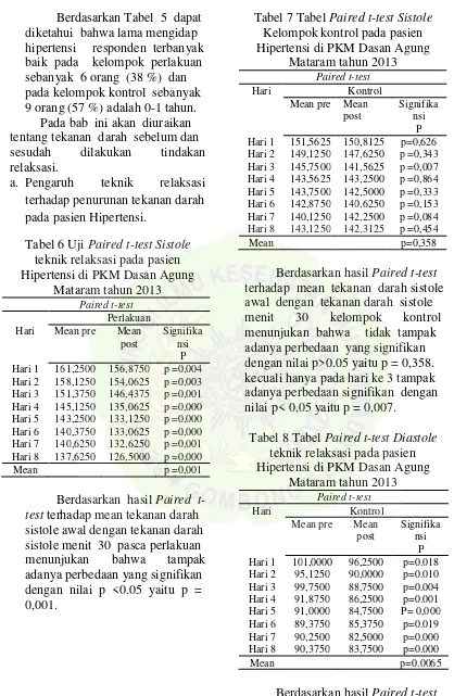
consecutive sampling. Pengumpulan data dilakukan dengan mengukur tekanan darah sebelum dan setelah diberikan intervensi. Uji statistik yang digunakan adalah Paired T-Test. Hasil penelitian menunjukkan ada perubahan yang signifikan pada tekanan darah sistole sebelum dan sesudah dilakukan teknik relaksasi pada kelompok perlakuan dengan nilai p < 0,05 yaitu p = 0,001 dibandingkan dengan kelompok kontrol dengan nilai p > 0,05 yaitu p = 0,358 dan ada perubahan yang signifikan pada tekanan darah diastole sebelum dan sesudah perlakuan dengan nilai p < 0,05 yaitu p=0.0065 dibandingkan dengan p > 0,05 yaitu p = 0,44375 Hasil penelitian ini menunjukkan bahwa relaksasi napas dalam dapat menurukan tekanan darah baik sistole dan diastole karena pada keadaan relaksasi mengakibatkan penurunan rangsangan emosional dan penurunan pada rangsangan pada area pengatur fungsi kardiovaskular seperti pada hipothalamus posterior dan nukleus perifornikel sehingga terjadi penurunan tekanan darah. Mengingat besarnya pengaruh teknik relaksasi maka dianjurkan pasien hipertensi agar petugas kesehatan mengajarkan teknik relaksasi. Hal tersebut untuk membantu untuk mengontrol tekanan darah sehingga membantu mempercepat kesembuhan dan menurukan biaya perawatan.
Kata Kunci: Hipertensi, Tekanan darah, Teknik relaksasi
Abstract
1 7 4
nerve activity of simpatis so that cause venous vasodilatation so that situation become relax become degradation of blood pressure.
Target of this research is to Analyse influence of technique of relaxation to degradation of blood pressure at hypertension patient in Publick Health Dasan
Agung Mataram. Design in research is Quasi experiment ( pre test and post test control group design). Big of sample 32 responder, taken pursuant to criterion of inclution. used by Sampling technique is cosecutive. Independent variable of this research is technique of relaxation and variable its him is degradation of blood pressure. Data collecting use blood pressure observation. Data analysis use statistical test of t-test paired.
Result showed that indicated the existence influence of technique relaxation to degradation of blood pressure at hypertension patient, test blood pressure meaning value paired t-test of systole p=0.001 for group intervention and p = 0,358 for group control., blood pressure of diastole p=0,0065 for group intervention and p = 0,44375 for group control. Technique of relaxation proven can degrade good blood pressure systole and diastole. At clinic test at hypertension patient, they
who conduct tardy exhalation during 15 minute per its day in the reality can degrade blood pressure 10-15 poin.
Keywords: Hypertension, Blood pressure, Technique of relaxation
Pendahuluan
Hipertensi merupakan faktor resiko, primer yang menyebabkan penyakit jantung dan stroke. Pada umumnya penderita hipertensi hampir tidak merasa dirinya sakit, namun hipertensi merupakan penyakit yang berbahaya karena organ tubuh terganggu di satu bidang yang amat penting yaitu peredaran darah. Hipertensi bahkan sering terabaikan karena tidak ada keluhan dan bila sudah mengeluh biasanya terlambat sehingga hipertensi sering disebut sebagai silent killer (Beevers, D.G., 2002)).
Sebenarnya hipertensi dapat dikontrol bila faktor resiko hipertensi mampu dikendalikan. Pengendalian ini meliputi upaya pemeliharaan kesehatan oleh petugas dan pemeliharaan kesehatan mandiri oleh individu yang bersangkutan. Upaya pengendalian ini melalui perawatan diri hipertensi meliputi: meminum
obat sesuai anjuran, memantau tekanan darah dan melakukan perubahan pola hidup (seperti olah raga, mengurangi konsumsi garam dan meningkatkan konsumsi buah dan sayuran) (Viera & Jamieson, 2007). Disisi lain perawat berperan besar dalam penanggulangan hipertensi melalui pendekatan non farmakologi. Intervensi yang termasuk dalam pendekatan non farmakologis salah satunya adalah dengan teknik relaksasi nafas dalam, namun ditatanan pelayanan intervensi ini jarang dilakukan atau mungkin tidak pernah dilakukan.
17 5 sudah melakukan diet yang benar,
berolah raga, keteraturan minum obat dan pengontrolan tekanan darah secara rutin serta 12 pasien hipertensi (100 %) tidak pernah melakukan teknik relaksasi napas dalam. Disamping itu juga kasus hipertensi berdasarkan rekam medis di PKM Dasan Agung juga mengalami peningkatan kasus serta urutan 10 penyakit terbanyak juga mengalami peningkatan tingkatan. Jumlah kasus hipertensi tahun 2010 sebanyak 1828 penderita dan urutan 8 dari sepuluh besar penyakit terbanyak, sedangkan tahun 2011 kasus hipertensi sebanyak 2336 dan urutan ke 7 dari sepuluh besar penyakit terbanyak.
Dampak yang ditimbulkan oleh hipertensi dapat menyebabkan frustasi, konflik dan krisis karena penyakit yang tidak segera sembuh. Stres dapat menimbulkan perubahan- perubahan yang meliputi tekanan darah yang meninggi, denyut jantung yang lebih cepat, irama pernafasan yang lebih cepat, karena metabolisme atau tingkat pembakaran dalam tubuh yang dipercepat dan aliran darah ke dalam otot lengan dan kaki meningkat (Benson, 2004).
Pada penderita hipertensi sudah terjadi peningkatan respon saraf simpatis, kemudian terjadi pengeluaran norepinefrin pada ujung saraf yang dapat meningkatkan frekuensi denyut jantung dan vasokonstriksi perifer yang selanjutnya menimbulkan peningkatan tekanan darah. Pada keadaan stress respon saraf simpatis meningkat sehingga peningkatan aktivitas saraf simpatis ini juga mengeluarkan norepinefrin yang dapat meningkatkan tekanan darah secara intermitten.
Aktivitas relaksasi bernafas yang dalam secara rutin akan dapat membantu mengatur tekanan darah. Para ahli menyimpulkan dengan melakukan pernafasan yang lambat selama beberapa menit sehari, sudah cukup dapat membantu seseorang menurunkan tekanan darahnya. Penanganan melalui metode farmakologi seperti penggunaan obat-obatan di antaranya jenis-jenis obat diuretik, _ EORFNHU, FDOFLXP channel blockers, atau angiotensin converting enzym inhibitor dapat menimbulkan berbagai efek samping yaitu menaikan kadar acidum uricum
dalam darah, hiperkalemi, meningkatkan pacuan terhadap susunan saraf simpatis sehingga mengakibatkan naiknya curah jantung dan bertambahnya denyut jantung (Benson, 2004).
Teknik relaksasi dipercaya dapat menurunkan tekanan darah. Ada tiga hal utama yang diperlukan dalam relaksasi yaitu posisi yang tepat, pikiran beristirahat, lingkungan yang tenang. (Priharjo, 1996). Konsep dasar pada relaksasi pada hakekatnya merupakan cara relaksasi yang diperlukan untuk menurunkan ketegangan otot yang dapat memperbaiki denyut nadi, tekanan darah dan pernafasan.( Martha D, 1995)
1 7 6 pengganti diet, olahraga ataupun obat-obatan. Bernafas dalam dan lambat merupakan tindakan relaksasi sehingga pembuluh darah mengalami dilatasi (William F.G, 2003).
Relaksasi merupakan pengaktifan dari saraf parasimpatis yang menstimulasi turunnya semua fungsi yang dinaikkan oleh sistem saraf simpatis, dan menstimulasi naiknya semua fungsi yang diturunkan oleh saraf simpatis. Masing-masing saraf parasimpatis dan simpatis saling berpengaruh maka dengan bertambahnya salah satu aktivitas sistem yang satu akan menghambat atau menekan fungsi yang lain (Utami, 1993). Latihan yang teratur melancarkan peredaran darah kemudian tekanan darah menjadi normal. Seperti diketahui, pembuluh darah yang tersumbat membuat tekanan darah meninggi, sehingga berisiko gangguan jantung. Adapun Pemberian teknik relaksasi nafas dalam untuk menurunkan tekanan darah, berarti telah memberikan penanganan alternatif pada pasien secara non farmakologi selain pengkonsumsian obat-obatan medika-mentosa dan dapat memberikan pengajaran pada pasien dalam mengatasi tekanan darah tinggi pada pasien hipertensi.
Berdasarkan latar belakang dan fenomena di atas, peneliti tertarik untuk melakukan penelitian tentang pengaruh pemberian teknik relaksasi nafas dalam terhadap tekanan darah pada pasien hipertensi
Metode
Dalam penelitian ini digunakan jenis penelitian Quasi eksperiment
dengan rancangan pretest posttest control group design. Pada rancangan ini terdapat kelompok kontrol. Sampel pada penelitian ini
sebesar 32 responden dengan rincian 16 orang kelompok kontrol dan 16 orang kelompok perlakuan diberikan relaksasi napas dalam yang diperoleh secara consecutive sampling.
Pengumpulan data
menggunakan instrument yang berupa observasi (pre test dan post test). Pre test dilakukan untuk mengetahui tekanan darah pada pasien yang berobat ke Puskesmas Dasan Agung. Kemudian peneliti memberikan intervensi teknik relaksasi nafas selama 15 menit. Setelah diberikan intervensi kemudian dilakukan post test berupa pengukuran tekanan darah pada klien yang dilakukan setiap hari selama 8 hari Kemudian semua data yang terkumpul di catat dalam lembar pengumpulan data.
Hasil
Gambaran umum responden pada penelitian ini, dilihat berdasarkan umur dan jenis kelamin. Adapun distribusi responden berdasarkan umur dan jenis kelamin adalah sebagai berikut:
a. Jenis kelamin
Distribusi Responden Berdasarkan Jenis kelamin
Tabel 1 Distribusi Responden Berdasarkan Jenis kelamin di PKM Dasan Agung Mataram Tahun 2013.
17 7 sebanyak 11 orang (68 %) adalah jenis kelamin perempuan.
b. Distribusi responden berdasarkan Pendidikan
Tabel 2 Distribusi Responden Berdasarkan Pendidikan di PKM
Dasan Agung Mataram Tahun 2013.
N o
Pendidikan Kelompok Perlakuan diketahui bahwa pendidikan responden terbanyak pada kelompok perlakuan sebanyak 5 orang (31%) adalah tidak bersekolah dan pada kelompok kontrol sebanyak 6 orang ( 38 %) adalah pendidikan SD
c. Umur
Pengelompokan data berdasarkan umur responden dapat dilihat pada tabel berikut :
Tabel 3 Distribusi Responden Berdasarkan Umur di PKM Dasan
Agung Mataram Tahun 2013.
N o
Usia Kelompok Perlakuan
Berdasarkan Tabel 3 dapat diketahui bahwa kelompok umur 56- 74 tahun, responden terbanyak pada kelompok perlakuan
sebanyak 11 orang (69 %) dan kelompok kontrol kelompok umur 45-55 tahun sebanyak 10 orang (63 %).
d. Jenis pekerjaan
Pengelompokan data berdasarkan jenis pekerjaan dapat dilihat pada tabel berikut :
Tabel 4 Distribusi Responden Berdasarkan Pekerjaan di PKM Dasan Agung Mataram Tahun
2013.
N o
Pekerjaan Kelompok Perlakuan
Berdasarkan Tabel 3.4 dapat diketahui bahwa pekerjaan responden terbanyak baik pada kelompok perlakuan sebanyak 13 orang (81 %) dan pada kelompok kontrol sebanyak 10 orang (64 %) adalah tidak bekerja.
e. Lama mengidap Hipertensi
Pengelompokan data berdasarkan lama mengidap Hipertensi dapat dilihat pada tabel berikut :
Tabel 5 Distribusi Responden Berdasarkan Lama mengidap Hipertensi di PKM Dasan Agung
Mataram Tahun 2013.