Indones. J. Chem., 2016, 16 (2), 117 - 123 117
Synthesis of ZnO Nanoparticles by Precipitation Method
with Their Antibacterial Effect
Muhammad Fajri Romadhan1, Nurgaha Edhi Suyatma1,2,*, and Fahim Muchammad Taqi1
1
Department of Food Science and Technology, Bogor Agricultural University, Bogor 16680, Indonesia
2Southeast Asian Food and Agricultural Science and Technology Center (SEAFAST Center)
Bogor Agricultural University, Bogor 16680, Indonesia
Received August 20, 2015; Accepted March 15, 2016
ABSTRACT
The aim of this study was to synthesize and characterize Zinc oxide nanoparticles (ZnO-NPs) prepared by precipitation method. Zinc nitrate and sodium hydroxide was used as starting materials with biopolymer pectin as capping agent. ZnO-NPs were synthesized at three levels of temperatures (60, 80 and 100 °C) without or with calcinations (500 °C). Particle size analyzer (PSA) analysis results showed that the samples without calcination (T60, T80 and T100) having an average particle size respectively 105.13, 78.53, and 76.43 nm, whereas at the samples by calcination (T60C, T80C and T100C) each have average particle size of 88.73, 44.30 and 543.77 nm. The results showed that preparation of ZnO-NPs by using heating at 80 °C followed with calcinations at 500 °C (T80C) produced the smallest size. T80C samples further were analyzed using XRD, SEM and the antimicrobial activity compared with the ZnO-NPs commercials. XRD analysis confirmed that ZnO-NPs were successfully obtained and have form of pure nanostructure. SEM analysis showed that ZnO-NPs obtained has a spherical shape. Furthermore, this ZnO-NPs (T80C) has a better antimicrobial activity compared than commercial ZnO-NPs in market.
Keywords: synthesis of nanoparticles; calcination; ZnO nanoparticles; antimicrobial
ABSTRAK
Tujuan dari penelitian ini adalah untuk mensintesis dan mengkarakterisasi nanopartikel ZnO (ZnO-NPs) yang disintesis dengan metode presipitasi. Seng nitrat digunakan sebagai bahan baku, natrium hidroksida digunakan sebagai reduktor dan biopolimer pektin sebagai capping agent. ZnO-NPs disintesis pada tiga variasi suhu berbeda (60, 80 dan 100 °C) dan dengan perlakuan kalsinasi (500 °C). Hasil pengukuran Particle size analyzer (PSA) menunjukkan bahwa sampel tanpa kalsinasi (T60, T80 dan T100) memiliki ukuran partikel rata-rata masing-masing 105,13, 78,53, dan 76,43 nm, sedangkan pada sampel dengan kalsinasi (T60C, T80C dan T100C) masing-masing memiliki ukuran partikel rata-rata 88,73, 44,30 dan 543,77 nm. Hasil penelitian menunjukkan bahwa sintesis ZnO-NPs dengan menggunakan pemanasan pada 80 °C diikuti dengan perlakuan kalsinasi pada (T80C) menghasilkan ukuran terkecil. Sampel T80C kemudian dianalisis lebih lanjut menggunakan XRD, SEM dan pengujian aktivitas antimikrobanya dibandingkan dengan ZnO-NPs komersial. Analisis XRD menunjukkan bahwa terbentuk nanostruktur ZnO-NPs murni. Hasil analisis SEM menunjukkan ZnO-NPs yang terbentuk mempunyai bentuk sperikal. Pengujian aktivitas antimikroba ZnO-NPs T80C dibandingkan dengan NPs komersial menunjukkan bahwa NPs T80C mempunyai aktivitas antimikroba yang lebih efektif dibandingkan dengan NPs komersial.
Kata Kunci: sintesis nanopartikel; kalsinasi; nanopatikel ZnO; antimikroba
INTRODUCTION
Nanotechnology is a new technology, which can bring a new revolution in every field of science [1]. It has been widely applied in many industrial sectors including pharmaceuticals [2], textiles [3] and packaging [4-5] as well as in food packaging. Nano-based food packaging possessed some advantages such as extension of shelf life, antimicrobial packaging and interactive packaging
[6]. Nanotechnology associated with nanoparticle aggregates of atoms or molecules characterized with a size less than 100 nm [7].
Zinc oxide (ZnO) in form of white powder is insoluble in a neutral solution, but it can be dissolved in an acid and alkali solution. ZnO compounds are classified as GRAS by the FDA [8]. Zn intake recommended by the FDA for an adult humans ranges
* Corresponding author. Tel/Fax : +62-251-8626725 Email address : [email protected]
Muhammad Fajri Romadhan et al.
8-13 mg/kg of body weight depending on gender and health condition.
ZnO in nano size (ZnO-NPs) can inhibit the growth
of Staphilococcus aureus, Listeria monocytogenes,
Salmonella Enteritidis, Escherichia coli, Botrytis cinerea
and Penicillium expansum [9-10]. This compound is
stronger antibacterial than other oxides like MgO, TiO2,
Al2O3, CuO and CeO2 [10]. Arabi et al. [11] reported that
interaction between ZnO and specific compounds like phosphorus and sulfur in DNA leads to destruction of cellular activities, i.e. replication and protein metabolism, thus preventing bacterial growth and multiplication. Moreover, ZnO induces the formation of hydrogen peroxide as a reactive oxygen species (ROS) that can interfere the microbial organelle. Antimicrobial effect of ZnO-NPs is depended on particle size, smaller the particle size stronger the antimicrobial effect [12].
There some techniques to synthesize ZnO-NPs, one of these is precipitation technique. This technique has many advantages i.e. more controllable, reproducible and the size of particle obtained can be control easily. The particle size is probably influenced by temperature and capping agent, the capping agent is used to prevent agglomeration between NPs [13]. Temperature and calcination process also affects the formation of ZnO nanoparticles [14-15]. At the high-temperature calcination obtained spherical shaped ZnO particles, a decrease in aggregate size and the
In precipitation technique, chemical synthesis of NPs is performed by adding adjusted concentration of reducing agent such as ascorbic acid, sodium hydroxide and sodium borohydrate to to raw material solution. To achieve reproducible size and stable nanoparticles, trial experiments are required [18]. Alwan et al. [19] reported that the synthesis of ZnO-NPs with precipitation technique using hydrogen peroxide as reducing agent resulting a pure ZnO-NPs with a size of 58.3 nm and a spherical shape. The chemical reaction of ZnO nanoparticles formation using sodium hydroxide as reducing agent can be shown below:
Zn(NO3)2.H2O + 2NaOH → Zn(OH)2 + 2Na(NO3) + effective process (reaction temperature and calcinations) for ZnO-NPs formation and then to c aracterize the
obtained ZnO-NPs properties, both physical and h
ntimicrobial. a
EXPERIMENTAL SECTION
Materials
All chemicals used in the experiment are
analytical grade. Zinc nitrate tetrahydrate
(Zn(NO3)2·4H2O) and sodium hydroxide (NaOH) was
purchased from Merck, Indonesia. ZnO-NPs commercial 1 was acquired from Wako Pure Chemicals Industries Ltd (Japan). ZnO-NPs commercial 2 was purchased from Xuancheng Jingrul New Material co. Ltd (China). Nutrient broth, potato dextrose broth, nutrient agar, potato dextrose agar was purchased
from Oxoid, Indonesia. Bacillus cereus (ATCC 16404),
Staphylococcus aureus (ATCC 25923), Eschericia coli
(ATCC 25922), Salmonella typhimurium (ATCC
14028), Saccharomyces cerevisiae (ATCC 9763),
Aspergillus niger (ATCC 16404), and Fusarium
oxysporum (local isolate) was obtained from the
microbiology laboratory, Department of Food Science IPB, Indonesia.
Instrumentation
Particle size analyzer (PSA), X-Ray
Diffractometer (XRD), Scanning Electron Microscope (SEM).
Procedure
Synthesis of ZnO nanoparticles
0.2 M Zinc nitrate and 4 M NaOH solution was prepared by dissolving zinc nitrate tetrahydrate
(Zn(NO3)2·4H2O) and NaOH respectively, in
demineralized water. To make ZnO nanoparticles, 25.0 mL of the alkali solution (4 M NaOH) was dropped by pipette into a 25 mL of 0.2M Zn(NO3)2 solution with
stirring. Still under stirring demineralized water was added to the mixture solution until reach a pH of 13. At this condition, a capping agent (pectin) was incorporated to the mixture, that to be heated at three different temperature levels (60, 80, and 100 °C) for 2 h. After heating, this solution was cooled to ambient temperature and centrifuged at 3000 rpm for 15 min. The obtained precipitate then washed by adding demineralized water and repeat centrifugation procedure. The clean precipitate then dried at 105 °C for over 5-6 h. ZnO-NPs powder then treated by calcinations in the furnace at a temperature of 500 °C for 5 h.
Characterization of ZnO nanoparticles
ZnO-NPs powder obtained were then
Indones. J. Chem., 2016, 16 (2), 117 - 123 119
Table 1. PSA test result ZnO-NPs of different temperature and calcinations
Treatment
Sample Temperature Calcination Average size (nm) Average PI
T60 60 °C 0 105.13c 0.32ab
a,b,c,d showed significant differences (p≤ 0.05) between means (Duncan test) in one column
Table 2. PSA results of sample and commercial NPs
Average size (nm)
Sample Manufacturers claim PSA result Average PI
ZnO-NPs Commercial 1 15-25 562.53 0.65
ZnO-NPs Commercial 2 30-40 762.77 0.59
ZnO-NPs T80C - 44.30 0.33
dispersed in demineralized water and put into a cuvette and analyzed at wavelength of 568 nm. ZnO powder which has the smallest size will be analyzed using XRD and SEM. X-ray diffraction pattern of ZnO NPs was recorded using an X-ray diffractometer (XRD GBC EMMA series) using Cu Kα (1.5406 Ǻ) radiation in the scan range 2θ = 20°-80°. Spectra analysis results can be used to determine the nanostructure of the sample. Morphology of the ZnO-NPs were investigated using scanning electron microscope (SEM Zeiss EVO MA10) with magnification range 5000-20000, resolution 200 Ǻ and acceleration voltage of 11kV. The sample has been coated with gold before observation.
Antimicrobial assay
The antimicrobial activity of ZnO-NPs committed against gram-positive bacteria (Bacillus cereus ATCC
16404and Staphylococcus aureus ATCC 25923),
Gram-negative bacteria (Eschericia coli ATCC 25922 and
Salmonella typhimurium ATCC 14028), yeast
(Saccharomyces cerevisiae ATCC 9763), and fungi
(Aspergillus niger ATCC 16404 and Fusarium
oxysporum (local isolate)). Test cultures prepared with
inoculated one loop pure cultures (NA and PDA) into a liquid medium (NB and PDB) aseptically. Test cultures were then incubated for 24 h at 37 °C. Antimicrobial activity was evaluated using agar well diffusion method [20], plate count method [21] and contact method [22].
RESULT AND DISCUSSION
Particle Size Analysis
ZnO-NPs was formed at temperature 60 °C, 80 °C and 100 °C, with and without calcination process. Non calcinations synthesis at 100 °C produced the smallest NPs size (average 76.43 nm), while the smallest size of
NPs with calcinations (average 44.30 nm) occurred at 80 °C (Table 1).
At this method, temperature has an important role in reducing the particle size. In the treatment without calcination, the higher temperature accounts for smaller nanoparticles. This pattern also occurs in the treatment with calcinations, except a temperature of 100 °C. This exception may be due to aggregation that occurs too fast. Kumar et al. [23] reported that at high temperatures can cause the small size of the NPs coalescence to form larger NPs. Decrease the size of crystallites and particles become smaller causing easy occurrence of agglomeration [24].
Radzimska and Jesionowski [25] reported that the particle size of ZnO-NPs synthesis using precipitation method is influenced by temperature, duration of calcination, and concentration of raw materials. In the sample T80C has a value of polydisperse index (PI) at 0.33. PI values below 0.7 (70%) showed an accurate measurement and indicates the particle size distribution is narrower [26]. Narrow size distribution indicates a more uniform particle size.
Table 2 shows the particle size measurement and PI for ZnO-NPs Commercial 1, ZnO-NPs Commercial 2 and ZnO-NPs T80C. ZnO-NPs Commercials have a larger size than the NPs T80C. Moreover, there is a considerable dissimilarity between claimed size and measured size. The difference may result from effect of storage period which leads to the aggregation. NPs have the possibility of larger aggregation during storage and transport [26]. Commercials NPs PI value is greater than NPs T80C PI, which means having a size range larger than NPs-T80C PI.
Results of statistical analysis using the one-way ANOVA with 95% significant level indicates that the calcination and temperature has the effect of particle size on the synthesis of ZnO NPs. Results of statistical analysis showed that the sample was significantly
Fig 1. XRD spectra of ZnO-NPs T80C
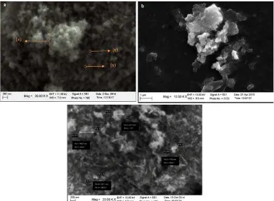
Fig 2. SEM result sample 2a. NPs T80C (x) aggregated NPs (y) single NPs ; b. NPs commercial 1;
ZnO-NPs commercial 2
different from the other samples and having the smallest size is T80C. Further characterization used T80C which has a size of NPs and PI result was the best between other treatments.
Nanostructure analysis
XRD analysis is used to determine the fingerprint which is typical of crystal structure. X-ray crystal
structure by XRD detects the distance between atoms in some angle. XRD spectra results such diffraction peaks are then compared with the reference standard in the database.
XRD patterns ZnO-NPs T80C is presented in Fig. 1. All diffraction peaks ZnO-NPs T80C indicate wurtzite structure of hexagonal phase ZnO crystal in accordance with the American Mineralogist Crystal Structure Database no. 5203 [27]. The diffraction peaks
Indones. J. Chem., 2016, 16 (2), 117 - 123 121
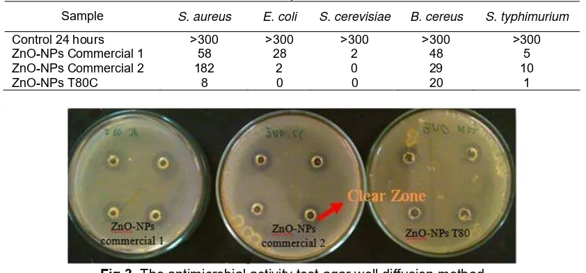
Table 3. Result of well diffusion method
ZnO-NPs zone of inhibition (mm)
No Microbes ZnO-NPs
Commercial 1 Commercial 2 ZnO-NPs ZnO-NPs T80C
1 S. aureus 7.125 6.13 6.63
2 Others microbes* No clear zone
*B. cereus; E. coli; S.thypimurium; A. niger; F. oxysporum; S. cerevisiae
Table 4. Result of plate count method
Sample S. aureus E. coli S. cerevisiae B. cereus S. typhimurium
Control 24 hours >300 >300 >300 >300 >300
ZnO-NPs Commercial 1 58 28 2 48 5
ZnO-NPs Commercial 2 182 2 0 29 10
ZnO-NPs T80C 8 0 0 20 1
Fig 3. The antimicrobial activity test agar well diffusion method
located at 31.88° (100), 34.54° (002), 36.40° (101), 47.74° (102), 56.60° (110), 63.10° (103), 66.70° (200), 68.00° (112), 69.16° (201), 72.9° (004) and 77.20° (202). The sharpness of the diffraction peaks shows that nanoparticles formed has a good crystallinity [28]. No other peaks were detected, diffraction peaks of crystalline ZnO NPs-T80C indicate that NPs are formed has a high purity level [29]. Residual pectin as a capping agent is not found in the XRD spectra because organic substances is reduced and disappear in the calcination process.
Morphology and Size Analysis
SEM analysis is used to determine the external morphology (structure) and size of the crystal particles. At a magnification of 20000x visible morphology ZnO-NPs tends to spherical shapes (Fig. 2a). Sign ‘x’ in the Fig. 2a shows aggregated NPs conditions. Single NPs indicated by signs ‘y’ are estimated to have a size less than 100 nanometers when compared with the scale. ZnO-NPs Commercial 1 has the form of spherical particles, whereas ZnO-NPs commercial 2 has a rod shape (Fig. 2b and 2c). Both of commercials NPs having a particle size larger than T80C ZnO-NPs. Differences in shape, size and spatial structure of the particles may be caused by differences in the method of synthesis of ZnO-NPs [25].
Antimicrobial Assay
Antimicrobial activity of all samples (ZnO NPs T80C, ZnO-NPs Commercial 1, ZnO-NPs Commercial 2) was examined using agar well diffusion method, plate count method and contact method. Table 3 shows the results of microbial inhibition using agar well diffusion method. This result showed that clear zone is only occur on S. aureus, and did not present in other
microbes tested. These results are slightly different from those obtained by other studies. Narayanan et al. [20] reported that the clear zone is also formed on the
testing of E. coli, Klebsiella pneumoniae and
Enterococcus faecalis. In this study ZnO-NPs
Commercial 1 has the largest inhibition zone compared to other ZnO samples. The presence of clear zone indicates that the ZnO-NPs have meaningful antimicrobial properties (Fig. 3). To determine the antimicrobial properties of ZnO-NPs, a further test using plate count method for antibacterial activity and contact method for antifungal activity were performed.
The number of microbes that can survive after treatment by ZnO-NPs using plate count method were presented at Table 4. The control, ZnO-NPs Commercial 1, and ZnO-NPs Commercial 2 have higher total number of viable bacterial tested than ZnO T80C. This indicates that ZnO T80C possesses the most effective antimicrobial effect.
Muhammad Fajri Romadhan et al.
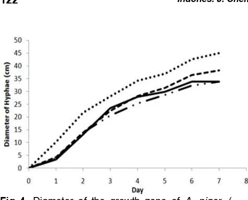
Fig 4. Diameter of the growth zone of A. niger, (˖˖˖˖˖)
control, (-˖˖-) ZnO-NPs Commercial 1, ( ) ZnO-NPs Commercial 2, (---) ZnO-NPs T80C
Fig 5. The diameter of the growth zone of F.
oxysporum, (˖˖˖˖˖) control, (-˖˖-) ZnO-NPs Commercial
1, ( ) ZnO-NPs Commercial 2, (---) ZnO-NPs T80C
In addition, the antimicrobial activity test results showed that the ZnO-NPs can be useful as a broad spectrum of antimicrobial. Narayanan et al. [20] reported that the antimicrobial effect of the ZnO-NPs increased at a smaller particle size. The present work successfully confirms the correlation between particle size and antimicrobial effects as previously reported in other studies [12,30-31].
Contact method was used to measure the effectiveness of NPs as an antifungal. Antifungal activity can be observed from the diameter of fungal growth on the media treated by ZnO-NPs. In Fig. 4 shows that the antifungal activity for A. niger are the most effective is
the ZnO-NPs Commercial 1. Antifungal activity the most effective for F. oxysporum is the ZnO NPs T80C (Fig. 5).
Diameter growth of A. niger in media treated fairly
quite large when compared to the diameter of F.
oxysporum. A. niger has a mechanism of adaptation to
cell wall stress given by forming more thick layer of chitin in their cell walls [32]. Antifungal activity of ZnO-NPs can be seen by comparing control and samples.
CONCLUSION
ZnO nanoparticles was successfully synthesized using precipitation method with NaOH as reducing agent. Characterization XRD showed that the method formed ZnO-NPs with desired purity. Best nanoparticle size was obtained at temperature of 80 °C and calcined with average particle size of 44.30 nm and uniform particle size (confirmed by PSA). Based on SEM analysis, morphology ZnO-NP T80C has a spherical shape and size smaller than the commercial NPs. Antimicrobial Test on NPs T80C showed excellent inhibitory effect, suggesting that it could potentially be used as a promising antimicrobial agent.
REFERENCES
1. Rico, C.M., Majumdar, S., Duarte-Gardea, M., Peralta-Videa, J.R., and Gardea-Torresdey, J.L., 2011, J. Agric. Food. Chem., 59 (8), 3485–3498.
2. Díaz, M.R., and Vivas-Mejia, P.E., 2013,
Pharmaceuticals, 6 (11), 1361–1380.
3. AbdElhady, M.M., 2012, Int. J. Carbohydr. Chem.,
2012, 1–6.
4. Li, X., Xing, Y., Jiang, Y., Ding, Y., and Li, W., 2009, Int. J. Food. Sci. Technol., 44, 2161–2168.
5. Chaudhry, Q., Scotter, M., Blackburn, J., Ross, B., Boxall, A., Castle, L., Aitken, R., and Watkins, R., 2008, Food Addit. Contam., 25 (3), 241–258.
Sci. Technol. Adv. Mater., 9 (3), 035004–035010.
13. Ravichandran, S., Franklin, D.R., and Kalyan, U., 2010, Natl. J. ChemBiosis, 1 (2), 4–6.
Indones. J. Chem., 2016, 16 (2), 117 - 123 123
Muhammad Fajri Romadhan et al.
15. Parra, M.R., and Haque, F.Z., 2014, J. Mater. Res.
Technol., 3 (4), 363–369.
16. Suganthi, K.S., and Rajan, K.S., 2012, Asian J. Sci. Res., 5 (4), 207–217.
17. Moharekar, S., Raskar, P., Wani, A., and Moharekar, S., 2014, World J. Pharm. Pharm. Sci., 3 (7), 1255–
1267.
18. Šileikaitė, A., Prosyčevas, I., Puišo, J., Juraitis, A., and Guobienė, A., 2006, Mater. Sci., 12 (4), 287–
291.
19. Alwan, R.M., Kadhim, Q.A., Sahan, K.M., Ali, R.A., Mahdi, R.J., Kassim, N.A., and Jassim, A.N., 2015,
Nanosci. Nanotechnol., 5 (1), 1–6.
20. Narayanan, P.M., Wilson, W.S., Abraham, A.T., and
Sevanan, M., 2012, BioNanoSci., 2, 329–335.
21. Jung W.K., Koo, H.C., Kim, K.W., Shin, S., Kim, S.H., and Park, Y.H., 2008, Appl. Environ. Microbiol.,
74 (7), 2172–2178.
22. Tullio, V., Nostro, A., Mandras, N., Dugo, P., Banche, G., Cannatelli, M.A., Cuffini, A.M., Alonzo, V., and Carlone, N.A., 2007, J. Appl. Microbiol., 102
(6), 1544–1550.
23. Kumar, S.S., Venkateswarlu, P., Rao, V.R., and Rao, G.N., 2013, Int. Nano Lett., 3 (1), 1–6.
24. Alias, S.S., Ismail, A.B., and Mohamad, A.A., 2010,
J. Alloys Compd., 499 (2), 231–237.
25. Kołodziejczak-Radzimska, A., and Jesionowski, T., 2014, Materials, 7 (4), 2833–2881.
26. Babaei, Z., Jahanshahi, M., and Sanati, M.H., 2008, Int. J. Nanosci. Nanotechnol., 4 (1), 23–30.
27. Downs, R.T., and Hall-Wallace, M., 2003, Am.
Mineral., 88, 247–250.
28. Nejati, K., Zolfaghar Rezvani, Z., and Pakizevand, R.,2011, Int. Nano Lett., 1, 75–81.
29. Wang, Y., Li J., and Hong, R., 2012, J. Cent.
South. Univ., 19 (4), 863–868.
30. Yousef , J.M., and Danial, E.N., 2012, J. Health Sci., 2 (4), 38–42.
31. Talebian, N., Amininezhad, S.M., and Doudi, M., 2013, J. Photochem. Photobiol., B, 120, 66–73.