A Spectrum of Prostate Cancer Developmentin
Transgenic Adenocarcinoma of the Mouse Prostate (TRAMP) Model
Krisna Murti
Anatomical Pathology Department, Medical Faculty, University of Sriwijaya, Indonesia
Abstract
Animal models that closely mimic the clinical disease can be exploited to facilitate translational research are needed. The Transgenic Adenocarcinoma of Mouse Prostate (TRAMP) model is uniquely suited to elucidate how a spectrum of prostate cancer development raised. Prostate cancer growths from premalignant stage, high grade prostatic intraepithelial neoplasia then progressed into highly dedifferentiated tumours that primarily metastasised to lymph nodes and lungs have similar pattern to that seen in humans.
Key words: animal model, prostate cancer, TRAMP, metastasis, high-grade prostatic intraepithelial neoplasia.
Abstrak
Hewan model yang sangat mirip dengan penyakit klinik yang dapat dieksploitasi dalam memfasilitasi penelitian sangat dibutuhkan. The Transgenic Adenocarcinoma of Mouse Prostate (TRAMP) model merupakan suatu model yang sangat cocok dalam menjabarkan suatu spektrum perkembangan keganasan prostat dari stadium pre-ganas, high grade prostatic intraepithelial neoplasia sampai terjadinya tumor prostat yang metastase ke kelenjar limfe dan paru-paru dengan pola yang sama seperti yang terjadi pada manusia.
Kata kata kunci: hewan model, kanker prostat, TRAMP, metastase, high grade prostatic intraepithelial neoplasia.
Prostate Cancer is the most common malignant disease in Western communities
and has caused the second cancer related deaths in man after lung cancer. In the USA this
cancer caused 27.000 death in 2007 1. In addition, the number of men having risk for
prostate cancer development is increasing rapidly as shown by demographic shift in
population 2.Prostate cancer (PCa) is an androgen dependent disease that can be treated
by androgen ablation therapy, and clinical trials are under way to prevent PCa through the
Extensive and adequate research is obviously necessary to identify and analyse
innovative modalities for prevention, intervention and regression of prostate cancer as
well as to study the potential relationship between molecular mechanism and clinical
progression. However, it is difficult to obtain human prostate cancer tissues, particularly,
at metastatic or very late stages of disease. Therefore, validation and establishment of a
model system that is suitable and analytical are in essential to accelerate the velocity of
translational research and into improving outcomes for patients with castrate resistant.
Animal models are ideal to fulfil these criteria. The use of transgenic mouse models
provides benefits as observers can manipulate its genome to analyse molecular events
related to the development of prostate cancer as well as milieu and genetic mutation can
be controlled 4
The transgenic adenocarcinoma of the mouse prostate (TRAMP) model developed
on C57BL/6 inbred strain by Professor Norman Greenberg. This model was generated by
using the prostate-specific rat probasin promoter to drive expression of the simian virus
40 large tumour antigen-coding region that acts as an oncoprotein through interactions
with the retinoblastoma (Rb) and p53, tumour suppressor gene products. The rat probasin
gene encodes an androgen and zinc-regulated protein specific to the epithelium of prostate
dorsolateral and ventral lobes. Cis-acting androgen–response regions within the 5’
flanking region have been identified and the ability of the prostate-specific rat probasin
gene promoter to target heterologous genes particularly in the prostate of transgenic mice
was established. In rats the gene promoter would be expected to act in the same manner
since it was isolated from this species .
5,6,7
A full spectrum of prostate cancer development was induced; from premalignant
stage, high grade prostatic intraepithelial neoplasia (HGPIN) then progressed into highly
cancers have similar patterns of the natural history that were observed in human prostate
carcinoma 5,7. Another study by Han et al. (2001), demonstrated that the TRAMP model
enabled to examine contributions of AR mutations, and the regulation of the androgen
signalling axis, in the development of castrate resistance prostate cancer 8. Other studies
have extensively investigated prostate cancer progression in the TRAMP model and
summarized that TRAMP model is suitable for the analysis of histopathobiology and
molecular events of progression of prostate cancer 4-10
In comparison of human and mouse prostate glands, some similarities as well as
the differences can be observed. The similarities can support the utilities of mouse models
to reveal the underlying molecular aspects that occurred in the development and
progression of prostate cancer. Nevertheless the differences between these two species
have influence into particular aspects on the analysis and the application of mouse models
to certain clinicopathological issue in human prostate cancer .
4,5 .
Normal Human Prostate
Normal human prostate is a single glandular organ divided into zones that are not
clearly demarcated (Figure 1A). A normal human prostate gland is assembled of the
periurethral transition zone (TZ), the peripheral zone (PZ), and the central zone (CZ). The
PZ is the major site for the incidence of prostatic intraepithelial neoplasia (PIN) and
prostate cancer 2,11. Normal prostate gland has a lobular formation and is surrounded by
abundance stroma consists of contracting spindle cells and collagen (Figure 2A). The
stroma expands further than glands’ outer boundary. In comparison with rodent, the
stroma in human is a much more plentiful. Benign prostate glands/acini are comprised of
two cell layers; a basal cell layer and a secretory cell layer. The basal cells are the
cells have not always been clearly seen by light microscopy, but can be identified by
immunostaining for high molecular weight cytokeratin (HMWCK). Between the two
layers there are a minor population of cells with neuroendocrine (NE) differentiation,
ultrastructurally characterized by dense core secretory granules. NE cells can be identified
by immunostaining for NE markers, such as chromogranin and Serotonin 11,12
Normal Mouse Prostate
.
The mouse prostate is divided into discreting lobes, the anterior prostate (AP) or
coagulating gland, the ventral prostate (VP), dorsal (DP) and lateral (LP) prostate, which
the DP and LP are grouped together as the dorso-lateral prostate (DLP) (Figure 1B).
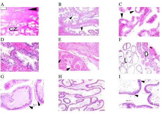
Figure 1. Comparison of human and mouse prostate anatomy. A, adult human prostate is divided into zones consisted of the periurethral transition zones (TZ), the peripheral zone (PZ), and the central zone (CZ). B, mouse prostate is categorized into antero-posterior prostate (AP), ventral (VP), dorsal and lateral lobes, which are grouped together as DLP 19.
The individual glands in each lobe are surrounded by very thin stroma, consisted
of a few layers of spindle cells among collagen fibers (Figure 2F, G, H, I) 15 A
.
B C
D E F
Figure 2. Comparison of human and mouse prostate histology. A-C, low, intermediate, and high magnification of normal CZ human glands. D, high magnification of human normal benign PZ glands that have a tufted or undulated luminal border and the sizes are larger than usual glands in prostate cancer. E-I, mouse prostate glands, with very thin stroma surrounds each gland in each lobe 15.
The glands of each lobe have cell populations that are homologous to the human
prostate, involving luminal secretory cells, a basal cell layer, and NE cells 12. Similar to
normal human prostate glands, basal cell layer in mouse prostate is not observable by
routine light microscopy. Moreover, the results of ultrastructural studies supported this
data as they found the lack of a continuous basal cell layer in normal mouse prostate
glands. These cells cannot be identified by antibodies to HMWCK (66kDa and 57KDa),
but they can be identified by immunostaining of a rabbit polyclonal antibody to mouse
cytokeratin 5 (CK5) and antibodies to CK14 13,14. In addition, nerve bundles, which are
laid within the prostate stroma in the postero-lateral of the human gland are not identified
within mouse stroma. However, in DLP sections thick nerve bundles and ganglia are often
seen in the peri-stromal loose connective tissue. Neuroendocrine (NE) cells which are
only few (< 1 %) can be observed by immunostaining with chromogranin and
synaptophysin 6
Prostate Pathology .
Several disorders have been reported in genetic engineered mouse (GEM) models
from development disorders until malignancy, however, in the present study the disorders
are restricted to hyperplasia, PIN, well differentiated (WD), Moderate differentiated (MD)
and poorly differentiated (PD) adenocarcinoma, and NE tumours 15
Non-malignancy
.
Non-neoplastic proliferation in prostate such as hyperplasia also occurs in
TRAMP model as well as in human, which is recognized as benign prostatic hyperplasia
increase in epithelial (glandular) tissue compared with age-matched wild-type control
mice” or “increase epithelial cells within normal appearing gland spaces”. The same
authors also identified that in GEM, proliferation patterns such as tufting, micropapillary
and cribriform are usually found. Hyperplasia in GEM should be defined as focal, in
which only a few glands are involved, and diffuse, in which more glands involved. Slight
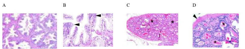
atypia can be present in hyperplasia (Figure 3 C, D) 15 A
.
B C D
Figure 3. Comparison of human and mouse hyperplasia. A and B, human benign prostate hyperplasia, showing the increase of gland numbers. C and D, low and high power magnification showing hyperplasia in mouse prostate glands with epithelial stratification due to proliferation with cribriform pattern (*) and stroma surround the hyperplastic glands (arrowhead) 15.
Several studies observed, premalignant lesions, PIN in TRAMP model which is
called mouse PIN (mPIN) described as “proliferation of atypical epithelial cells within
pre-existing glands” are characterized by nuclear stratification, epithelial tufting,
micropapillary projections, cribriform structures, elongated nuclei, increased mitoses and
apoptosis and the nuclei showed nuclear atypia. This spontaneous lesion can be observed
in all lobes by 8 weeks of age. In non-transgenic mice, PIN is observed in 40-60% mice
and was observed mostly in VP. In human, PIN is classified into low and high grade
(LGPIN and HGPIN). HGPIN is associated with invasive prostate cancer with the glands
in this stage shows nuclear stratification, enlargement and distinctly atypical epithelial
cells with nuclear enlargement and prominently enlarged nucleoli (macronucleoli) as the
human HGPIN characteristic feature (Figure 4A, B). The same authors, also classified
mPIN into with documented and without documented progression to invasive carcinoma
Classification of Prostate Disorders of Genetic Engineered Mouse (GEM) models
Non-neoplastic Disorders
1. Hyperplasia
Neoplastic Disorders
1. Prostatic intraepithelial neoplasia/neoplastic proliferation of premalignant potential
1.1. With documented progression to invasive carcinoma 1.2. Without documented progression to invasive carcinoma 2. Carcinoma (invasive)
2.1. Microinvasive carcinoma 2.2. Invasive carcinoma 2.2.1. Adenocarcinoma
2.2.1.1. Well differentiated 2.2.1.2. Moderately differentiated 2.2.1.3. Poorly differentiated 2.2.2. Neuroendocrine carcinoma
2.2.2.1. Small cell carcinoma 2.3.1. Squamous Cell carcinoma
2.3.2. Spindle cell/Sarcomatoid carcinoma 2.3.3. Undifferentiated carcinoma
2.3.4. Mixed carcinoma (specify component: Adenosquamous carcinoma)
Furthermore, mPIN with documented progression is naturally comparable to
human HGPIN, while PIN without documented is tentatively categorized at the beginning
of identifying an mPIN lesion in a new tissue, hence, the classification should be
converted if the invasion is identified afterwards. mPIN should be recognized either
focality or/and progression, in which, the lesion should begin focally, rather than
homogenous throughout the prostate and shows progression either increased of glands
involvement or increased nuclear atypia or both (Figure 4 C, D) 10,15
Malignancy and Neuroendocrine Differentiation
.
In human, adenocarcinoma is the majority type of prostate malignancies and
histologically it is classified into WD, MD and PD based on gland appearances. WD
adenocarcinoma shows well defined glands either small or medium sized lined with a
nuclei basically located (Figure 4E). In PD adenocarcinoma, gland formation is difficult
to observe, because the merge of glands creating more solid appearance, consequently
cells grow in cords, nest or sheets patterns (Figure 4G). MD is the mixture of WD and PD
appearances, where gland formation can be found side by side with solid sheets (Figure
4F)16. In TRAMP model the stages of cancer can be identified from premalignant lesions,
PIN, WD, MD, and PD adenocarcinoma, and NE carcinoma can be observed 15
Well differenciated PCa is characterized by increased quantity of small glands
(Figure 4A)
.
15
. There is often an associated desmoplastic response or stromal thickening.
The cells have round nuclei with fewer hyperchromatic nuclei than in PIN lesions.
Increased mitoses and apoptosis are apparent and may be associated with inflammation.
MD is characterized by nearly anaplastic sheets of cells that may contain remaining of
glandular architecture or the many glands fused and the others preserve individual
outlines but are closely packed with surrounding glands(Figure 4B), however, this stage
is not frequently observed 16. PD is characterized by anaplastic sheets of cells containing
pleomorphic (many and different shapes) cells with irregular nuclei, very little cytoplasm
surrounding the nuclei. There are often normal glands trapped within sheets of cells.
These lesions are often highly vascularized, hemorrhagic, and in the larger lesions can be
accompanied by necrotic (Figure 4C) 15 A
.
B C D E
F G H I J
Prostate cancer of human often demonstrates focal NE differentiation cells as, but
these cells are not neoplastic and the neoplastic type is found only 5% of all prostatic
neoplasms 10. This type of prostate cancer can also be found in several transgenic mouse
models including TRAMP as observed by some studies 4,5
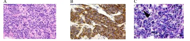
The description of this tumour as a NE carcinoma is based on its histological and
cytological characteristics that form solid and cribriform pattern with rosette-like
formation (Figure 5C)
. They identified the
transformation into an NE phenotype in PD tumours in untreated and castrated mice,
which indicates that NE differentiation is associated with advanced prostate cancer.
15,17
. Some authors proposed the spectrum of NE differentiation in
prostate cancer from NE with large eosinophilic granules to carcinoid-like pattern to small
cell carcinoma 16,18. The focal NE differentiation can be observed with the spread of
individual cells or cell nests in conventional prostatic adenocarcinoma. NE appearance
can also be observed as the entirely spread of small tumour cells indicated by nuclear
hyperchromasia, nuclear molding, small punctate nucleoli, and vigorous mitotic activity,
and this is called the aggressive small cell carcinoma or carcinoid/carcinoid-like tumour
(Figure 5A, B). These features should be verified by immunostaining (Figure 5B) for the
markers such as chromogranin and synaptophysin 6 A
.
B C
CONCLUSION
The TRAMP mouse model provides a consistent resource of primary and
metastatic tumours representative of those seen in clinical prostate cancer for
histopathobiological and molecular analyses to elucidate and further characterise the
molecular events involved in the development, progression and metastasis of prostate
cancer. The use of TRAMP tissues at different stages of disease progression and treatment
response will facilitate molecular and cellular analyses.
REFFERENCES
1. Taichman RS, Loberg RD, Mehra R, Pienta KJ. The Evolving Biology and Treatment of Prostate Cancer. J Clin Invest 2007; 117: 2351–61.
2. De Marzo AM, DeWeese TL, Platz EA, Meeker AK, Nakayama M, Epstein JI, Isaacs WB, Nelson WG. Pathological and Molecular Mechanisms of Prostate Carcinogenesis: Implications for Diagnosis, Detection, Prevention, and Treatment. J Cell Biochemist 2004; 91: 459-77.
3. Stanbrough M, Leav I, Kwan PWL, Bubley GJ, Balk SP. Prostatic Intraepithelial Neoplasia in Mice Expressing an Androgen Receptor Transgene in Prostate Epithelium. PNAS 2001; 98: 10823–8.
4. Kasper S, Smith JA. Genetically Modified Mice and Their Use in Developing Therapeutic Strategies for Prostate Cancer. J Urol 2004; 172: 12-9.
5. Kaplan-Lefco PJ, Chen TM, Ittmann MM, Barrios RJ, Ayala GE, Huss WJ, Maddison LA, Foster BA, Greenberg NM. Pathobiology of Autochthonous Prostate Cancer in a Pre-clinical Transgenic Mouse Model. Prostate 2003; 55: 219-37.
6. Masumori N, Thomas TZ, Chaurand P, Case T, Paul M, Kasper S, Caprioli RM, Tsukamote T, Shappell SB, Matusik RJ. A Probasin-Large T Antigen Transgenic Mouse Line Develops Prostate Adenocarcinoma and Neuroendocrine Carcinoma with Metastatic Potential. Cancer Res 2001; 61: 2230-49.
7. Haram KM, Peltier HJ, Lu B, Bhasin M, Otu HH, Choy B, Regan M, Libermann TA, Latham GJ, Sanda MG, Arredouani MS. Gene Expression Profile of Mouse Prostate Tumors Reveals Dysregulations in Major Biological Processes and Identifies Potential Murine Targets for Preclinical Development of Human Prostate Cancer Therapy. Prostate 2008; 68:1517-30.
8. Han G, Foster BA, Mistry S, Buchanan G, Harris JM, Tilley WD, Greenberg NM. Hormone Status Selects for Spontaneous Somatic Androgen Receptor Variant that Demonstrate Specific Ligand and Cofactor Dependent Activities in Autochronous Prostate Cancer. J Biol Chemist 2001; 276: 11204-13.
10. Burman PR, Wu H, Powell WC, Hagenkord J, Cohen MB. Genetically Defined Mouse Models That Mimic Natural Aspects of Human Prostate Cancer Development. Endoc Rel Cancer 2004; 11: 225-54.
11. Laczko’ I, Hudson DL, Freeman A, Feneley MR, Masters JR. Comparison of the Zones of The Human Prostate with Seminal Vesicle: Morphology, Immunohistochemistry, And Cell Kinetics. Prostate, 2005; 62: 260-6.
12. Sciarra A, Mariotti G, Gentile V, Voria G, Pastore A, Monti S, Di Silverio F. Neuroendocrine Differentiation in Human Prostate Tissue: Is It Detectable and Treatable? B J Urol Int 2003; 91: 438-45.
13. DiGiovanni J, Kiguchi K, Frijhoff A, Wilker E, Bol DK, Beltran L, Moats S, Ramorez A, Jorcano J, Conti C. Deregulated Expression of Insulin-Like Growth Factor 1 in Prostate Epithelium Leads To Neoplasia in Transgenic Mice
14. Kim MJ, Cardiff RD, Desai N, Petrosky WAB, Parsons R, Shen MM, Shen CA. Cooperativity of Nkx3.1 and Pten Loss of Function in A Mouse Model of Prostate Carcinogenesis 99: 2884-9.
15. Shappell SB, Thomas GV, Roberts RL, Herbert R, Ittmaan MM, Rubin MA, Humphrey PA, Sundberg JP, Rozengurt N, Barries R, Ward JM, Cardiff RD. Prostate Pathology of Genetically Engineered Mice: Definitions and Classification. The Consensus Report from the Bar Harbor Meeting of The Mouse Models of Human Cancer Consortium Prostate Pathology Committee. Cancer Res 2004; 64: 2270-305.
16. Ramnani, DM. A Visual Survey of Urologic Pathology. 2005. Viewed on 14 June 2005. Available on: <http
17. Evangelou AI, Winter SF, Huss WJ, Bok RA, Greenberg NM. Steroid Hormones, Polypeptide Growth Factors, Hormone Refractory Prostate Cancer, and The neuroendocrine Phenotype. J Cell Biochemist 2004; 91: 671-83.
18. Di Sant'Agnese PA. Divergent Neuroendocrine Differentiation in Prostatic Carcinoma. Sem Diagn Pathol 2000; 17: 149-61.
19. Cardiff R, Paulus M, Johnson A, Griffey S, Henderson G, Jue T. Comparative Prostate in: Anatomy Quicktime Movies of Histologic Anatomy. Viewed on 06 June 2005. Available on:
20. The Norman Greenberg Lab TRAMP: Genitourinary Histopathology 2001. Viewed on 06 June 2005. Available on:
Author:
Krisna Murti, MD., M.Biotech., SpPA : Anatomical Pathology Department, Medical Faculty, University of Sriwijaya, Indonesia.
Current Address: Department of Molecular Pathology, Institute of Pathology, University of Wuerzburg, Germany
Synopsis