Indonesian Journal of Biotechnology
VOLUME 22(2), 2017, 107–113 | RESEARCH ARTICLE
Genera on of recombinant scFv an body fragments against
Ochratoxin A (OTA)
Ranya Pranomphon1, Witsanu Srila1, and Montarop Yamabhai1,∗
1School of Biotechnology: Ins tute of Agricultural Technology, Suranaree University of Technology, 111 University Avenue, Nakhon
Ratchasima 30000, Thailand
∗Corresponding author:[email protected]
ABSTRACTOchratoxin A (OTA) is a mycotoxin commonly found in agricultural products and can accumulate in the blood and ssues a er contaminated food is consumed. Recombinant single-chain an body fragments (scFv) against OTA were selected from phage display libraries. A er one round of biopanning against BSA-conjugated OTA (OTA-BSA), 52 and 6 phage clones displaying scFv an bodies were isolated from human (Yamo I.3) and rabbit (Bozmix I.2) libraries. Two phage clones (one from each library, i.e. yOTA1e3 and bOTA2a9) showed binding to free toxin by compe ve ELISA. The soluble scFv an bodies were produced by superinfec ng phage clones intoEscherichia colisuppressor strain HB2151. The scFv genes from these two phage clones were sub-cloned into pKP300∆III vectors to generate scFv-AP fusions. The binding affinity
(IC50) of an bodies derived from the human library were higher than those from the rabbit library. The binding property of
recombinant an bodies in the form of scFv-AP was be er than those in soluble scFv form. Cross-reac vity analysis indicated that the two recombinant an bodies did not cross-react with other soluble toxins, namely AFB1, DON, ZEN, and FB. The ability to use the recombinant scFv-AP to detect contaminated toxins in an agricultural product (corn) was demonstrated. These an bodies can be used as a template for further engineering and op miza on for the detec on of contaminated OTAs in the future.
KEYWORDSalkaline phosphatase fusion; ochratoxin A; phage display; recombinant an body; single chain fragments
1. Introduc on
Ochratoxin A (OTA) is a mycotoxin, produced by fungi
such asAspergillus ochraceousandPenicillium
verruco-sum(Pfohl-Leszkowicz and Manderville 2007). It is com-monly found during the processing and storage of
agri-cultural products (Zinedine and Mañes 2009). OTA has
been found in a wide variety of agricultural commodities, including coffee, cereals, dried fruits, and wine (Giraudi et al. 2007). In addition to being nephrotoxic, teratogenic, neurotoxic and immunotoxic, OTA displays carcinogenic and immunosuppressive properties, and has been classi-fied as a Group 2B carcinogen by the International Agency for Research Cancer (Yang et al. 2015;Zou et al. 2016). This toxin is hazardous to both humans and animals and is very harmful because it is stable and can accumulate in the circulatory system, liver and other tissues (Turner et al. 2009; Peraica et al. 2010). Analytical methods such as thin layer chromatography (TLC), high perfor-mance liquid chromatography (HPLC) and gas chromatog-raphy (GC) (Turner et al. 2009) have been used to con-trol OTA that contaminates both food and feed in agricul-tural products but these techniques are expensive and
time-consuming (Zou et al. 2016). Immunological analysis,
especially enzyme-linked immunosorbent assay (ELISA), which uses monoclonal or polyclonal antibodies, has sev-eral advantages including high sensitivity, high specificity and higher simplicity compared with instrumental
meth-ods (Rangnoi et al. 2011; Qiu et al. 2015). However,
the cost for the production of antibodies is still relatively high. Phage display technology, which was established by George P. Smith in 1985 (Smith 1985), is an alterna-tive approach to the generation of recombinant antibod-ies. This technology uses filamentous bacteriophage, e.g., M13 to displayed antibody fragment, such as single-chain Fragment variable (scFv) on its surface. The key advan-tage of this technology is the direct linkage between
geno-types and phenogeno-types (Hoogenboom et al. 1998),
allow-ing further engineerallow-ing or large-scale production of
re-combinant antibodies from Escherichia colior other
cre-ate scFv-alkaline phosphatase fusions (scFv-AP). Binding properties of these recombinant antibodies obtained from these two libraries were compared and analyzed.
2. Materials and methods
2.1. MaterialsYamo I.3, a naïve human scFv library, was constructed in our laboratory from B cells in the plasma of 140 non-immunized human donors in Northeastern Thailand as pre-viously described (Pansri et al. 2009). KM13 helper phage was obtained with the Tomlinson libraries and was prop-agated as described in the MRC phage display protocols. Ochratoxin A (OTA) conjugated with BSA (OTA-BSA), OVA (OTA-OVA) and KLH (OTA-KLH), and soluble aflatoxins (AFs) B1, deoxynivalenol (DON), zearalenone (ZEN) and fumonisins (FB) toxins were purchased from
Sigma, USA, and Aokin, Germany. E. coli TG1 and
HB2151 were obtained from MRC, Cambridge, UK. His prob-HRP was purchased from Sigma. Reference corn material were purchased from Trilogy, USA. All exper-iments were conducted under the Ethical Principles and Guidelines of Suranaree University of Technology.
2.2. Construc on of rabbit scFv an body library
The immunized rabbit scFv antibody library was con-structed as previously described (Vu et al. 2017). The spleens from two rabbits (White New Zealand), immu-nized with OTA-BSA and ZON-BSA, were used. The rab-bit immune responses were determined by indirect ELISA, using OTA-BSA as a target. Peripheral blood mononu-clear cell (PBMC) was collected from ear vein and total RNA was extracted according to the TRIZOL extraction method. Three µg of total RNA from each rabbit was used as template to generate cDNA, using M-MuLV Reverse
Transcriptase. Then, the light and heavy chain (VL and
VH) genes were amplified with separate sets of 29 PCR
primers, using a Taq DNA polymerase. The amplified VH and VL sequences were linked via a polynucleotide linker by pull-through PCR and cloned into phagemids vector (pMOD) between the5’SfiI or 3’NotI restriction sites, as previously described (Pansri et al. 2009). This library was designated Bozmix I.2
2.3. Biopanning
Biopanning was performed according to Rangnoi et al.
(2011) using Yamo I.3 (Pansri et al. 2009) and rabbit Bozmix library, which were constructed as described above. One well of Immuno 96 microWellTM plate (Nunc, Denmark) was coated with 25 µg of OTA-BSA conjugate in PBS buffer and incubated at 4°C for overnight. The next day, the well was blocked with PBS supplemented with skimmed milk (2%, w/v, MPBS) and incubated for 1 h at room temperature. Following that, the well was washed three times with PBS. The libraries were pre-incubated with 2% (w/v) MPBS and 1% (w/v)
BSA for 30 min at room temperature before being added into wells of immunized toxin and then incubated at room temperature for 2 h. Then, the well was washed 10 times with PBST (PBS supplemented with 0.05% (v/v) Tween 20) and with PBS 10 times to remove the unbound phage. Bound phage was eluted by adding 100 µL of 1 mg/mL trypsin buffer (in PBS) and incubated at room tempera-ture for 10 min with rotation. Acid elution was subse-quently performed by adding 100 µL of 100 mM glycine– HCl, pH 2.0 and incubated for 10 min, followed by neu-tralization with 100 µL of neuneu-tralization buffer (200 mM NaHPO4, pH 7.5). To isolate individual phage clones, the log phaseE. coliTG1 cells were infected with the eluted
phage by adding 2 mL ofE. coliTG1 log phase with 150
µL eluted phage and incubated at 37°C for 30 min without shaking. Follow this, 10-fold serial dilutions of infected cells were performed using 2xYT media and the cells were spread onto 2xYT agar containing 100 µg/mL ampicillin and 1% w/v glucose. The plates were then incubated at 37°C overnight.
2.4. Individual phage rescue
Individual phage clones were picked into 100 µL of 2xYT supplemented 100 µg/mL ampicillin and 1% (w/v) glu-cose in each well of a 96-well plate (Nunc, Denmark), and incubated overnight at 37°C with shaking. After that, 10 µL from each well were transferred to a new deep-well mi-croculture plate (Nunc, Denmark), containing 400 µL of 2xYT plus 100 µg/mL ampicillin and 1% (w/v) glucose. The first plate was kept as the glycerol stock by adding glycerol to a final concentration of 15% (v/v) and kept at -70°C. The deep-well micro culture plate was then
incu-bated at 37°C for 3 h with vigorous shaking. After that, phage was rescued by adding 1010of helper phage to each well and incubated at 37°C for 1 h without shaking. The pellet was collected by spinning at 3,000xg for 15 min, and re-suspended in 400 µL of 2xYT containing 100 µg/mL of ampicillin and 50 µg/mL of kanamycin. After incubation with shaking at 30°C for 20 h, the supernatant was col-lected for phage ELISA after centrifugation at 4,000 rpm for 10 min.
2.5. Phage ELISA
The 96-well microtiter plates (Immuno 96 microWellTM,
wells were washed again, as described above. Then 200 µL of HRP substrate (ABTS, Sigma) were added to each well. After 30 min of incubation at room temperature, the optical density (OD) at 405 nm was measured using a mi-crotiter plate reader (Tecan, Austria).
2.6. Expression and Purifica on of soluble scFv
2.6.1. Expression using E. coli HB2151
The expression of soluble scFv antibody was performed as previously described (Rangnoi et al. 2011). Approx-imately 10-20 µL of phage particles were mixed with
500 µL of log-phase (OD600= 0.4)E. coliHB2151
(non-suppressor host), and incubated at 37°C for 30 min with-out shaking. Infected bacteria were then streaked onto 2xYT agar containing 100 µg/mL ampicillin and 1% (w/v) glucose. After that, the plates were incubated at 37°C overnight. A single colony was picked and cultured in 5 mL of 2xYT plus 100 µg/mL ampicillin and 2% (w/v) glucose, and incubated at 30°C with vigorous shaking overnight. Fifty µL of overnight culture was added into 5 mL of 2xYT containing 100 µg/mL ampicillin and 0.1% (w/v) glucose, and incubated with shaking at 30°C until the OD600reached 0.9. Then, 1 mM of isopropylthiogalac-toside (IPTG) was added to induce the scFv expression. The incubation was continued at 30°C for 20 h. The se-creted antibody could be found in the supernatant. 2.6.2. Expression using E. coli HMS174
For large-scale expression, the scFv genes of clones Y1E3 and RB2A9 were sub-cloned into pET27b vectors
be-tween Nco I and Not I restriction enzymes. The
plas-mids were purified using commercial kit (QIAgen spin
Mini-prepkit, USA) and transformed intoE. coliHMS174
pLysS (Novagen). The transformed bacteria were grown in 5 mL of M9ZB media [Dissolve 10 g Bacto tryptone, 5 g NaCl, 6 g Na2HP04, 3 g KH2P04and 1 g NH4Cl in 1 liter of water, after autoclaving add 20 ml sterile 20% glucose and 1 ml sterile 1 M MgS04] (Kunze et al., 1995), contain-ing 50 µg/mL kanamycin and 20% w/v glucose. The cul-ture was incubated at 30°C, 250 rpm for overnight. Then, 1% (v/v) of each overnight culture was added into M9ZB media supplemented with 20% w/v glucose and 50 µg/mL kanamycin. The culture was then incubated with shaking at 30°C for 4 h. Later, the cells were centrifuged at 16°C, 5000 rpm for 15 min and re-suspended with M9ZB media containing 10% w/v glycerol, 50 µg/mL kanamycin and 1mM IPTG (Amresco, USA). The incubation was contin-ued at 16°C, 250 rpm for 20 h, then the cell pellet was collected by centrifugation at 4°C, 8000 rpm for 10 min. Periplasmic extraction was performed to collect scFv an-tibodies as previously described (Yamabhai et al. 2008). Briefly, 50 mL of cell culture were spun down to col-lect the cell. The bacterial cells were then re-suspended in 2.5 mL of cold spheroplast buffer [1M Tris–HCl, pH 8.0, 0.5M EDTA, 20% sucrose, containing 0.1 M phenyl-methylsulfonyl fluoride (PMSF)] and incubated for 5 min
on ice. Then, the cells were centrifuged at 8000×g for 10 min at 4°C, and re-suspended in 1.0 mL of ice-cold sterile
water containing 1mM MgCl2and incubated on ice with
frequent shaking for 5 min. Periplasmic fractions were collected by centrifugation at 8000×g for 15 min at 4°C. The extracted scFv antibodies were detected by sodium do-decyl sulphate polyacrylamide gel electrophoresis (SDS-PAGE).
2.7. ScFv ELISA
The scFv ELISA was similar to the phage ELISA method as describe above except that 50 µL of supernatant con-taining scFv antibody and 50 µL of PBST were added into each well instead. The bound scFv was detected by addition of 100 µL of His prob-HRP (1:5000 dilution in PBS), using ABTS as substrate. The average optical den-sity (OD) at 405 nm with standard deviation of triplicate wells was reported.
2.8. Cloning, expression, and purifica on of scFv-AP fusion
The scFv-AP was constructed as previously described (Rangnoi et al. 2011). Briefly, the scFv gene from the phagemid vector was digested withNcoI andNotI restric-tion enzymes and sub-cloned into pKP300∆III, an alkaline
phosphatase fusion vector (Kay and Hoess 1996). The in-tegrity of these constructs were performed by automated DNA sequencing (Macrogen, Korea). To express the scFv-alkaline phosphatase (scFv-AP) fusions the recombinant plasmids (pKP300∆III vector) were extracted and
trans-formed intoE. coliTG1 competent cells. A single colony was picked and cultured in 5 mL of LB (Luria-Bertani) medium containing 100 mg/mL ampicillin at 37°C with shaking at 250 rpm, overnight. After that, 100 µL of overnight culture was inoculated into 100 mL of low-phos media [3.75 g ammonium sulphate, 0.71 g sodium citrate dehydrate, 1.07 g KCl, 5.36 g Yeast extract, 5.36 g
Hy-case SF Hy-casein hydrolysate, 7 mM MgSO4, 14 mM
glu-cose per liter, pH 7.3 (adjusted with KOH) containing 100
mg/mL ampicillin, 1 M MgSO4 and 1 M glucose]. The
culture was incubated at 30°C with shaking at 250 rpm for 20 h. Then, the cultures were centrifuged at 8,000 rpm for 10 min at 4°C. The cell pellets were re-suspended in 10 mL of fresh lysis buffer (10 mM imidazole (pH 8.0), BugBuster 10X, 100 mM PMSF). The cell debris was re-moved using centrifugation at 8,000 rpm for 15 min at 4°C. The recombinant proteins were purified using a Ni-NTA resin, according to the manufacturer’s instructions. Sub-sequently, the eluted proteins were dialyzed with TBS by Amicon Ultra-15 filter device (MW cutoff at 10 kDa, Mil-lipore), then MgCl2was added to the purified protein to a final concentration of 1mM.
2.9. ScFv-AP ELISA
microtiter plate (Immuno 96 WellsTM, Nunc, Denmark) were immobilized with 2 µg of OTA-BSA in 100 µL PBS or 1% (w/v) BSA in PBS (a negative control). The plate was kept at 4°C overnight. The next day, the plate was blocked with 2% (w/v) fat-free powdered milk (MPBS) in TBS. After incubation for 1 h at room temperature, the wells were washed 3 times with TBST. Then 50 µL of TBST and 50 µL of scFv-AP fusion was added into each well and incubated at room temperature for 1–2 h. After that, the plate was washed three times with 0.05% (v/v) Tween 20 (TBST) and two times with TBS. Finally, 200 µL of p-Nitrophenyl phosphate substrate (pNPP; SIGMA, USA) was added to each well, and the optical density (OD) at 405nm was measured after 30 min of incubation at room temperature.
2.10. Compe ve ELISA
The wells of 96-well microtiter plate were coated with 2 µg of OTA-BSA. The optimal amount of antibody was first determined by serial dilution ELISA and the amount of the antibody that showed the binding at the log-phase of the binding curve was used. The appropriate concentra-tion of antibody was pre-incubated with varying concen-trations of soluble OTA in TBS containing 0.05% Tween 20 in the wells of the micro-titer plates at 37°C for 30 min with shaking at 300 rpm. After that, the mixture was transferred into wells of the ELISA plate that had been coated and blocked as described previously. After incuba-tion for 1 h, non-specific adsorpincuba-tion was washed away 3 times with TBS containing 0.05% Tween 20, followed by 2 times with TBS. After that, 200 µL of substrate solution (pNPP, Sigma, USA) was added into each well. The OD at 405 nm of triplicate wells was measure using microtiter plate reader (Tecan, Austria).
2.11. DNA sequence analysis
The phage clones that showed positive binding by compet-itive ELISA were collected and the plasmids was prepared using plasmid miniprep kit (Qiagen), and then the DNA se-quences were determined by automated DNA sequencing (Macrogen, Korea). The DNA sequences were analyzed with IgBLAST and the complementarity determining re-gions (CDRs) were identified using Imgt software. The 3D structures were predicted by Phyre2 software.
2.12. Cross-reac vity analysis
Cross-reactivity of the isolated antibody against ochra-toxin A (OTA), aflaochra-toxin B1 (AFB1), deoxynivalenol (DON), zearalenone (ZEN) and fumonisins (FB) toxins, was evaluated by competitive ELISA, using scFv-AP an-tibody format. The toxins were pre-incubated with appro-priate concentration of antibody at 37°C for 30 min, with shaking at 450 rpm. Then, the plate was washed 5 times with TBS containing 0.05% Tween 20. The OD values were obtained as described above.
2.13. Spike analysis of OTA in corn sample
Three gram of toxin-free reference corn material (Trilogy, USA) was added into 15 mL of 70% methanol. The ex-tract was shaken for 3 min at room temperature and left to stand for 10 min to collect the clear solution. Then the extract was filtered through Whatman filter paper No.1. The spiked solutions with known amounts of OTA (10– 30,000 ng/mL) were prepared by adding various concen-trations of OTA standards to the extraction solution. Af-ter that the spiked solutions were diluted 1:3 with 0.05% (v/v) Tween 20 (TBST). The diluted solution was added into plate instead of OTA standard by competitive ELISA as described before. Data were collected to plot the curve between A/A0 and concentration of spiked OTA. Limit of detection (LOD) and IC50was evaluated from the graph.
3. Results and discussion
After one round of biopanning using OTA-BSA as a tar-get, 52 clones from human library (Yamo I.3) and 6 clones from rabbit library (Bozmix I.2) were obtained. Amino acid sequence analysis indicated that only 2 unique se-quences, one from human and one from rabbit libraries, were affinity selected. These two clones were designated RB2A9 and Y1E3, for scFv derived from phage displayed-rabbit, and human libraries, respectively. The reason why we only obtained 2 unique clones could be because the sizes of the two libraries were relatively small and these were the clones that could grow very well. The nucleotide sequences of clone RB2A9 from rabbit library and clone Y1E3 from human library were translated into
(a)
(b)
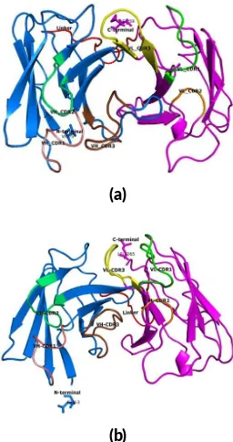
FIGURE 1Three dimensional structure predic on of scFv-AP clone RB2A9 (a) and clone Y1E3 (b). The VHand VLare shown in blue
FIGURE 2Compe ve ELISA analysis of scFv clone RB2A9 from rabbit and clone Y1E3 from human against OTA. The results are shown as A/A0, where A is the absorbance of an body in the pres-ence of varying OTA concentra on, and A0 is the absorbance of an body in the absence of soluble OTA.
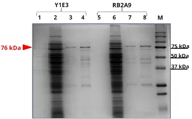
FIGURE 3SDS-PAGE of purified scFv-AP clone Y1E3 (human) and clone RB2A9 (Rabbit) inE. coliTG1. Lane 1 and 5, culture super-natants; lane 2 and 6, crude extract of cell lysate; lane 3 and 7, pu-rified scFv an body; lane 4 and 8, pupu-rified and concentrated scFv an body. M: kaleidoscope protein standard (Bio-Rad). The scFv-AP band of approximately 76 kDa is indicated.
amino acid sequences, and the schematic ribbon cartoon
of scFv clone RB2A9 and Y1E3 were shown in Figure1.
The complementarity-determining region (CDR1, CDR2
and CDR3) of the VH and VL were indicated by pymol
program. To express soluble scFv antibody formats, the scFv genes were sub-cloned into pET27b vector and
ex-pressed inE. coliHMS174, pLysS BL21. After that,
com-petitive ELISA was performed to detect the binding ability between soluble scFv and free OTA (Figure2). The results indicated that scFv clone Y1E3 from human library could bind to the target better than the clone RB2A9 from
rab-bit library. The IC50of clone RB2A9 and Y1E3 were 1.0
µg/mL and 0.3 µg/mL, respectively. Previous study has shown that rabbit scFv was relatively difficult to express inE. coli (Ayyar et al. 2015), therefore the rabbit clone that could bind better to OTA might have been selected out because it could not be expressed inE. coli.
Since we have previously shown that the recombinant antibody in the form of scFv-AP format is more efficient to be used as one-step detection probe for the detection
of mycotoxin contaminations (Rangnoi et al. 2011), in
FIGURE 4Western blot analysis of purified scFv-AP clone Y1E3 (human) and clone RB2A9 (rabbit). M: kaleidoscope protein stan-dard (Bio-Rad). The scFv-AP band of approximately 76 kDa is indi-cated.
FIGURE 5Compe ve ELISA of scFv-AP clone RB2A9 from rab-bit and clone Y1E3 from human. The results are shown as A/A0, where A is the absorbance of an body in presence of varying con-centra on of OTA, and A0 is the absorbance of an body in the absence of soluble OTA.
the next step, the scFv-alkaline phosphatase (scFv-AP) fu-sions were generated by insertion of the scFv gene into recombinant plasmid (pKP300∆III vector) (Pershad et al. 2011). After that, the recombinant scFv-AP fusions were purified from cell lysate by immobilized metal affinity chromatography (IMAC) using Ni-NTA resin. The
pu-rified fractions were detected by SDS-PAGE (Figure 3)
and western blot analysis (Figure4). The expected size of scFv-AP fusion was about 76 kDa. The reason why the protein could not be purified to apparent homogene-ity could be because the His-tag was not very efficient for protein purification (Berkman, M., personal commu-nication). After that, competitive ELISA was performed by one-step detection procedure to evaluate the binding
of scFv-AP antibody to soluble OTA (Figure4). The
A
/A
0
Concentration of soluble OTA (μg/mL) (a)
A
/A
0
Concentration of soluble OTA (μg/mL)
(b)
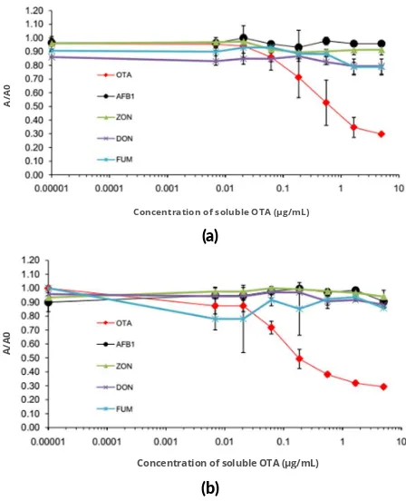
FIGURE 6Cross-reac vity assay. Compe ve ELISA of scFv-AP clone RB2A9 from rabbit (a) and clone Y1E3 from human (b) with different common mycotoxins are shown.
A
/A
0
Concentration of soluble OTA (μg/mL)
FIGURE 7Spike analysis. Various concentra on of OTA were spiked on corn sample extract and tested against scFv-AP clone RB2A9 from rabbit, and clone Y1E3 from human. The inhibi on curved are compared with the standard curves (dashed lines).
In the next step, cross-reactivity analysis of scFv-AP clone RB2A9 and Y1E3 were performed with various sol-uble mycotoxins; namely, ochratoxin A (OTA), aflatoxins B1 (AFB1), deoxynivalenol (DON), zearalenone (ZEN),
and fumonisins (FB), by competitive ELISA (Figure 5).
The results showed that both of the scFv-AP clones did not cross-react with the other tested mycotoxins, indicat-ing that specific scFv-AP antibodies against OTA could be generated.
Finally, to demonstrate the potential application of scFv-AP for the detection of mycotoxins from agricultural samples, the spike analysis of OTA was performed. Ref-erence corn material containing no toxin (Trilogy, USA)
was extracted and spiked with various concentration of
OTA before subjecting to competitive ELISA (Figure6).
The result indicated that the limit of detection (LOD) of scFv-AP clones RB2A9 and Y1E3 were 400 ng/mL and 200 ng/mL, respectively. The detection limit was much higher than the values obtained from competitive ELISA using standard OTA.
These results indicated that the corn matrix could in-terfere with the binding of scFv-AP to OTA, using the ex-traction method as described in this work. Therefore, fur-ther optimizations of various factors are required before recombinant scFv-AP antibody can be adapted for OTA detection in the field. These include 1) enhancing of affin-ity of scFv-AP, 2) optimizing competitive ELISA assay, and 3) optimizing sample extraction procedure to fit dif-ferent agricultural products.
4. Conclusions
In this study, OTA-specific scFv antibodies were gener-ated from rabbit and human phage display antibody library. Potential application of scFv-AP format as one-step detec-tion probe for the detecdetec-tion of free OTA was demonstrated.
Acknowledgments
This work was supported by Grants from Suranaree Uni-versity of Technology (SUT) and Agricultural Research Development (Public Organization) [ARDA] grants No. CRP550701085. RP was supported by the Royal Golden Jubilee scholarship from Thailand Research Fund (TRF).
Authors’ contribu ons
RP performed most of the experiment and draft the manuscript. WS constructed the phage display library, supervised laboratory work and drafted the manuscript. MY conceived of the study and edited the manuscript. All authors read and approved the final version of the manuscript.
Compe ng interests
MY is on the editorial board of theIndonesian Journal of Biotechnology, and was recused from this article’s review and decision. The authors declare no other competing in-terests.
References
Ayyar BV, Hearty S, O’Kennedy R. 2015. Facile domain rearrangement abrogates expression recalcitrance in a rabbit scFv. Appl Microbiol Biotechnol. 99(6):2693– 2703. doi:10.1007/s00253-014-6268-4.
Better M, Chang CP, Robinson RR, Horwitz AH. 1988.
Escherichia coli secretion of an active chimeric
an-tibody fragment. Science 240(4855):1041–1043.
Giraudi G, Anfossi L, Baggiani C, Giovannoli C, Tozzi C. 2007. Solid-phase extraction of ochratoxin A from wine based on a binding hexapeptide prepared by com-binatorial synthesis. J Chromatogr A 1175(2):174– 180. doi:10.1016/j.chroma.2007.10.057.
Hoogenboom HR, de Bruı̈ne AP, Hufton SE, Hoet RM, Arends JW, Roovers RC. 1998. Antibody phage dis-play technology and its applications.
Immunotechnol-ogy 4(1):1–20. doi:10.1016/S1380-2933(98)00007-4.
Kay BK, Hoess RH. 1996. Principles and applications of phage display. In: Phage display of peptides and proteins. Burlington: Academic Press. p. 21–34. doi:10.1016/B978-012402380-2/50004-6.
Pansri P, Jaruseranee N, Rangnoi K, Kristensen P, Yam-abhai M. 2009. A compact phage display human scFv library for selection of antibodies to a wide variety of antigens. BMC Biotechnol. 9(1):6. doi: 10.1186/1472-6750-9-6.
Peraica M, Flajs D, Domijan AM, Ivić D,
Cv-jetković B. 2010. Ochratoxin A contamination
of food from Croatia. Toxins 2(8):2098–2105.
doi:10.3390/toxins2082098.
Pershad K, Sullivan MA, Kay BK. 2011. Drop-out phagemid vector for switching from phage displayed affinity reagents to expression formats. Anal Biochem. 412(2):210–216. doi:10.1016/j.ab.2011.02.006. Pfohl-Leszkowicz A, Manderville RA. 2007. Ochratoxin
A: an overview on toxicity and carcinogenicity in an-imals and humans. Mol Nutr Food Res. 51(1):61–99. doi:10.1002/mnfr.200600137.
Qiu YL, He QH, Xu Y, Bhunia AK, Tu Z, Chen B, Liu YY. 2015. Deoxynivalenol-mimic nanobody isolated from a naïve phage display nanobody library and its applica-tion in immunoassay. Anal Chim Acta 887:201–208. doi:10.1016/j.aca.2015.06.033.
Rangnoi K, Jaruseranee N, O’Kennedy R, Pansri P, Yam-abhai M. 2011. One-step detection of aflatoxin-b1 using scFv-alkaline phosphatase-fusion selected from human phage display antibody library. Mol Biotech-nol. 49(3):240–249. doi:10.1007/s12033-011-9398-2.
Smith GP. 1985. Filamentous fusion phage: novel
expression vectors that display cloned antigens on the virion surface. Science 228(4705):1315–1317. doi:10.1126/science.4001944.
Turner NW, Subrahmanyam S, Piletsky SA. 2009. Analytical methods for determination of mycotox-ins: a review. Anal Chim Acta 632(2):168–180. doi:10.1016/j.aca.2008.11.010.
Vu NX, Pruksametanan N, Srila W, Yuttavanichakul W, Teamtisong K, Teaumroong N, Boonkerd N, Tit-tabutr P, Yamabhai M. 2017. Generation of a rab-bit single-chain fragment variable (scFv) antibody for
specific detection of Bradyrhizobium sp. DOA9 in
both free-living and bacteroid forms. PLoS One 12(6):e0179983. doi:10.1371/journal.pone.0179983.
Yamabhai M, Emrat S, Sukasem S, Pesatcha P, Jaruser-anee N, Buranabanyat B. 2008. Secretion of recom-binantBacillushydrolytic enzymes usingEscherichia coliexpression systems. J Biotechnol. 133(1):50–57. doi:10.1016/j.jbiotec.2007.09.005.
Yang L, Zhang Y, Li R, Lin C, Guo L, Qiu B, Lin Z, Chen G. 2015. Electrochemiluminescence biosensor for ultrasensitive determination of ochratoxin A in corn samples based on aptamer and hyperbranched rolling circle amplification. Biosens Bioelectron. 70:268– 274. doi:10.1016/j.bios.2015.03.067.
Zinedine A, Mañes J. 2009. Occurrence and
legislation of mycotoxins in food and feed
from Morocco. Food Control 20(4):334–344.
doi:10.1016/j.foodcont.2008.07.002.
Zou X, Chen C, Huang X, Chen X, Wang L, Xiong
Y. 2016. Phage-free peptide ELISA for
ochra-toxin A detection based on biotinylated mimotope
as a competing antigen. Talanta 146:394–400.