Vicente Sanchis-Alfonso (Ed)
Anterior Knee Pain
and Patellar Instability
With 240 Figures
Vicente Sanchis-Alfonso, MD, PhD (Member of the International Patellofemoral Study Group/Member of the ACL Study Group)
Department of Orthopaedic Surgery Hospital Arnau de Vilanova
Valencia Spain
British Library Cataloguing in Publication Data Anterior knee pain and patellar instability
1. Patellofemoral joint - Dislocation 2. Patella -
Dislocation 3. Knee - Diseases 4. Knee - Wounds and injuries 5. Knee - Surgery 6. Pain - Physiological aspects
I. Sanchis-Alfonso, Vicente 617.5′82
ISBN-10: 1846280036
Library of Congress Control Number: 2005925983
ISBN-10: 1-84628-003-6 e-ISBN 1-84628-143-1 ISBN-13: 978-1-84628-003-0
Printed on acid-free paper
© Springer-Verlag London Limited 2006
First published in 2003 asDolor anterior de rodilla e inestabilidad rotuliana en el paciente joven. This English-language edition published by arrangement with Editorial Médica Panamericana S.A.
Apart from any fair dealing for the purposes of research or private study, or criticism or review, as permitted under the Copyright, Designs and Patents Act 1988, this publication may only be reproduced, stored or trans-mitted, in any form or by any means, with the prior permission in writing of the publishers, or in the case of reprographic reproduction in accordance with the terms of licences issued by the Copyright Licensing Agency. Enquiries concerning reproduction outside those terms should be sent to the publishers.
The use of registered names, trademarks, etc., in this publication does not imply, even in the absence of a specific statement, that such names are exempt from the relevant laws and regulations and therefore free for general use.
Product liability: The publisher can give no guarantee for information about drug dosage and application thereof contained in this book. In every individual case, the respective user must check its accuracy by consulting other pharmaceutical literature.
Printed in Singapore (SPI/KYO)
9 8 7 6 5 4 3 2 1
Foreword
Anterior knee pain is one of the really big problems in my specialty, sports orthopaedic surgery, but also in all other types of orthopaedic surgery. Many years ago Sakkari Orava in Finland showed that among some 1311 Finnish runners, anterior knee pain was the second most common complaint. In young school girls around 15 years of age, anterior knee pain is a common complaint. In ballet classes of the same age, as much as 60-70% of the students complain of anterior knee pain. It is therefore an excellent idea of Dr. Sanchis-Alfonso to publish a book about anterior knee pain and patello-femoral insta-bility in the active young.
He has been able to gather a group of extremely talented experts to help him write this book. I am particularly happy that he has devoted so much space to the non-operative treatment of anterior knee pain. During my active years as a knee surgeon, one of my worst problems was young girls referred to me for surgery of anterior knee pain. Girls that had already had 8-12 surgeries for their knee problem — surgeries that had ren-dered them more and more incapacitated after each operation. They now came to me for another operation. In all these cases, I referred them to our pain clinic for careful analy-sis, and pain treatment followed by physical therapy. All recovered but had been the vic-tims of lots of unnecessary knee surgery before they came to me.
I am also happy that Suzanne Werner in her chapter refers to our study on the per-sonality of these anterior knee patients. She found that the patients differ from a normal control group of the same age. I think this is very important to keep in mind when you treat young patients with anterior knee pain.
In my mind physical therapy should always be the first choice of treatment. Not until this treatment has completely failed and a pain clinic recommends surgery, do I think surgery should be considered.
In patello-femoral instability the situation is different. When young patients suffer from frank dislocations of the patella, surgery should be considered. From my many years of treating these types of patients, I recommend that the patients undergo an arthroscopy before any attempts to treat the instability begin. The reason is that I have seen so many cases with normal X-rays that have 10-15 loose bodies in their knees. If these pieces consist of just cartilage, they cannot be seen on X-ray. When a dislocated patella jumps back, it often hits the lateral femoral condyle with considerable force. Small cartilage pieces are blasted away as well from femur as from the patella. If they are overlooked they will eventually lead to blockings of the knee in the future.
The role of the medial patello-femoral ligament can also not be overstressed. When I was taught to operate on these cases, this ligament was not even known.
I also feel that when patellar instability is going to be operated on, it is extremely important that the surgeon carefully controls in what direction the instability takes place. All instability is not in lateral direction. Some patellae have medial instability. If someone performs a routine lateral release in a case of medial instability, he will end up
having to repair the lateral retinaculum in order to treat the medial dislocation that eventually occurs. Hughston and also Teitge have warned against this in the past.
It is a pleasure for me to recommend this excellent textbook by Dr.Vicente Sanchis-Alfonso.
Ejnar Eriksson, MD, PhD Professor Emeritus of Sports Medicine Karolinska Institute, Stockholm, Sweden
Preface
This book reflects my deep interest in the pathology of the knee, particularly that of the extensor mechanism, and to bring to the fore the great importance I give to the concept of subspecialization, this being the only way to confront the deterioration and medioc-rity of our speciality, Orthopaedic Surgery; and to provide our patients with better care. In line with the concept of subspecialization, this book necessarily required the partici-pation of various authors. In spite of this, I do not think there is a lack of cohesion between the chapters. Now, there are certain variations in form, but not in basic content, regarding some topics dealt with by different authors. It is thus evident that a few aspects remain unclear, and the controversy continues.
With this work, we draw upon the most common pathology of the knee, even though the most neglected, the least known and the most problematic (Black Hole of Orthopaedics). To begin with, the terminology is confusing (The Tower of Babel). Our knowledge of its etiopathogeny is also limited, with the consequence that its treatment is of the most complex among the different pathologies of the knee. On the other hand, we also face the problem of frequent and serious diagnostic errors that can lead to unnecessary interventions. The following data reflect this problem: 11% of patients in my series underwent unnecessary arthroscopy, and 10% were referred to a psychiatrist by physicians who had previously been consulted.
Unlike other publications, this work gives great weight to etiopathogeny; the latest theories are presented regarding the pathogeny of anterior knee pain and patellar insta-bility, although in an eminently clinical and practical manner. In agreement with John Hunter, I think that to know the effects of an illness is to know very little; to know the cause of the effects is what is important. Nonetheless, we forget neither the diagnostic methods nor therapeutic alternatives, both surgical and non-surgical, emphasizing min-imal intervention and non-surgical methods. Similarly, much importance is given to anterior knee pain following ACL reconstruction. Further, the participation of diverse specialists (orthopaedic surgeons, physiotherapists, radiologists, biologists, patholo-gists, bioengineers, and plastic surgeons), that is, their multidisciplinary approach, assures us of a wider vision of this pathology. The second part of this monograph is given over to discussion of complex clinical cases that are presented. I reckon we learn far more from our own errors, and those of other specialists, than from our successes. We deal with oft-operated patients with sequelae due to interventions, adequate or oth-erwise, but which have become complicated. The diagnoses arrived at are explained, and how the cases were resolved (“Good results come from experience, experience from bad results”, Professor Erwin Morscher).
Nowadays we are plunged into the “Bone and Joint Decade”(2000-2010). The WHO’s declared aim is to make people aware of the great incidence of musculoskeletal pathol-ogy and to reduce both economic and social costs. These same goals I have laid out in this book. Firstly, we are mindful of the soaring incidence of this pathology, and the impact on young people, athletes, workers, and the economy. Secondly, to improve prevention and diagnosis in order to reduce the economic and social costs of this
pathology. The final objective is to improve health care in these patients. This, rather than being an objective, should point the way forward.
Anterior Knee Pain and Patellar Instability is addressed to orthopaedic surgeons (both general and those specialized in knee surgery), specialists in sports medicine and physiotherapists.
We feel thus that with this approach, this monograph will fill an important gap in the literature of pathology of the extensor mechanism of the knee. However, we do not intend to substitute any work on patellofemoral pathology, but rather to complement existing literature (“All in all, you’re just another brick in the wall”, Pink Floyd, The Wall). Although the information contained herein will evidently require future revision, it serves as an authoritative reference on one of the most problematic entities current in pathology of the knee. We trust that the reader will find the work useful, and conse-quently, be indirectly valuable for patients.
Vicente Sanchis-Alfonso, MD, PhD Valencia, Spain February 2005
Acknowledgments
I wish to express my sincere gratitude to my friend and colleague, Dr Donald Fithian, who I met in 1992 during my stay in San Diego CA, for all I learned, together with his help, for which I will be forever grateful; to Professor Ejnar Eriksson for writing the fore-word; to Dr Scott Dye for writing the epilogue, to Nicolás Fernández for his valuable photographic work, and also to Stan Perkins for his inestimable collaboration, without whom I would not have managed to realize a considerable part of my projects. My grat-itude also goes out to all members of the International Patellofemoral Study Group for their constant encouragement and inspiration.
Further, I have had the privilege and honor to count on the participation of outstand-ing specialists who have lent prestige to this monograph. I thank all of them for their time, effort, dedication, amiability, as well as for the excellent quality of their contribut-ing chapters. All have demonstrated generosity in sharcontribut-ing their great clinical experience in clear and concise form. I am in debt to you all. Personally, and on behalf of those patients who will undoubtedly benefit from this work, thank you.
Last but not least, I am extremely grateful to both Springer in London for the confi-dence shown in this project, and to Barbara Chernow and her team for completing this project with excellence from the time the cover is opened until the final chapter is presented.
Vicente Sanchis-Alfonso, MD, PhD
Contents
Foreword
Ejnar Eriksson . . . .. . . vii
Preface
Vicente Sanchis-Alfonso . . . ix
Acknowledgments
Vicente Sanchis-Alfonso . . . xi
Contributors . . . xvii
Section I
Etiopathogenic Bases and Therapeutic Implications
1 Background: Patellofemoral Malalignment versus Tissue Homeostasis. Myths and Truths about Patellofemoral Disease
Vicente Sanchis-Alfonso . . . . 3 2 Pathogenesis of Anterior Knee Pain and Patellar Instability in the Active Young.
What Have we Learned from Realignment Surgery?
Vicente Sanchis-Alfonso, Fermín Ordoño,
Alfredo Subías-López, and Carmen Monserrat . . . 21 3 Neuroanatomical Bases for Anterior Knee Pain in the Young Patient:
“Neural Model”
Vicente Sanchis-Alfonso, Esther Roselló-Sastre,
Juan Saus-Mas, and Fernando Revert-Ros . . . 33 4 Biomechanical Bases for Anterior Knee Pain and Patellar
Instability in the Young Patient
Vicente Sanchis-Alfonso, Jaime M. Prat-Pastor, Carlos M. Atienza-Vicente, Carlos Puig-Abbs,
and Mario Comín-Clavijo . . . . 55 5 Anatomy of Patellar Dislocation
Donald C. Fithian and Eiki Nomura . . . 77 6 Evaluation of the Patient with Anterior Knee Pain
and Patellar Instability
Vicente Sanchis-Alfonso, Carlos Puig-Abbs,
and Vicente Martínez-Sanjuan . . . 93
7 Uncommon Causes of Anterior Knee Pain
Vicente Sanchis-Alfonso, Erik Montesinos-Berry,
and Francisco Aparisi-Rodriguez . . . 115 8 Risk Factors and Prevention of Anterior Knee Pain
Erik Witvrouw, Damien Van Tiggelen, and Tine Willems . . . 135 9 Conservative Treatment of Athletes with Anterior Knee Pain.
Science: Classical and New Ideas
Suzanne Werner . . . 147 10 Conservative Management of Anterior Knee Pain:
The McConnell Program
Jenny McConnell and Kim Bennell . . . 167 11 Skeletal Malalignment and Anterior Knee Pain: Rationale,
Diagnosis, and Management
Robert A. Teitge and Roger Torga-Spak . . . 185 12 Treatment of Symptomatic Deep Cartilage Defects of the Patella
and Trochlea with and without Patellofemoral Malalignment: Basic Science and Treatment
László Hangody and Ivan Udvarhelyi . . . 201 13 Autologous Periosteum Transplantation to Treat Full-Thickness
Patellar Cartilage Defects Associated with Severe Anterior Knee Pain
Håkan Alfredson and Ronny Lorentzon . . . . . . . 227 14 Patella Plica Syndrome
Sung-Jae Kim . . . 239 15 Patellar Tendinopathy: Where Does the Pain Come From?
Karim M. Khan and Jill L. Cook . . . 257 16 Patellar Tendinopathy: The Science Behind Treatment
Karim M. Khan, Jill L. Cook, and Mark A. Young . . . 269 17 Prevention of Anterior Knee Pain after Anterior Cruciate
Ligament Reconstruction
K. Donald Shelbourne, Scott Lawrance, and Ron Noy . . . 283 18 Lysis of Pretibial Patellar Tendon Adhesions (Anterior Interval
Release) to Treat Anterior Knee Pain after ACL Reconstruction
Sumant G. Krishnan, J. Richard Steadman, Peter J. Millett,
Kimberly Hydeman, and Matthew Close . . . 295 19 Donor-Site Morbidity after Anterior Cruciate Ligament
Reconstruction Using Autografts
Clinical, Radiographic, Histological, and Ultrastructural Aspects
Jüri Kartus, Tomas Movin, and Jon Karlsson . . . 305
Section II
Clinical Cases Commented
20 Complicated Case Studies
Roland M. Biedert . . . 323 21 Failure of Patellofemoral Surgery: Analysis of Clinical Cases
Robert A. Teitge and Roger Torga-Spak . . . 337
22 Arthrofibrosis and Patella Infera
Christopher D. Harner, Tracy M. Vogrin,
and Kenneth J. Westerheide . . . 353
23 Neuromatous Knee Pain: Evaluation and Management
Maurice Nahabedian . . . 363
Epilogue
Scott F Dye . . . 373
Contributors
xvii
Håkan Alfredson, MD, PhD
Associate Professor Umeå University Sports Medicine Unit Department of Surgical and Perioperative Science Umeå, Sweden
Francisco Aparisi-Rodriguez, MD, PhD
Department of Radiology Hospital Universitario La Fe Valencia, Spain
Carlos M. Atienza-Vicente, Mch Eng, PhD
Orthopaedic Biomechanics Group Instituto de Biomecánica de Valencia (IBV)
Universidad Politécnica de Valencia Valencia, Spain
Kim Bennell, BAppSc(physio), PhD
Centre for Health, Exercise and Sports Medicine
School of Physiotherapy
Faculty of Medicine, Dentistry and Health Sciences
University of Melbourne Australia
Roland M. Biedert, MD
Member of the “International Patellofemoral Study Group”
Associate Professor, University of Basle Swiss Federal Institute of Sports Orthopaedics & Sport Traumatology Magglingen, Switzerland
Matthew Close, BA
Steadman Hawkins Sports Medicine Foundation
Vail, Colorado, USA
Jill L. Cook
Musculoskeletal Research Centre La Trobe University School of Physiotherapy
Melbourne, Australia
Mario Comín-Clavijo, Mch Eng, PhD
Orthopaedic Biomechanics Group Instituto de Biomecánica de Valencia (IBV)
Universidad Politécnica de Valencia Valencia, Spain
Scott F. Dye, MD
Member of the “International Patellofemoral Study Group” Associate Clinical Professor of Orthopaedic Surgery
University of San Francisco San Francisco, California, USA
Ejnar Eriksson, MD, PhD
Professor Emeritus of Sports Medicine Karolinska Institute
Stockholm, Sweden
Donald C. Fithian, MD
László Hangody, MD, PhD, DSc
Uzsoki Hospital
Orthopaedic & Trauma Department Budapest, Hungary
Christopher D. Harner, MD
Medical Director
Center for Sports Medicine
Department of Orthopaedic Surgery University of Pittsburgh Medical Center Pittsburgh, PA, USA
Kimberly Hydeman, BA
Steadman Hawkins Sports Medicine Foundation
Vail, Colorado, USA
Jon Karlsson, MD, PhD
Department of Orthopaedics Sahlgrenska University Hospital Göteborg, Sweden
Karim M. Khan
Department of Family Practice & School of Human Kinetics
University of British Columbia Vancouver, Canada
Jüri Kartus, MD, PhD
Department of Orthopaedics NÄL-Hospital
Trollhättan, Sweden
Sung-Jae Kim, MD, PhD, FACS
Arthroscopy and Joint Research Institute Department of Orthopaedic Surgery Yonsei University College of Medicine Seoul, Korea
Sumant G. Krishnan, MD
W.B. Carrell Memorial Clinic Dallas, Texas, USA
Scott Lawrance, PT, ATC
The Shelbourne Clinic at Methodist Hospital
Indianapolis, Indiana, USA
Ronny Lorentzon, MD, PhD
Professor Umeå University Sports Medicine Unit Department of Surgical and Perioperative Science Umeå, Sweden
Vicente Martinez-Sanjuan, MD, PhD
Profesor of Radiology
Universidad Cardenal Herrera
ERESA-Hospital General Universitario MR and CT Unit
Valencia, Spain
Jenny McConnell, Grad Dip Manip Ther, MBiomedEng
Centre for Health, Exercise and Sports Medicine
School of Physiotherapy
Faculty of Medicine, Dentistry and Health Sciences
University of Melbourne Australia
McConnell and Clements Physiotherapy Sydney, Australia
Peter J. Millett, MD, MSc
Harvard Medical School Brigham & Women’s Hospital Boston, MA, USA
Eric Montesinos-Berry, MD
Department of Orthopaedics Hospital Arnau de Vilanova Valencia, Spain
Carmen Monserrat
Department of Radiology Hospital Arnau de Vilanova Valencia, Spain
Tomas Movin, MD, PhD
Department of Orthopaedics Karolinska University Hospital Karolinska Institutet
Stockholm, Sweden
Maurice Y. Nahabedian, MD, FACS
Associate Professor of Plastic Surgery Georgetown University Hospital
The Shelbourne Clinic at Methodist Hospital
Indianapolis, Indiana, USA
Fermín Ordoño, MD, PhD
Department of Neurophysiology Hospital Arnau de Vilanova Valencia, Spain
Jaime M. Prat-Pastor, MD, PhD
Orthopaedic Biomechanics Group Instituto de Biomecánica de Valencia (IBV)
Universidad Politécnica de Valencia Valencia, Spain
Carlos Puig-Abbs, MD
Orthopaedic Surgeon Department of Orthopaedics Hospital Universitario Dr Peset Valencia, Spain
Esther Roselló-Sastre, MD, PhD
Pathologist
Department of Pathology Hospital Universitario Dr. Peset Valencia, Spain
Vicente Sanchis-Alfonso, MD, PhD
Member of the International
Patellofemoral Study Group and Member of the ACL Study Group
Staff Orthopaedic Surgeon Department of Orthopaedics Hospital Arnau de Vilanova Valencia, Spain
K. Donald Shelbourne, MD
The Shelbourne Clinic at Methodist Hospital
Indianapolis, Indiana, USA
J. Richard Steadman, MD
Steadman Hawkins Sports Medicine Foundation
Robert A. Teitge, MD
Member of the “International Patellofemoral Study Group” Department of Orthopaedics Wayne State University School of Medicine
Orthopaedic & Trauma Department Budapest, Hungary
Damien Van Tiggelen, PT
Department of Rehabilitation Sciences and Physical Therapy
Faculty of Medicine University of Gent Gent, Belgium
Department of Traumatology and Rehabilitation
Military Hospital of Base Queen Astrid Brussels, Belgium
Tracy M. Vogrin
Center for Sports Medicine
Department of Orthopaedic Surgery University of Pittsburgh Medical Center
Pittsburgh, PA, USA
Suzanne Werner, PT, PhD
Associated Professor Dpt Physical Therapy
Karolinska Institutet & Section Sports Medicine
Karolinska Hospital Stockholm, Sweden
Kenneth J. Westerheide, MD
Center for Sports Medicine
Department of Orthopaedic Surgery University of Pittsburgh Medical Center
Tine Willems
Department of Rehabilitation Sciences and Physical Therapy
Faculty of Medicine University of Gent Gent, Belgium
Erik Witvrouw, PT, PhD
Department of Rehabilitation Sciences and Physical Therapy
Faculty of Medicine University of Gent Gent, Belgium
Mark A. Young
Musculoskeletal Research Centre La Trobe University School of Physiotherapy
Melbourne, Australia
I
Introduction
Anterior knee painais the most common knee complaint seen in adolescents and young adults, in both the athletic and nonathletic population, although in the former, its incidence is higher. The rate is around 9% in young active adults.69 Its incidence is 5.4% of the total injuries and as high as a quarter of all knee problems treated at a sports injury clinic.16 Nonetheless, I am con-vinced that not all cases are diagnosed and hence the figure is bound to be even higher. Furthermore, it is to be expected that the num-ber of patients with this complaint will increase because of the increasing popularity of sport practice. On the other hand, a better under-standing of this pathology by orthopedic sur-geons and general practitioners should lead to this condition being diagnosed more and more frequently. Females are particularly predisposed to it.14Anatomic factors such as increased pelvic width and resulting excessive lateral thrust on the patella, and postural and sociological factors such as wearing high heels and sitting with legs adducted can influence the incidence and sever-ity of this condition in women.29Moreover, it is a nemesis to both the patient and the treating physician, creating chronic disability, limitation from participation in sports, sick leave, and gen-erally diminished quality of life.
Special mention should be made of the term “patellar tendonitis,” closely related to anterior knee pain. In 1998, Arthroscopy published an article by Nicola Maffulli and colleagues52that bore the title “Overuse tendon conditions: Time to change a confusing terminology.” Very aptly, these authors concluded that the clinical syndrome characterized by pain (diffuse or localized), tumefaction, and a lower sports per-formance should be called “tendinopathy.”52The terms tendinitis, paratendinitis, and tendinosis should be used solely when in possession of the results of an excision biopsy. Therefore the per-vasive clinical diagnosis of patellar tendinitis, which has become the paradigm of overuse ten-don injuries, would be incorrect. Furthermore, biopsies in these types of pathologies do not prove the existence of chronic or acute inflam-matory infiltrates, which clearly indicate the presence of tendinitis. Patellar tendinopathy is a frequent cause for anterior knee pain, which can turn out to be frustrating for physicians as well as for athletes, for whom this lesion can well mean the end of their sports career. This means that in this monograph we cannot leave out a discussion of this clinical entity, which is dealt with in depth in Chapters 15 and 16.
Finally, anterior knee pain is also a well-documented complication and the most com-mon complaint after anterior cruciate ligament (ACL) reconstruction. Because of the upsurge of all kinds of sports, ACL injuries have become increasingly common and therefore their surgical a Term that describes pain in which the source is either
within the patellofemoral joint or in the support structures around it.
3
1
Background: Patellofemoral Malalignment versus
Tissue Homeostasis
Myths and Truths about Patellofemoral Disease
treatment is currently commonplace.bThe inci-dence of anterior knee pain after ACL recon-struction with bone-patellar tendon-bone (B-PT-B) autografts is from 4% to 40% .24In this sense, we must remember that the tissue most commonly used for ACL reconstruction, accord-ing to the last survey of the ACL Study Group (May 29–June 4, 2004, Forte Village Resort, Sardinia, Italy), is the B-PT-B.9Moreover, ante-rior knee pain is also a common complaint, from 6% to 12.5% after 2 years, with the use of hamstring grafts.4,11,48,65 For the reasons men-tioned above, we believe it is interesting to carry out a detailed analysis in this book of the appearance of anterior knee pain secondary to ACL reconstructive surgery, underscoring the importance of treatment, and especially, pre-vention. In order not to fall into the trap of dog-matism, the problem is analyzed by different authors from different perspectives (see Chapters 17 to 19).
The Problem
In spite of its high incidence, anterior knee pain syndrome is the most neglected, the least known, and the most problematic pathological knee condition. This is why the expression “Black Hole of Orthopedics” that Stanley James used to refer to this condition is extremely apt to describe the current situation. On the other hand, our knowledge of the causative mecha-nisms of anterior knee pain is limited, with the consequence that its treatment is one of the most complex among the different pathologies of the knee. As occurs with any pathological condition, and this is not an exception, for the correct application of conservative as well as operative therapy, it is essential to have a thor-ough understanding of the pathogenesis of the same (see Chapters 2, 3, 4, 8, and 11). This is the only way to prevent the all-too-frequent stories of multiple failed surgeries and demoralized patients, a fact that is relatively common for the clinical entity under scrutiny in this book as compared with other pathological processes affecting the knee (see Chapters 20 and 21).
Finally, diagnostic errors, which can lead to unnecessary interventions, are relatively frequent in this pathologic condition. As early as 1922, in the German literature, Georg Axhausen5stated that chondromalacia can simulate a meniscal lesion resulting in the removal of normal menisci. In this connection, Tapper and Hoover,66in 1969, suspected that over 20% of women who did badly after an open meniscectomy had a patellofemoral pathology. Likewise, John Insall,41in 1984, stated that patellofemoral pathology was the most com-mon cause of meniscectomy failure in young patients, especially women. Obviously, this fail-ure was a result of an erred diagnosis and, conse-quently, of a mistakenly indicated surgery. At present, the problem of diagnostic confusion is still the order of the day. The following data reflect this problem. In my surgical series 11% of patients underwent unnecessary arthroscopic meniscal surgery, which, far from eradicating the symptoms, had worsened them. An improvement was obtained, however, after realignment surgery of the extensor mechanism. Finally, 10% of patients in my surgical series were referred to a psychiatrist by physicians who had previously been consulted.
The question we ask ourselves is: Why is there less knowledge about this kind of pathology than about other knee conditions? According to the International Patellofemoral Study Group (IPSG),42there are several explanations: (1) The biomechanics of the patellofemoral joint is more complex than that of other structures in the knee; (2) the pathology of the patella arouses less clinical interest than that of the menisci or the cruciate ligaments; (3) there are various causes for anterior knee pain; (4) there is often no correlation between symptoms, physical findings, and radiological findings; (5) there are discrepancies regarding what is regarded as “normal;” and (6) there is widespread termino-logical confusion (“the Tower of Babel”). As regards what is considered “normal” or “abnor-mal” it is interesting to mention the work by Johnson and colleagues,45who makes a gender-dependent analysis of the clinical assessment of asymptomatic knees. We discuss some of the conclusions of this interesting study below.
In 1995, the prevailing confusion led to the foundation by John Fulkerson of the United States and Jean-Yves Dupont of France of the IPSG in order to advance in the knowledge of the patellofemoral joint disorders by intercul-tural exchange of information and ideas. The
4 Etiopathogenic Bases and Therapeutic Implications
condition is of such high complexity that even within this group there are antagonistic approaches and theories often holding dogmatic positions. Moreover, to stimulate research efforts and education regarding patellofemoral problems John Fulkerson created in 2003 the Patellofemoral Foundation. The Patellofemoral Foundation sponsors the “Patellofemoral Research Excellence Award” to encourage outstanding research leading to improved understanding, prevention, and treatment of patellofemoral pain or instability. I want to emphasize the importance to improve preven-tion and diagnosis in order to reduce the economic and social costs of this pathology (see Chapters 6, 8, and 17). Moreover this foundation sponsors the “Patellofemoral Traveling Fellowship” to promote better under-standing and communication regarding patello-femoral pain, permitting visits to several centers, worldwide, that offer opportunities to learn about the complexities of patellofemoral pain.
This chapter provides an overview of the most important aspects of etiopathogenesis of ante-rior knee pain and analyzes some myths and truths about patellofemoral disease.
Historical Background: Internal
Derangement of the Knee and
Chondromalacia Patellae; Actual
Meaning of Patellar Chondral Injury
Anterior knee pain in young patients has histor-ically been associated with the terms “internal derangement of the knee” and “chondromalacia patellae.” In 1986, Schutzer and colleagues63 pub-lished a paper in the Orthopedic Clinics of North America about the CT-assisted classification of patellofemoral pain. The authors of that paper highlight the lack of knowledge that besets this clinical entity when they associate the initials of internal derangement of the knee (IDK) with those of the phrase “I Don’t Know,” and those of chondromalacia patellae (CMP) with those of “Could be – May be – Possibly be.” Although we think that nowadays this is certainly an exagger-ation, it is true that the analogy helps us under-score the controversies around this clinical entity, or at least draw people’s attention to it.
The expression “internal derangement of the knee” was coined in 1784 by British surgeon William Hey.50This term was later discredited by the German school surgeon Konrad Büdinger, Dr. Billroth’s assistant in Vienna, who in 1906
described fissuring and degeneration of the patel-lar articupatel-lar cartilage of spontaneous origin,7and in 1908 in another paper described similar lesions of traumatic origin.8Although Büdinger was the first to describe chondromalacia, this term was not used by Büdinger himself. Apparently it was Koenig who in 1924 used the term “chondroma-lacia patellae” for the first time, although accord-ing to Karlson this term had already been used in Aleman’s clinic since 1917.1,28 What does seem clear is that it was Koenig who popularized the term. Büdinger considered that the expression “internal derangement of the knee” was a “wastebasket” term. And he was right since the expression lacks any etiological, therapeutic, or prognostic implication.
source of pain in itself, damage of articular carti-lage can lead to excessive loading of the subchon-dral bone, which, due to its rich innervation, could be a potential source of pain. Therefore, a possible indication for very selected cases could be a resurfacing procedure such as mosaicplasty (see Chapter 12) or periostic autologous trans-plants (see Chapter 13).
According to the IPSG,42 the term chondro-malacia should not be used to describe a clinical condition; it is merely a descriptive term for morphologic softening of the patellar articular cartilage. In conclusion, this is a diagnosis that can be made only with visual inspection and pal-pation by open or arthroscopic means and it is
irrelevant. In short, chrondromalacia patellae is not synonymous with patellofemoral pain. Thus, the term chondromalacia, is also, using Büdinger’s own words, a wastebasket term as it is lacking in practical utility. In this way, the fol-lowing ominous 1908 comment from Büdinger about “internal derangement of the knee” could be applied to chondromalacia:22“[It] will simply not disappear from the surgical literature. It is the symbol of our helplessness in regards to a diagnosis and our ignorance of the pathology.”
Although I am aware of the fact that traditions die hard, the term “chondromalacia patellae” should be excluded from the clinical terminol-ogy of current orthopedics for the reasons I have expressed. Almost one century has elapsed and this term is still used today, at least in Spain, by clinicians, by the staff in charge of codifying the different pathologies for our hospitals’ data-bases, as well as by private health insurers’ lists of covered services.
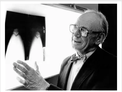
Patellofemoral Malalignment
In the 1970s anterior knee pain was related to the presence of patellofemoral malalignment (PFM).c In 1968, Jack C. Hughston (Figure 1.2) published an article on subluxation of the patella, which represented a major turning point in the recogni-tion and treatment of patellofemoral disorders.35 In 1974, Al Merchant, in an attempt to better understand patellofemoral biomechanics,
intro-6 Etiopathogenic Bases and Therapeutic Implications
Figure 1.2.Jack C. Hughston, MD (1917–2004). One of the founding fathers of Sports Medicine. (Reproduced with permission from the Journal of Athletic Training, 2004; 39: 309.)
c We define PFM as an abnormality of patellar tracking that involves lateral displacement or lateral tilt of the patella, or both, in extension, that reduces in flexion.
duced the axial radiograph of the patellofemoral joint.54The same author suggested, also in 1974, the lateral retinacular release as a way of treating recurrent patellar subluxation.55 In 1975, Paul Ficat, from France, popularized the concept of patellar tilt, always associated with increased tightness of the lateral retinaculum, which caused excessive pressure on the lateral facet of the patella, leading to the “lateral patellar compres-sion syndrome” (“Syndrome d’Hyperpression Externe de la Rotule”).21According to Ficat lateral patellar compression syndrome would cause hyperpressure in the lateral patellofemoral com-partment and hypopressure in the medial patellofemoral compartment. Hypopressure and the disuse of the medial patellar facet would cause malnutrition and early degenerative changes in the articular cartilage because of the lack of nor-mal pressure and function. This may explain why early chondromalacia patellae is generally found in the medial patellar facet. Hyperpression also would favor cartilage degeneration, which might explain the injury of the lateral facet. Two years later, in 1977, Ficat and Hungerford22published
Disorders of the Patellofemoral Joint,a classic of
knee extensor mechanism surgery and the first book in English devoted exclusively to the exten-sor mechanism of the knee. In the preface of the book these authors refer to the patellofemoral joint as “the forgotten compartment of the knee.” This shows what the state of affairs was in those days. In fact, before the 1970s only two diagnoses were used relating to anterior knee pain or patel-lar instability: chondromalacia patellae and recurrent dislocation of the patella. What is more, the initial designs for knee arthroplasties ignored the patellofemoral joint. In 1979, John Insall pub-lished a paper on “patellar malalignment syn-drome”38and his technique for proximal patellar realignment, used to treat this syndrome.39 According to Insall lateral loading of the patella is increased in malalignment syndrome. In some cases, this causes chondromalacia patellae, but it does not necessarily mean that chondromalacia is the cause of pain.41In this way, in 1983 Insall and colleagues reported that anterior knee pain corre-lates better to malalignment rather than with the severity of chondromalacia found during sur-gery.40 Fulkerson and colleagues have also emphasized the importance of PFM and exces-sively tight lateral retinaculum as a source of anterior knee pain.25,26,63Finally, in 2000, Ronald Grelsamer,31from the IPSG, stated that malalign-ment appears to be a necessary but not sufficient
condition for the onset of anterior knee pain.d According to Grelsamer,31 pain seems to be set off by a trigger (i.e., traumatism). In this sense, Grelsamer30 tells his patients that “people with malaligned knees are akin to someone riding a bicycle on the edge of a cliff. All is well until a strong wind blows them off the cliff, which may or may not ever happen.” Although it is more common to use the term malalignment as a mal-position of the patella on the femur some authors, as Robert A Teitge, from the IPSG, use the term malalignment as a malposition of the knee joint between the body and the foot with the subse-quent effect on the patellofemoral mechanics (see Chapter 11).
In a previous paper61we postulated that PFM, in some patients with patellofemoral pain, pro-duces a favorable environment for the onset of symptoms, and neural damage would be the main “provoking factor” or “triggering factor.” Overload or overuse may be another triggering factor. In this sense, in our surgical experience, we have found that in patients with symptoms in both knees, when the more symptomatic knee is operated on, the symptoms in the contralateral less symptomatic malaligned knee disappear or decrease in many cases, perhaps because we have reduced the load in this knee; that is, it allows us to restore joint homeostasis. In this connection, Thomee and colleagues suggested that chronic overloading and temporary overuse of the patellofemoral joint, rather than malalignment, contribute to patellofemoral pain.68
For many years, PFM has been widely accepted as an explanation for the genesis of anterior knee pain and patellar instability in the young patient. Moreover, this theory had a great influence on orthopedic surgeons, who devel-oped several surgical procedures to “correct the malalignment.” Unfortunately, when PFM was diagnosed it was treated too often by means of surgery. A large amount of surgical treatments has been described, yielding extremely variable results. Currently, however, the PFM concept is questioned by many, and is not universally accepted to account for the presence of anterior knee pain and/or patellar instability.
At present, most of the authors agree that only a small percentage of patients with patellofemoral pain have truly malalignment and are candidates for surgical correction of malalignment for resolution of symptoms. In fact, the number of realignment surgeries has dropped dramatically in recent years, due to a reassessment of the paradigm of PFM. Moreover, we know that such procedures are, in many cases, unpredictable and even danger-ous; they may lead to reflex sympathetic dys-trophy, medial patellar dislocations, and iatrogenous osteoarthrosis (see Chapters 20 and 21). We should recall here a phrase by doc-tor Jack Hughston, who said: “There is no problem that cannot be made worse by sur-gery” (see Chapters 20 to 23). Among problems with the knee, this statement has never been more relevant than when approaching the extensor mechanism. Therefore, we must emphasize the importance of a correct diagno-sis (see Chapters 6 and 7) and nonoperative treatment (see Chapters 9 and 10).
Criticism
The great problem of the PFM concept is that not all malalignments, even of significant propor-tions, are symptomatic. Even more, one knee may be symptomatic and the other not, even though the underlying malalignment is entirely symmetrical (Figure 1.3). On the other hand,
patients with normal patellofemoral alignment on computed tomography (CT) can also suffer from anterior knee pain (Figure 1.4). Therefore, PFM cannot explain all the cases of anterior knee pain, so other pathophysiological processes must exist. Moreover, PFM theory cannot adequately explain the variability of symptoms experienced by patients with anterior knee pain syndrome.
Finally, we must also remember that it has been demonstrated that there are significant differ-ences between subchondral bone morphology and geometry of the articular cartilage surface of the patellofemoral joint, both in the axial and sagittal planes6 (Figure 1.5). Therefore, a radi-ographical PFM may not be real and it could induce us to indicate a realignment surgery than could provoke involuntarily an iatrogenic PFM leading to a worsening of preoperative symptoms. This would be another point against the universal acceptance of the PFM theory. Moreover, this could explain also the lack of predictability of operative results of realignment surgery.
Critical Analysis of Long-term Follow-up
of Insall’s Proximal Realignment for
PFM: What Have We Learned?
In agreement with W.S. Halsted, I think that the operating room is “a laboratory of the highest order.” As occurs with many surgical techniques, and realignment surgery is not an exception,
8 Etiopathogenic Bases and Therapeutic Implications
after wide usage, surgeons may question the basic tenets and may devise clinical research to test the underlying hypothesis, in our case the PFM concept.
In this way we have evaluated retrospectively 40 Insall’s proximal realignments (IPR) per-formed on 29 patients with isolated sympto-matic PFM.eThe average follow-up after surgery was 8 years (range 5–13 years). The whole study is presented in detail in Chapter 2.
One of the objectives of this study was to ana-lyze whether there is a relationship between the presence of PFM and the presence of anterior knee pain or patellar instability.
In my experience IPR provides a satisfactory centralization of the patella into the femoral trochlea in the short-term follow-up.60However, this satisfactory centralization of the patella is lost in the CT scans performed in the long-term follow-up in almost 57% of the cases. That is, IPR does not provide a permanent correction in all the cases. Nonetheless, this loss of centralization does not correlate with a worsening of clinical
results. Furthermore, I have not found, in the long-term follow-up, a relation between the result, satisfactory versus nonsatisfactory, and the presence or absence of postoperative PFM. I postulate that PFM could influence the home-ostasis negatively, and that realignment surgery could allow the restoring of joint homeostasis when nonoperative treatment of symptomatic PFM fails. Realignment surgery temporarily would unload inflamed peripatellar tissues, rather than permanently modify PFM. Moreover, according to Dye, rest and physical therapy are most important in symptoms resolution than realignment itself. Once we have achieved joint homeostasis, these PFM knees can exist happily within the envelope of function without symp-toms. Moreover, in my series, 12 patients pre-sented with unilateral symptoms. In 9 of them the contralateral asymptomatic knee presented a PFM and only in 3 cases was there a satisfactory cen-tralization of the patella into the femoral trochlea. We can conclude that not all patellofemoral malaligned knees show symptoms, which is not surprising, as there are numerous examples of asymptomatic anatomic variations. Therefore, PFM is not a sufficient condition for the onset of symptoms, at least in postoperative patients. Thus, no imaging study should give us an indica-tion for surgery. History and physical exam must
10 Etiopathogenic Bases and Therapeutic Implications
Figure 1.5.Scheme of gadolinium-enhanced MR arthrotomogram of the left knee in the axial plane. Note perfect patellofemoral congruence (a). Note patellofemoral incongruence of the osseous contours (b). (Reprinted from Clin Sports Med, 21, HU Staeubli, C Bosshard, P Porcellini, et al., Magnetic resonance imaging for articular cartilage: Cartilage-bone mismatch, pp. 417–433, 2002, with permission from Elsevier.)
point toward surgery and imaging only to allow us to confirm clinical impression (see Chapter 6).
Relevance of our Findings
To think of anterior knee pain or patellar insta-bility as somehow being necessarily tied to PFM is an oversimplification that has posi-tively stultified progress toward better diagno-sis and treatment. The great danger in using PFM as a diagnosis is that the unsophisticated or unwary orthopedic surgeon may think that he or she has a license or “green light” to cor-rect it with misguided surgical procedures that very often make the patients’ pain worse (see Chapters 20 and 21).
Tissue Homeostasis Theory
In the 1990s, Scott F. Dye, of the University of California, San Francisco, and his research group, came up with the tissue homeostasis theory.17,19 The initial observation that led to the develop-ment of the tissue homeostasis theory of patellofemoral pain was made by Dye, when a patient with complaints of anterior knee pain without evidence of chondromalacia or malalign-ment underwent a technetium 99m methylene diphosphonate bone scan evaluation of the knees in an attempt to assess the possible presence of covert osseous pathology. The bone scan of that individual manifested an intense diffuse patellar uptake in the presence of normal radiographic images. This finding revealed the presence of a covert osseous metabolic process of the patella in a symptomatic patient with anterior knee pain and normal radiographic findings.
The tissue homeostasis theory is in agree-ment with the ideas exposed by John Hilton (1807–1876) in his famous book Rest and
Pain:50“The surgeon will be compelled to admit
that he has no power to repair directly any injury . . . it is the prerogative of Nature alone to repair . . . his chief duty consists of ascertaining and removing those impediments with thwart the effort of Nature.” Moreover, this is in agree-ment with the ideas exposed by Thomas Sydenhan (1624–1689), “the father of English Medicine,” and a cardinal figure in orthopedics in Britain and the world, who looked back to Hippocrates, who taught that Nature was the physician of our diseases. According to Sydenhan the doctor’s task was to supplement, not to supplant Nature.50
The tissue homeostasis theory states that joints are more than mechanical structures –
they are living, metabolically active systems. This theory attributes pain to a physiopatholog-ical mosaic of causes such as increase of osseous remodeling, increase of intraosseous pressure, or peripatellar synovitis that lead to a decrease of what he called “Envelope of Function” (or “Envelope of Load Acceptance”).
According to Dye,17the Envelope of Function describes a range of loading/energy absorption that is compatible with tissue homeostasis of an entire joint system, that is, with the mechanisms of healing and maintenance of normal tissues. Obviously, the Envelope of Function for a young athlete will be greater than that of sedentary eld-erly individual. Within the Envelope of Function is the region termed Zone of Homeostasis (Figure 1.6A). Loads that exceed the Envelope of Function but are insufficient to cause a macrostructural failure are termed the Zone of Supraphysiological Overload (Figure 1.6A). If sufficiently high forces are placed across the patellofemoral system, macrostructural failure can occur (Figure 1.6A).
For Dye17 the following four factors deter-mine the Envelope of Function or Zone of Homeostasis: (1) anatomic factors (morphol-ogy, structural integrity and biomechanical characteristics of tissue); (2) kinematic factors (dynamic control of the joint involving propri-oceptive sensory output, cerebral and cerebellar sequencing of motor units, spinal reflex mecha-nisms, and muscle strength and motor control); (3) physiological factors (the genetically deter-mined mechanisms of molecular and cellular homeostasis that determine the quality and rate of repair of damaged tissues); and (4) treatment factors (type of rehabilitation or surgery received).
where many activities of daily living previously well tolerated (e.g., stair climbing, sitting down in and arising out of chairs, pushing the clutch of a car) become sufficiently high (supraphysio-logical loads for that patient) to lead to subver-sion of tissue healing and continued symptoms (Figure 1.6B). Decreasing loading to within the newly diminished Envelope of Function allows normal tissue healing processes (Figure 1.6C).
Finally, according to Dye many instances of giving way, in patients with patellofemoral pain, could represent reflex inhibition of the quadri-ceps, which results from transient impingement
of swollen, innervated peripatellar soft tissues, such as inflamed synovium in patients with nor-mal alignment.
Clinical Relevance
Patients with an initial presentation of anterior knee pain frequently will respond positively to load restriction within their Envelope of Function and pain-free rehabilitation program. Moreover, Dye believes that enforced rest after realignment surgery could also be important in symptom resolution. Even if patients, parents, and trainers are apt to stubbornly reject any
12 Etiopathogenic Bases and Therapeutic Implications
suggestion to introduce changes into the patient’s activities and training routine demand-ing an urgent surgical procedure, orthopedic surgeons should under no circumstances alter their opinions and recommendations, however strong the pressure exerted upon them may be. Trainers, physical therapists, and physicians all have a high degree of responsibility and need to behave in an ethical way.
Patellofemoral Malalignment Theory
versus Tissue Homeostasis Theory
In essence, the proponents of tissue homeostasis theory look at PFM as representing internal load shifting within the patellofemoral joint that may lower the threshold (i.e., decrease of the Envelope of Function) for the initiation and per-sistence of loss of tissue homeostasis leading to the perception of patellofemoral pain. Pain always denotes loss of tissue homeostasis. From this perspective, there is no inherent conflict between both theories. However, these are not two co-equal theories. Tissue homeostasis the-ory easily incorporates and properly assesses the clinical importance of possible factors of PFM, whereas the opposite is not true.
In conclusion, I truly believe that both theo-ries are not exclusive, but complementary. In my experience, a knee with PFM can exist hap-pily within its envelope of function, but once it is out, for example by overuse, training error, pat-terns of faulty sports movements, or trauma-tism, it can be harder to get back within it, and realignment surgery could be necessary in very selected cases.
Myths and Truths about
Patellofemoral Disease
Myth:Anterior knee pain and patellar instability are always self-limited and therefore active treatment is unnecessary. The natural history of this pathological entity is always benign.
Traditionally, anterior knee pain syndrome is considered to be a self-limited condition with-out long-term sequelae. This is true of many cases but cannot be regarded as a golden rule. A large percentage of patients experience spon-taneous recoveries; indeed, many patients remain asymptomatic even without specific treatment. In the case of some of our patients, 10 years elapsed from the onset of symptoms until the time of surgery; their symptoms not only
failed to improve but they worsened in spite of the passage of time and of the patient’s restrict-ing or even abandonrestrict-ing sports practice. These same patients obtained excellent or good results after correction of their symptomatic PFM, which persisted in the long-term follow-up (see Chapter 2). Milgrom and colleagues57performed a prospective study to determine the natural history of anterior knee pain caused by over-activity. At six years’ follow-up, half of the knees originally with anterior knee pain were still symptomatic, but in only 8% of the originally symptomatic knees was the pain severe, hinder-ing physical activity. Clinical experience shows that a prolonged and controlled active conserva-tive treatment generally solves the problem. On the other hand, trying to negligently ignore the problem causes disability in some patients. Unfortunately, the patients’ own ambition, as well as that of their parents and coaches, pre-vails over their doctor’s judgment, which is nec-essarily based on avoiding for at least 3 to 6 months any sports movement that could cause pain. That is, the fact that this process is on occasion self-limited should not make us forget the need to indicate active treatment in all cases. This means that the process we are studying is reversible at least until a certain point has been reached. The question we ask ourselves is: Where is the point of no return?
Primary patellar dislocation is not a trivial condition either. It is true that with the passage of time the frequency of recurrent dislocations tends to diminish, but each episode is a potential source for a chondral injury.31 A long-term assessment of patients (mean follow-up of 13 years) reveals that conservative treatment of patellar dislocation results in 44% of redisloca-tions and 19% of late patellofemoral pain.51
(p < 0.001) in the patients who underwent patellofemoral replacement for isolated patellofemoral osteoarthrosis (22% and 14% respectively) than in those who underwent uni-compartmental replacement for isolated medial compartment osteoarthrosis (6% and 1% respectively). They conclude that anterior knee pain syndrome is not always a self-limiting condition given that it may lead to patellofemoral osteoarthrosis. On the other hand, Arnbjörnsson and colleagues3 found a high incidence of patellofemoral degenerative changes (29%) after nonoperative treatment of recurrent dislocation of the patella (average fol-low-up time 14 years with a minimum folfol-low-up time of 11 years and a maximum follow-up time of 19 years (range 11–19 years)). Bearing in mind that the mean age of the patients at follow-up was 39 years they conclude that recurrent dislocation of the patella seems to cause patello-femoral osteoarthrosis. In conclusion, PFM’s natural history is not always benign.
Quite often, symptomatic PFM is associated with a patellar tendinopathy.2The latter has also been called a self-limited pathology. It has been shown that it is not a benign condition that sub-sides with time; that is, it is not a self-limited process in athletes.53 Normally, the injury pro-gresses and when it gets to Blazina’s stage III it generally becomes irreversible and leads to the failure of conservative treatment.53
Myth:Anterior knee pain is related to growth and, therefore, once the patient has fully grown symptoms will disappear.
Anterior knee pain has also been related to growing pains. It is true that in young athletes during their maximum growth phase (“growth spurt”) there can be an increase in the tension of the extensor mechanism as a consequence of some “shortcoming” or “delay” in its develop-ment vis-à-vis bone growth. There may exist also a delay in the development of the VMO with regard to other muscles in the knee and there-fore a transient muscle imbalance may ensue. But it is also true that quite often parents tell us that the doctor their child saw told them that when the child stopped growing the symptoms would go away and that, nevertheless, these per-sist once the child has fully grown.
Myth:Anterior knee pain in adolescents is an expres-sion of psychological problems.
Many physicians believe that anterior knee pain is a sign of psychological problems.
Consequently this condition has been associated with a moderate elevation of hysteria and, to a lesser degree, hypochondria with the problem in the knee being considered an unconscious strat-egy to confront an emotional conflict.44Likewise, it has been shown that, on some occasions, in adolescent women anterior knee pain with no evident somatic cause can represent a way to control solicitous or complacent parents.44 What cannot be questioned is that anybody at whatever age can somatize or try to attract other people’s attention through some disease. In spite of this, one should be very cautious when it comes to suggesting to parents that their child’s problem is wholly psychological. Nonetheless, it has to be recognized that these types of patients present with a very particular psychological pro-file (see Chapter 6). Furthermore, there are patients with objective somatic problems who disproportionately exaggerate their pain because of some associate psychological compo-nent or secondary emotional or financial gains.
Unfortunately, in my personal current surgi-cal series (84 patients, 102 knees) there are 8 patients (7 females and 1 male) who had been referred to a mental health unit. Strangely enough, these patients’ problem was satisfacto-rily addressed by surgery, which shows that the problem was not psychological. In addition, both the histological and the immunohisto-chemical and immunoimmunohisto-chemical techniques– based studies of the lateral patellar retinacula of these patients showed objective alterations that made it possible for us to detect that the pain had a neuroanatomic base. In short, the ortho-pedic surgeon has the duty to rule out mechani-cal problems as well as other pathologies that may cause anterior knee pain before blaming the pain on emotional problems or feigning.
Myth:Patellofemoral crepitation is in itself an indica-tion of disfuncindica-tion.
A very common symptom that worries patients very much is patellofemoral crepitation. Crepitation is indicative of an articular cartilage lesion in the patella or in the femoral trochlea. Nonetheless, some patients who present with crepitation have a macroscopically intact cartilage at the moment of performing the arthroscopy.30 The crepitation could be caused by alterations in the synovial or in other soft tissues.
The International Knee Documentation Committee (IKDC)33stated: “The knee is normal when crepitation is absent.” However, this statement cannot be upheld after Johnson and
colleagues45 published their 1998 paper in
Arthroscopyon the assessment of asymptomatic
knees. Indeed, patellofemoral crepitation has a great incidence in asymptomatic women (94% in females versus 45% in males).45 Patellofemoral crepitation has been associated with the lateral subluxation of the patella, but Johnson and col-leagues45have observed that lateral subluxation of the patella in asymptomatic persons is more common in males than in females (35% vs. 19%). Crepitation is not always present in patients with significant pain. Furthermore, when it is present is does not necessarily cause anterior knee pain. In short, since crepitation is frequent in asympto-matic knees, its presence is more significant when it is absent from the contralateral knee or when there is some kind of asymmetry.
Myth:VMO is responsible for patellar stability. It has been stated that the vastus medialis obliquus (VMO) is responsible for patellar sta-bility, but we have not found convincing evi-dence in the literature for this belief; and, as ligaments are the joint stabilizers, this premise would appear to be faulty. In theory, the VMO resists lateral patellar motion, either by active contraction or by passive muscle resistance. In this way, in Farahmand’s study,20lateral patel-lar force-displacement behavior was not affected by simulated muscle forces at any flexion angle from 15 to 75°. On the other hand, the orientation of the VMO varies greatly during knee flexion. The VMO’s line of pull most efficiently resists lateral patellar motion when the knee is in deep flexion, at which time trochlear containment of the patella is independent of soft tissues influ-ences (see Chapter 5).
It seems likely that operations that advance the VMO include tightening of the underlying medial patellofemoral ligament (MPFL), and it would be responsible for the success of the surgical technique (see Chapter 2). In this sense, we must note that the VMO tendon becomes confluent with the MPFL in the region of patellar attachment. Therefore, it would be more logical to protect the VMO and address the ligament deficiency surgically as needed (see Chapter 5).
Controversy:Should the Q angle be measured? If so,
how should it be measured? Is this of any use?31,58
Another aspect that normally receives great importance in the physical examination of these
patients is their Q angle, to the extent that some authors regard it as one of the criteria to be used for indicating a realignment surgery. Nonetheless, values considered to be normal vary greatly across the different studies carried out. In addition, there are no scientific criteria that correlate the incidence of patellofemoral pathology with the Q angle measure. Nowadays, some believe that the Q angle, as it is calculated, is not a very accurate way of measuring the patella’s alignment since the measurement is made in extension and a laterally subluxating patella would lead to a falsely low measurement. In sum, even if Q angle measurement has tradi-tionally been used in the clinical assessment of patients with a patellofemoral pathology, cur-rently the usefulness of this measurement is uncertain in spite of the multiple studies per-formed to date. A realignment surgery must never be justified on the basis of a high Q angle (see Chapter 20, clinical case 1). The real contro-versy at present is how to measure the Q angle.
Myth: Lateral release is a minor risk-free surgical procedure.
Over the years, lateral retinacular release has been recommended for a number of specific patellofemoral conditions:23 recurrent lateral patellar dislocations or subluxations, chronic lat-eral subluxation – fixed latlat-eral position, excessive lateral pressure syndrome, lateral retinacular tightness, and retinacular neuromata. A possible explanation for this wide range of surgical indica-tions could be that some orthopedic surgeons consider the lateral release as a minor risk-free surgical procedure. However, I believe in agree-ment with Ronald Grelsamer that “There is no such thing as minor surgery – only minor sur-geons.” Surprisingly, in a survey of the IPSG23on isolated lateral retinacular release, published in 2004 in Arthroscopy, most respondents (89%) indicated that this surgical procedure is a legiti-mate treatment, but only on rare occasions (1% to 2% of surgeries performed, less than 5 lateral releases a year). Furthermore, strong consensus (78%) existed that objective evidence should show lateral retinacular tension if a lateral release is to be performed.
on CT scans,27(2) to increase passive medial dis-placement of the patella,64,67and (3) to increase passive lateral displacement of the patella.15 Finally, in cadaver knees without preexisting lateral retinacular tightness, lateral release had no effect on articular pressures when the quadri-ceps were loaded.34
In conclusion, indiscriminate use of lateral release is of little benefit and can often cause increased symptoms. That is the reason why lengthening of the lateral retinaculum is the therapy chosen by authors such as Roland Biedert (see Chapter 20).
Reality:Patellofemoral pathology leads to diagnostic error and, therefore, to inappropriate treatments and to patients being subjected to multiple procedures and to a great deal of frustration.
All myths and controversies analyzed through-out the present chapter could lead the reader to attribute importance to things that in actual fact are unimportant (i.e., crepitation) or, on the contrary, to underrate or cast aside complaints like anterior knee pain or functional patellar instability, considering them to be either a psy-chological problem or a condition bound to sub-side with time. Sometimes we do not go far enough, which may lead us to overlook other pathologies (diagnostic errors leading to thera-peutic errors). In other cases we overdo it and treat cases of malalignment that are not sympto-matic. So we have seen patients with symptoms of instability who were treated for malalignment when what they really had was instability caused by a tear in their ACL.
We have also seen patients treated for a meniscal injury who really had isolated sympto-matic PFM. In this connection it is important to point out that McMurray’s test, traditionally associated with meniscal pathology, can lead to a medial-lateral displacement of the patella and also cause pain in patients with PFM. Finally, it is worrying to see how many patients are referred to outpatient orthopedic surgery prac-tices in our hospitals with an MRI-based diag-nosis of a tear in the posterior horn of the medial meniscus who during clinical examina-tion present with anterior knee pain and no meniscal symptoms. It is a proven fact that given the overcrowding of outpatient units’ orthopedic services and because of social pres-sure, as time passes doctors tend to conduct more superficial physical examinations and to order more MRIs. In this way we must
remem-ber the statement by Dr. Casscells:10“Technology: a good servant, but a bad master.” According to Augusto Sarmiento, former Chairman of the American Academy of Orthopedic Surgeons (AAOS), MRIs are unfortunately replacing the physical examination when it comes to assessing a painful joint.62 MRI is not a panacea and, what’s more, it gives rise to false positives. Patients’ great faith in technology and their skepticism regarding their doctors and an increasingly dehumanized medical practice has resulted in the failure of partial arthroscopic meniscectomies owing to a bad indication, in frustrated patients, and in the squandering of resources. In 1940, Karlson46wrote the following about chondromalacia patellae: “The diagnosis is difficult to make and the differential diagnosis of injury to the meniscus . . . causes special diffi-culties, as in both these ailments [meniscal and patellar pathology] there is a pressure tenderness over the medial joint space.” Hughston endorsed these words when he stated, first in 1960 and then in 1984:36“The orthopedic surgeon who has not mistaken a recurrent subluxation of the patella for a torn meniscus has undoubtedly had a very limited and fortunate experience with knees and meniscectomies.” Just think of the sheer amount of arthroscopies performed unnecessarily on the basis of a complaint of anterior knee pain!
Nowadays this problem has been magnified because of the relative ease with which meniscec-tomies are indicated and performed thanks to the benefits of arthroscopy. In a lecture delivered at the Conference of the Nordic Orthopaedic Federation held in Finland in 2000, Augusto Sarmiento stated that the number of unnecessary surgeries (including arthroscopies) carried out in our field in the United States is extremely high.62It is therefore essential to underscore the importance of physically examining the patient (see Chapter 6).
Finally, another source of frustration for the patient is the lack of communication with his or her doctor (dehumanized medicine), which may lead to unrealistic expectations. It is essential for the patient to understand the difficulties inher-ent in treating patellofemoral problems. This is the only way in which patients can be satisfied after surgery even if their symptoms do not dis-appear completely.
Reality: “Treatment should be customized.”
It is very important to identify the pathological alteration responsible for the clinical aspect of