Microleakage in class V gingiva-shaded
composite resin restorations
Claudio Poggio, MD, DDS Marco Chiesa, DDS Alberto Dagna, DDS Marco Colombo, DDS, PhD Andrea Scribante, DDS, PhD1
University of Pavia, Italy
Department of Operative Dentistry 1Department of Orthodontics
Corresponding author:
The purpose of this study was to evaluate the microleakage in Class V cavities restored with a new gingiva-shaded microhybrid composite resin and with a conventional microhybrid composite resin using three different dentin bonding systems (DBS). Class V cavities were prepared in sixty freshly extracted human teeth with the incisal margin in enamel and the apical margin in dentin/cementum. Restored specimens, after thermocycling, were placed in 2% methylene blue solution for 24 hours. Longitudinal sections were obtained and studied with a stereomicroscope for assessment of the microleakage according to degree of dye penetration (scale 0-3). Data were analyzed with Kruskal-Wallis test and with Mann-Whitney U-test.
In this study there was no leakage in enamel: all the cavities showed no dye penetration at the incisal margins (located in enamel). None of the DBS used eliminated microleakage in apical margins (located in dentin or cementum): three-step total-etch and single-step self-etch were more effective in reducing microleakage in dentin margins when compared with two-step total-etch. This in vitro study concluded that microleakage in Class V cavities restored with the composite resins tested is similar.
Key words: gingiva-shaded composite resin, dentin bonding systems, microleakage.
Introduction
Cervical lesions are frequently found in daily practice. Degenerative processes and gingival recession, as
result of chronic periodontal inlammation or aggressive
periodontal therapy, expose root dentin predominantly in older patients (1). The aetiology of cervical lesions is multifactorial (2). Non-carious cervical lesions are characterized by the loss of dental hard tissue at cementum-enamel junction as a result of abrasive
or erosive effects (3); additionally cuspal lexure and
tensile stresses in cervical region of the teeth are hypothesized to cause disruption of the bonds among the hydroxyapatite crystals, leading to cracks and loss of enamel and underlying dentin (4). Food acids and inappropriate tooth brushing procedures promote the development of saucer- or wedge-shaped lesions in the cervical area of the teeth (1). In such patients, low dental care and non-effective preventive programs lead to rapid development of caries. The treatment regimen depends on the individual cause of the cervical lesion (5). Instructions for adequate oral hygiene, dietary advices, occlusal adjustment and restoration of the defect are recommended (6). Small non-carious cervical defects with no hypersensitivity, especially in the posterior region, may require only dietary consulting and oral hygiene instruction in order to avoid further progression (1,5). Pain-associated or caries-affected Class V lesions need to be restored. Particular attention is required in anterior teeth, both for small non-carious and for big carious lesions, where aesthetic reasons are perdominant. Glass ionomer cements, compomers and composite resins are being mostly used to restore class V defects (1,3,6-8). Amalgam and gold restoration were used in the past but today the increased demand for aesthetic decreased their utilization. Composite resins combined with dentin bonding systems have already substituted glass ionomer cements and compomers in Class V restorations because of their excellent aesthetics, superior mechanical and physical properties and higher bond strength to enamel and dentin (1,9,10). The complex morphology of Class V defects with margins partly in enamel as well as in root dentin presents challenging task for the restorative material (1). Bond strength and sealing ability of adhesive systems to dentin is still inferior compared to enamel cavity segments (11). In Class V the polymerization of composite resins competes with the bond strength of the adhesive systems and challenges marginal integrity and sealing ability especially in dentin segments. Marginal gap formation leads to leakage, responsible for marginal discoloration, secondary caries and partial or total loss of the restoration (6,12). However, newly formulated improved bonding systems provide a better adhesion of the composite resin to dentin, although this is an unresolved problem. The exposed and frequently worn cervical areas of teeth often lack a pleasing appearance
when restored with conventional materials such as glass ionomer cements or tooth coloured composite resins, especially when associated with aging and loss of gingiva. Today the interest is directed toward the development of new restorative materials that permit chair side gingival shade matching, in order to restore aesthetic and function of cervical areas exposed after gingival recession (discoloured or hypersensitive necks of teeth) and cervical V-shaped defects (13).
The purpose of this in vitro study was to investigate the microleakage in Class V cavities restored with a new gingiva-shaded microhybrid composite resin (Amaris Gingiva) and with a conventional microhybrid composite resin (Amaris) using three different dentin bonding systems (three-step total-etch, two-step total-etch and single-step self-etch). The null hypothesis of the study
was that there is no signiicant difference in microleakage
scores among the various groups.
Materials and methods
Specimen preparation
Sixty caries-free vital human teeth freshly extracted for periodontic or orthodontic reasons were used in this study. The teeth were cleaned with dental scalers, polished with pumice and stored in a 0.25% mixture of sodium azide in Ringer solution until the date of use. In each tooth two standardized Class V cavities (on buccal and on lingual surfaces) were prepared with a round-nosed no.245 carbide bur (Dentsply/Caulk, Milford, DE, USA) at high-speed with air/water spray, according to procedure described in Corona et al. (14): the cavities were prepared with the incisal margin located in enamel and the apical margin located in dentin/cementum (3 mm beyond the cementum-enamel junction); the dimensions of Class V cavities were similar, with mesiodistal width, incisal-apical measure and depth of 4 mm. The same trained operator prepared all the cavities. The teeth were randomly assigned to six experimental groups (of 10 specimens and 20 cavities each); Class V cavities were
illed with two composite resins and with three dentin
bonding systems (DBS) as follows: group 1: Solobond Plus + Amaris Gingiva, group 2: Solobond Plus + Amaris, group 3: Solobond M + Amaris Gingiva, group 4:
After application of DBS (according to Manufacturers’ instructions) cavities were restored with composite resin in three increments. Each increment of composite resin was light-cured for 40 seconds with a curing light in softstart-polymerization mode (Celalux 2 High-Power LED curing-light, Voco GmbH, Cuxhaven, Germany).
The restorations were inished and polished with inishing/polishing disks (Sof-Lex Pop-On, 3M ESPE,
St. Paul, MN, USA) in decreasing granulation. All teeth were coated with two layers of nail varnish up to 1 mm from the restorations margins, while the apical part was sealed with wax. The restored teeth were then subjected
to artiicial aging by thermocycling. All specimens were
immersed alternately in water baths at 5 and 60°C for 1000 cycles, with at dwell time of 60 seconds in each bath and a transfer time of 15 seconds. After thermocycling, the specimens were immersed in a 2% methylene blue dye solution and incubated at 37°C for 24 hours.
Microleakage analysis
The teeth were rinsed with distilled water, dried for 10 minutes, and sectioned longitudinally in a buccolingual direction with a low-speed water-cooled diamond cutter. All specimens were examined at 25´ in a stereomicroscope (Inspective 4Geek, Serravalle, RSM) and standardized digital images were obtained. Two observers scored each section blindly; consensus was forced if disagreements occurred. An ordinal scale from 0 to 3 was used to score microleakage separately at incisal (enamel) and apical (dentin/cementum) margins of each section based on the following criteria, as described in Osorio et al.(15) and in Moldes et al.(16): 0 = no leakage visible at tooth/restoration interface, 1 = dye penetration along the interface up to one-half of the cavity depth, 2 = dye penetration greater than one-half of the cavity depth, 3 = dye penetration to and along axial wall.
Statistical analysis
The results of microleakage evaluation were submitted to statistical analysis using “Stata 7.0” computer software (Stata Corp., Station College, TX). A Kruskal-Wallis test and a Mann-Whitney U-test were performed.
Signiicance was predetermined at p<0.05.
IC
Ed
izi
o
n
i I
n
te
rn
a
zi
o
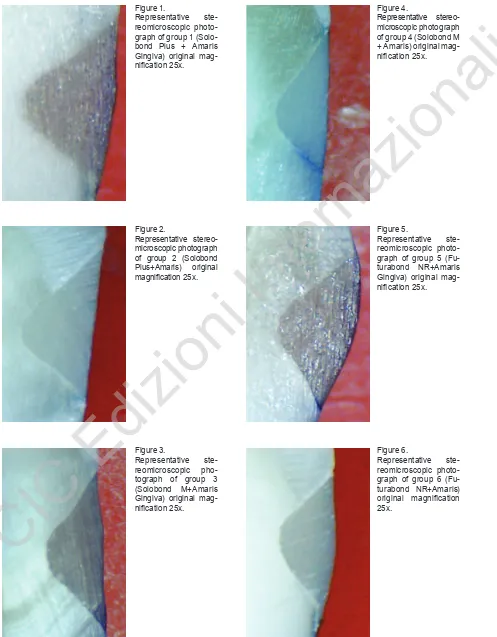
Figure 1.
Representative ste-reomicroscopic photo-graph of group 1 (Solo-bond Plus + Amaris Gingiva) original
mag-niication 25x.
Figure 4.
Representative stereo-microscopic photograph of group 4 (Solobond M + Amaris) original
mag-niication 25x.
Figure 2.
Representative stereo-microscopic photograph of group 2 (Solobond Plus+Amaris) original
magniication 25x.
Figure 5.
Representative ste-reomicroscopic photo-graph of group 5 (Fu-turabond NR+Amaris Gingiva) original
mag-niication 25x.
Figure 3.
Representative ste-reomicroscopic pho-tograph of group 3 (Solobond M+Amaris Gingiva) original
mag-niication 25x.
Figure 6.
Representative ste-reomicroscopic photo-graph of group 6 (Fu-turabond NR+Amaris)
original magniication
25x.
©
C
IC
Ed
izi
o
n
i I
n
te
rn
a
zi
o
occlusal margin scores, no signiicant differences were
showed among the three different adhesives tested (p>0.05). On the other hand, when analyzing gingival
margin scores, groups 3 and 4 showed signiicant higher
frequency of microleakage scores of “1” (P<0.05) than the other groups, that both showed higher frequency of score “0”.
Discussion
The null hypothesis of the study was partially rejected. The results of the present investigation showed no
signiicant differences in microleakage values among
the gingiva-shaded microhybrid composite resin (Amaris Gingiva) and the conventional microhybrid composite resin (Amaris). The microleakage of the DBS tested in
enamel is signiicantly reduced or null. The results of this study are in agreement with indings in Literature
(14,15,17,20): all the specimens showed less dye penetration at incisal margins (in enamel), with no differences emerging between the different adhesive systems. On the other side, the results revealed that
the priming and the bonding steps (also deined
total-etch systems); two-step systems include self-priming adhesives (that require a separate etching step) or self-etching primers (that require a separate bonding step);
inally, the recently introduced single-step (self-etching)
adhesives combine all bonding procedures in a single application, consisting of a mixture of acid monomers that etch enamel and dentin as well as primers that permits penetration of resin into the demineralized dentin (27). No DBS currently available achieved a perfect seal in dentin/cementum (15,19,20,28,30-34). The causes of microleakage are usually associated with the composite resin used, the occlusal load, the location of the prepared margins and the polymerization shrinkage (23): DBS have the aim to minimize those effects.
The restoration of cavities with margins located in dentin/ cementum is an unresolved problem in operative dentistry; microleakage has clinical effects and causes failure of resin restorations. The reason for this difference between apical and incisal leakage scores is that bonding to dentin/cementum is much more technique-sensitive and substrate-sensitive than bonding to enamel (25,30-34).
Conclusion
This study showed no differences between the microleakage in class V cavities restored with the composite resins tested: Amaris Gingiva (gingiva-shaded microhybrid composite resin) and Amaris (conventional microhybrid composite resin). Within the limitations of this study it may be concluded that microleakage among two composite resins tested is similar. The microleakage
of all DBS tested in enamel is signiicantly reduced or
null, but three-step total-etch and single-step self-etch were more effective in reducing microleakage in dentin/ cementum margins when compared with a two-step total-etch.
References
1. Manhart J, Chen HY, Mehl A, Weber K, Nickel R. Marginal quality and microleakage of adhesive class V restorations. J Dent 2001;29:123-130.
Table 2 - Occlusal margin-microleakage scores obtained for each experimental group (n=20).
Table 3 - Gingival margin-microleakage scores obtained for each experimental group (n=20).
groups Score 0 Score 1 Score 2 Score 3
1 18 (90%) 2 (10%) 0 (0%) 0 (0%)
2 19 (95%) 1 (5%) 0 (0%) 0 (0%)
3 17 (85%) 2 (10%) 1 (5%) 0 (0%)
4 17 (85%) 3 (15%) 0 (0%) 0 (0%)
5 17 (85%) 3 (15%) 0 (0%) 0 (0%)
6 16 (80%) 3 (15%) 1 (5%) 0 (0%)
groups Score 0 Score 1 Score 2 Score 3
1 13 (65%) 5 (25%) 2 (10%) 0 (0%)
2 12 (60%) 7 (35%) 1 (5%) 0 (0%)
3 3 (15%) 9 (45%) 5 (25%) 3 (15%)
4 2 (10%) 11 (55%) 6 (30%) 1 (5%)
5 14 (70%) 4 (20%) 2 (10%) 0 (0%)
6 12 (60%) 5 (25%) 3 (15%) 0 (0%)
IC
Ed
izi
o
n
i I
n
te
rn
a
zi
o
8. Van Meerbek B, Peumans M, Gladys S, Braem M, Lam-brechts P, Vanherle G. Three-year clinical effectiveness of four total-etch dentinal adhesive systems in cervical le-sions. Quintessence Int 1996;27:775-84.
9. Abdalia AI, Alhadainy HA, Garcia-Godoy F. Clinical evalu-ation of glass ionomers and compomers in class V carious lesions. Am J Dent 1997;10:18-20.
10. Gladys S, Van Meerbek B, Braem M, Lambrechts P, Van-herle G. Comparative physico-mechanical characteriza-tion of new hybrid restorative materials with convencharacteriza-tional glass-ionomer and resin composite restorative materials. J Dent Res 1997;76:883-94.
11. Dietrich T, Losche AC, Losche GM, Roulet JF. Marginal ad-aptation of direct composite and sandwich restorations in class II cavities with cervical margins in dentine. J Dent 1999;27:119-28.
12. Sparrius O, Grossman ES. Marginal leakage of composite resin restorations in combination with dentinal and enamel bonding agents. J Prosthet Dent 1989;61:678-84. 13. Günay H, Geurtsen W, Lührs AK. Conservative treatment
of periodontal recessions with class V-defects using gingiva-shaded composite--A systematic treatment con-cept. Dent Update. 2011;38:124-32.
14. Corona SAM, Borsatto MC, Pecora JD, De Sa Rocha RAS, Ramos TS, Palma-Dibb RG. Assessing microleakage of different class V restorations after Er:Yag laser and bur preparation. J Oral Rehabil 2003;30:1008-14.
15. Osorio R, Toledano M, de Leonardi G, Tay F. Microleak-age and interfacial morphology of self-etching adhesives in class V resin composite restorations. J Biomed Mater Res B Appl Biomater 2003; 66:399-409.
16. Moldes VL, Capp CI, Navarro RS, Matos AB, Youssef MN, Cassoni A. In vitro microleakage of composite restorations prepared by Er:YAG/Er,Cr:YSGG lasers and conventional drills associated with two adhesive systems. J Adhes Dent 2009;11:221-9.
17. Ferrari M, Goracci G, Garcia-Godoy F. Bonding mechanism of three “one bottle” system to conditioned and uncondi-tioned enamel and dentin. Am J Dent 1997;10:224-30. 18. Hannig M, Reinhardt KJ, Bott B. Self-etching primer vs
phosphoric acid : an alternative concept for composite-to-enamel bonding. Oper Dent 1999;24:172-80.
19. Shigetani Y, Tate Y, Okamoto A, Iwaku M, Abu-Bakr N. A study of cavity preparation by Er :YAG laser effects on the marginal leakage of composite resin restoration. Dent Ma-ter J 2002;21:238-49.
20. Kolinotou-Koumpa E, Dionysopoulos P, Koupia E. In vivo evaluation of microleakage from composites with new den-tine adhesives. J Oral Rehabil 2004;31:1014-22.
21. Haller B. Recent development in dentin bonding. Am J Dent 2000;13:44-50.
22. Amaral CM. Microleakage of hydrophilic adhesive systems in class V composite restorations. Am J Dent 2001;14:31-3.
23. Arias VG, Campos IT, Pimenta LAF. Microleakage study of three adhesive systems. Braz Dent J 2004;15:194-8. 24. Fusayama T, Nakamura M, Kurosaki N, Iwaku M.
Non-pressure adhesion of a new adhesive restorative resin. J Dent Res 1979;58:1364-70.
25. Tay FR, Pashley DH. Permeability of single-step, self-etch adhesives: the cost of saving time. In: Proceedings of the International symposium’ 01 in Tokio June 26,2001 Edited by Junji Tagami
26. Gladys S, Van Meerbeek B, Lambrechts P, Vanherle G. Microleakage of adhesive restorative materials. Am J Dent 2001;14:170-6.
27. Nikaido T, Nakajima M, Higashi T, Kanemura M, Pereira Pn, Tagami J. Shear bond strength of a single-step bonding system to enamel and dentin. Dent Mater J 1997;16:40-7. 28. Prati C, Nucci C, Davidson CL, Montanari G. Early
mar-ginal leakage and shear bond strength of dentin bonding systems. Dent Mater 1990;6:195-200.
29. Abo T, Uno S, Sano H. Comparison of bonding eficacy of
an all-in-one adhesive with a self-etching primer system. Eur J Oral Sci 2004;112:286-92.
30. Ateyah NZ, Elhejazi AA. Shear bond strengths and micro-leakage of four types of dentin adhesive materials. J Con-temp Dent Pract 2004;5:63-73.
31. Fortin D, Swift EJ, Denehy GE, Reinhardt JW. Bond strength and microleakage of current dentine adhesives. Dent Mater 1994;10:253-8.
32. Pashley EL, Agee KA, Pashley DH, Tay FR. Effects of one
versus two applications of an unilled, all-in-one adhesive
on dentine bonding. J Dent 2002;30:83-90.
33. Yaseen SM, Subba Reddy VV. Comparative evaluation of microleakage of two self-etching dentin bonding agents on primary and permanent teeth. An in vitro study. Eur J Paediatr Dent. 2010;11:127-31.
34. Kirk PC, Fitchie JG, Phillips SM, Puckett AD. Microleak-age evaluation of four self-etching adhesive systems. Gen Dent. 2010;58:104-9.