S C I E N T I F I C A R T I C L E
Australian Dental Journal2008; 53: 172–175doi: 10.1111/j.1834-7819.2008.00028.x
Microleakage of composite resin restorations in cervical
cavities prepared by Er,Cr:YSGG laser radiation
S Shahabi,* L Ebrahimpour,
LJ Walshà
*Department of Dental Materials, School of Dentistry and Dental Research Center, Tehran University of Medical Sciences, Iran.
General Dental Practice, Tehran, Iran.
àSchool of Dentistry, The University of Queensland, Brisbane, Australia.
ABSTRACT
Background: Evaluation of microleakage is important for assessing the success of new methods for surface preparation and new adhesive restorative materials. The aim of this laboratory study was to assess microleakage at the margins of composite restorations in Er,Cr:YSGG laser prepared cavities on the cervical aspects of teeth by means of dye penetration, and compare this with conventionally prepared and conditioned cavities.
Methods: Class V cavities were produced on sound extracted human teeth, which had been assigned randomly to one of three groups (N = 10 each), as follows: Group 1 – prepared using a diamond cylindrical bur and then treated with 37% phosphoric acid; Group 2 – irradiated with an Er,Cr:YSGG laser (Biolase Waterlase) and then treated with 37% phosphoric acid; Group 3 – irradiated only with the laser. After application of bonding agent (Excite, Ivoclar Vivadent), all cavities were restored with composite resin (Heliomolar). After polishing the restorations, the teeth were thermocycled from 5–50C for 500 cycles. Dye leakage was assessed after immersion in methylene blue, by examining longitudinal sections in a stereomicroscope at·30 magnification.
Results: The extent of dye penetration was lowest in the laser only group (Group 3). Penetration of dye to dentine and axial walls occurred in 80 per cent of conventionally prepared (bur + acid) specimens, but in the laser group, dye penetration to the axial wall occurred in only 30 per cent of cases. There was a strong statistical association between treatment group and the distribution of microleakage scores (Chi-square, P = 0.0023).
Conclusions: For Class V cavities, with the adhesive materials employed, higher microleakage occurs with phosphoric acid etching of bur- or laser-cut surfaces, than with the surface created by use of the laser alone without additional conditioning.
Key words:Er,Cr:YSGG, laser, composite resin, tooth preparation, microleakage.
(Accepted for publication 19 October 2007.)
INTRODUCTION
In recent years, there has been growing interest in the use of lasers for routine cavity preparation and for conditioning enamel and dentine surfaces, the latter as an alternative to conventional acid etch methods.1,2 While not a panacea for all restorative dentistry procedures, caries removal and cavity preparation with middle infrared lasers (Er:YAG and Er,Cr:YSGG) has replaced conventional high and low-speed dental drills in many situations, providing the same clinical effec-tiveness with freedom from pain and discomfort by eliminating pressure, intense vibration, noise and in most cases, the need for injected local anaesthetic.3
Given the unique topography created by laser interactions with dentine and enamel, it is possible that the surface alterations caused by laser irradiation may affect the microleakage of adhesive restorative
mater-ials. The integrity and durability of the marginal seal is an important factor in the longevity of adhesive dental restorative materials, particularly for composite resins. The absence of a seal at restoration margins permits the entry of oral bacteria and fluids, which can result in postoperative sensitivity, adverse pulpal responses and recurrent caries. There is a two-way interaction in that the potential for leakage is influenced not only by the surface texture of the prepared tissues, but also by the composition and physical properties of the restorative materials applied to it.4
Effective ablation of dental hard tissues by means of Er,Cr:YSGG laser systems has been reported.5,6 This laser system uses a pulsed irradiation mode, and the energy is delivered through a proprietary flexible fibre to a handpiece, to which is attached sapphire tips of 0.4 or 0.6 mm diameter. During irradiation and between pulses, the tissues are bathed in a water mist spray, and
this spray is employed for most soft tissue surgical procedures as well as when cutting enamel, dentine and bone. When dental hard tissues are irradiated by the Er,Cr:YSGG laser accompanied with a water spray, a net negative thermal effect occurs, with the tissues becoming cooler. Use of the water mist spray also increases the cutting efficiency of the laser.5–7 Histo-logical studies have reported minimal pulpal inflamma-tory responses when hard tissues are treated by the Er,Cr:YSGG laser with water mist spray,5,8,9 with effective cutting and no adverse clinical side effects.10-13 The ability of lasers to alter the surface of enamel and dentine has been studied comprehensively for the past 15 years. Studies on surface alterations of enamel and dentine after Er:YAG and Er,Cr:YSGG laser irradiation demonstrate micro-irregularities on both tissues, and lack of a smear layer. Such alterations have both macro-and micro-roughness. Laser-induced changes in the surface texture of enamel and dentine could potentially affect microleakage of adhesive restorative materials.5,7 In fact, given the increasing use of composite resin materials in restorative dentistry, the quality of the margins of composite restorations in terms of leakage is an important issue for clinicians when considering the use of a laser for hard tooth tissue preparation.
The aim of the present study was to examine the quality of the margin of composite restorations in Er,Cr:YSGG laser prepared cavities by means of the well established dye penetration test, and to compare this with the conventional bur⁄etch method.
MATERIALS AND METHODS
Thirty freshly extracted human caries- and restoration-free permanent posterior teeth were cleaned and stored in distilled water at 4C. Prior to the study commenc-ing, the teeth were placed in 0.5% chloramine solution for one week at 4C. The apices of the roots were sealed completely with composite resin in order to prevent subsequent penetration of dye into the pulp chamber during testing. The teeth were randomly allocated into three groups of 10 each. A standard reproducible class V cavity (4 mm wide, 2 mm high and 2 mm deep) was prepared on the buccal or lingual surfaces of the teeth, with a periodontal probe used during the procedure to measure the preparations. The cervical margin of the cavities was placed onto the root surface (in the cementum), while the occlusal margins were in enamel. The samples were prepared as follows: Group 1 – the cavities were prepared using a diamond straight cylin-der (008) bur in an air turbine handpiece, and conditioned with 37% phosphoric acid (Total Etch, Vivadent, Liechtenstein) for 30 seconds; Group 2 – the cavities were prepared using an Er,Cr:YSGG laser (Waterlase, Biolase, USA), with no acid etching step. The laser parameters were: wavelength 2.78lm, pulse
frequency 20 Hz, pulse duration 140ls, sapphire fibre diameter 400lm, tip to target distance 1.5 to 2 mm. For enamel and dentine cutting, the manufacturerÕs recommended settings were used, namely, for enamel 5.5 W power, 275 mJ⁄pulse, 95% air flow, 80% water flow, and for dentine 3.5 W power, 175 mJ⁄pulse, 75% air flow, and 65% water flow; Group 3 – the cavities were prepared by the laser as in Group 2, but then etched with phosphoric acid for 30 seconds.
After washing for 30 seconds, all cavities were treated with a resin-based adhesive system in accor-dance with the manufacturerÕs instructions. The adhe-sive used was Excite (Vivadent, Liechtenstein), which is a fifth generation acetfree (alcohol-based) one-component light-activated bonding agent. This was light cured for 20 seconds, and the cavities then restored with Heliomolar restorative composite resin (Vivadent, Lichtenstein), which was light cured for 40 seconds. All specimens were then stored in distilled water at room temperature for 24 hours and after this time, the surfaces of the restorations were polished using flexible polishing disks.
The teeth were then subjected to a thermal cycling regimen of 500 cycles, between 5 and 55C, using water baths. They were then dried superficially and the roots embedded in chemically activated acrylic (PMMA) resin, while the exposed crown and root structure was covered with two coats of nail varnish, leaving a 1 mm window around the cavity margins. The samples were then immersed in 2% methylene blue solution for 24 hours. After this, any surface-adhered dye was carefully rinsed away in tap water.
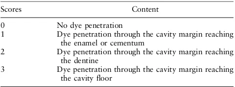
To measure the vertical extent of microleakage, the teeth were bisected longitudinally in a mesiodistal direction with a low speed diamond saw. The sectioned teeth were evaluated in a stereomicroscope at·30 final magnification. The degree of microleakage using dye penetration was scored in a blinded manner using a four-point qualitative scale (Table 1). Differences in the frequency distribution of scores between groups were assessed using the Chi-squared test.
RESULTS
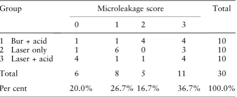
The frequency of different microleakage degree in three groups is shown in Table 2. In conventionally prepared Table 1. Qualitative scale for dye penetration
Scores Content
0 No dye penetration
1 Dye penetration through the cavity margin reaching the enamel or cementum
2 Dye penetration through the cavity margin reaching the dentine
3 Dye penetration through the cavity margin reaching the cavity floor
ª2008 Australian Dental Association 173
cavities (bur + acid, group 1), there was dye penetra-tion into the dentine and axial wall (scores of 2 and 3) in 8 of the 10 restorations. Only in one cavity was dye penetrated limited to enamel (score 1). In one cavity, no dye penetration could be seen (score 0). In the laser-prepared group (Group 2), there was dye penetration into the enamel and dentine in 7 of the 10 restorations. However, dye was detected reaching the axial wall in only three samples. In the laser plus acid etch group (Group 3), there was a bimodal distribution with half the samples showing low leakage and half high leakage. The Chi-square test showed a strong statistical associ-ation between treatment group and the distribution of microleakage scores (P = 0.0023), the higher micro-leakage scores being associated with acid etching.
DISCUSSION
The purpose of this laboratory study was to compare the extent of microleakage which occurs with laser irradiation alone, when used for cavity preparation, and when followed with conventional acid etching, as compared to diamond bur preparation followed by acid etching. The extent of leakage after thermal cycling is relevant to clinical practice since microleakage of saliva, oral fluids and bacteria at the tooth-restoration interface has been linked causally to a range of problems, including marginal staining, postoperative sensitivity, and secondary caries.4
In the present study, standardized methods were used to minimize confounding factors. The dye penetration test is the most widely used laboratory method for assessing leakage, and for the purposes of this study a simple grading system was used.1 The apical extent of the test cavities was intentionally placed into the root surface because leakage at this site is known to be a clinical concern when Class II and Class V cavities are restored with composite resin materials.
The results of the present study indicate that laser cavity preparation for these Class V cavities results in less microleakage than if phosphoric acid is used on laser-cut surfaces or if the same acid is used after cavity prepara-tion with a bur. There are no comparable laboratory studies of microleakage using the Er,Cr:YSGG laser as
earlier work has been conducted exclusively by using the Er:YAG laser.1Both lasers rely upon a water mist for optimal hard tissue ablation in terms of both thermal safety and the efficiency of ablation.6,13The presence of water also appears to have a positive effect on the caries-preventive effect of irradiation.14 When these middle infrared lasers are used for cavity preparation, sudden evaporation of the water contained in the dental hard tissues causes photomechanical disruption. Loss of particles from the tooth surface gives it a macro- and micro-roughened topography, which can substitute for conditioning with acid.15,16
The surface energy of the lased surface and the presence of moisture may contribute to optimal wet-ting by contemporary, hydrophilic bonding agents. Although bond strength tests were not undertaken as part of the present study, the results for microleakage infer that of the three methods, the Er,Cr:YSGG laser provided good adhesion with the restorative materials used. A previous study which examined cross-sections of Er,Cr:YSGG laser prepared cavities by SEM reported excellent adhesion between the restorative materials and lased tooth structure, and noted the lack of gaps at the interfaces.5The highly irregular surfaces without a smear layer thus appear to provide a suitable surface for adhesion, at least in terms of cervical restorations.
It remains to be determined whether use of the Er,Cr:YSGG laser affects microleakage for cavities whose margins are only in enamel, such as occlusal preparations. It is not possible to extrapolate the present findings to this situation since there are differences in the ablation properties between the cervical, buccal and oral regions of the enamel.17With regard to these other sites, or the use of other materials such as glass ionomers, several studies using the Er:YAG18 or Er,Cr:YSGG1 laser have not found significant differences between the combination of classical preparation technique with acid etching and the Er,Cr:YSGG laser preparation with additional acid etching.
CONCLUSIONS
The present findings suggest that in the challenging situation of the cervical restoration which extends onto the root surface, use of the laser may result in less microleakage than conventional methods of cavity preparation. If the laser is used, the need for an acid etch step can now be questioned.5
REFERENCES
1. Gutknecht N, Apel C, Schafer C, Lampert F. Microleakage of composite filling in Er,Cr:YSGG laser-prepared class II cavities. Lasers Surg Med 2001;28:371–374.
Table 2. Frequency distribution of microleakage in the three groups
Per cent 20.0% 26.7% 16.7% 36.7% 100.0%
174 ª2008 Australian Dental Association
2. Apel C, Gutknecht N. Bond strength of composites on Er:YAG and Er,Cr:YSGG laser-irradiated enamel. In: SPIE proceedings Vol. 3564, Medical Application of Lasers in Dermatology, Cardiology, Ophthalmology and Dentistry II 1999;197–200. 3. Corona SA, Borsatto MC, Pecora JD,et al. Assessing
micro-leakage of different class V restorations after Er:YAG laser and bur preparation. J Oral Rehabil 2003;30:1008–1014.
4. Armengol V, Jean A, Enkel B, Assoumou M, Hamel H. Micro-leakage of class V composite restorations following Er:YAG and Nd:YAG laser irradiation compared to acid-etch: an in vitro study. Laser Med Sci 2002;17:93–100.
5. Hossain M, Nakamura Y, Yamada Y, Murakami Y, Matsumoto K. Microleakage of composite resin restoration in cavities prepared by Er,Cr:YSGG laser irradiation and etched bur cavities in primary teeth. J Clin Pediatr Dent 2002;26:263– 268.
6. Eversole LR, Rizoiu IM. Preliminary investigations on the utility of an erbium, chromium YSGG laser. J Calif Dent Assoc 1995;23:41–47.
7. Hossain M, Nakamura Y, Yamada Y, Kimura Y, Matsumoto N, Matsumoto K. Effect of Er,Cr;YSGG laser irradiation in human enamel and dentin ablation and morphological studies. J Clin Laser Med Surg 1999;17:155–159.
8. Eversole LR, Rizoiu I, Kimmel AI. Pulpal response to cavity preparation by an erbium, chromium: YSGG laser-powered hydrokinetic system. J Am Dent Assoc 1997;128:1099–1106. 9. Rizoiu I, Kohanghadosh F, Kimmel AI, Eversole LR. Pulpal
thermal responses to an erbium, chromium: YSGG pulsed laser hydrokinetic system. Oral Surg Oral Med Oral Pathol Oral Radiol Endod 1998;86:220–223.
10. Harashima T, Kinoshita J, Kimura Y, et al. Morphological comparative study on ablation of dental hard tissues at cavity preparation by Er:YAG and Er,Cr:YSGG lasers. Photomed Laser Surg 2005;23:52–55.
11. Hossain M, Nakamura Y, Yamada Y, Kimura Y, Matsumoto N, Matsumoto K. Effect of Er,Cr:YSGG laser irradiation in human
enamel and dentin ablation and morphological studies. J Clin Laser Med Surg 1999;17:155–159.
12. Yu DG, Kimura Y, Kinoshita J, Matsumoto K. Morphological and atomic analytical studies on enamel and dentin irradiated by an erbium, chromium: YSGG laser. J Clin Laser Med Surg 2000;18:139–143.
13. Matsumoto K, Hossain M, Hossain MM, Kawano H, Kimura Y. Clinical assessment of Er,Cr:YSGG laser application for cavity preparation. J Clin Laser Med Surg 2002;20:17–21.
14. Hossain M, Nakamura Y, Kimura Y, Yamada Y, Ito M, Mat-sumoto K. Caries-preventive effect of Er:YAG laser irradiation with or without water mist. J Clin Laser Med Surg 2000;18:61– 65.
15. Apel C, Schafer C, Gutknecht N. Demineralization of Er:YAG and Er,Cr:YSGG laser-prepared enamel cavities in vitro. Caries Res 2003;37:34–37.
16. Hibst R, Keller U. Experimental studies of the application of the Er:YAG laser on dental hard substances: I. Measurement of the ablation rate.. Lasers Surg Med 1989;9:338–344.
17. Apel C, Meister J, Ioana RS, Franzen R, Hering P, Gutknecht N. The ablation threshold of Er:YAG and Er:YSGG laser radiation in dental enamel. Lasers Med Sci 2002;17:246–252.
18. Quo BC, Drummond JL, Koerber A, Fadavi S, Punwani I. Glass ionomer microleakage from preparations by an Er:YAG laser or a high-speed handpiece. J Dent 2002;30:141–146.
Address for correspondence: Professor Laurence J Walsh School of Dentistry The University of Queensland 200 Turbot Street Brisbane, Queensland 4000 Email: [email protected]
ª2008 Australian Dental Association 175