AbSTRACT Objecive:
To compare the microleakage at the enamel and denine/cemen-tum margins of three nanocomposites and a microhybrid com-posite in Class II restoraions.
Materials and Methods:
Four light-cured dental resin restoraive materials in combinaion with their respecive bonding agents were invesigated. Eighty non-carious, extracted human molars were divided into 4 groups of 20 teeth each. The apices of the teeth were sealed with a resin modi-ied glass ionomer cement. Standardized Class II slot caviies were prepared on the proximal surfaces of each tooth. Each group had an equal number of caviies with gingival margins on enamel and on denine/cementum. Restoraions were placed as indicated: Group 1 (G1): Ceram-X mono/Prime & Bond NT (Dentsply), G2: Premise/ OpiBond Solo Plus (Kerr), G3: Grandio/Admira Bond (VOCO), G4: Z100/Adper Scotchbond Muli-Purpose (3M ESPE). Ater thermocy-cling and immersion in 0.5% methylene blue dye soluion, the teeth were secioned and dye penetraion was scored on a scale of 0 to 3 on both the enamel and denine/cementum margins. The data were analyzed using a Kruskal-Wallis one way ANOVA and Mann-Whitney U test of ranks (signiicance at p<0.05).
Results:
In the enamel group: Z100/Adper Scotchbond Muli-Purpose showed signiicantly higher leakage when compared to the other three groups (p<0.05). In the denine/cementum group: Grandio/ Admira Bond showed signiicantly lower microleakage when com-pared to the other materials tested while Z100/Adper Scotchbond Muli-Purpose showed the largest microleakage (p<0.05). Conclusion:
No material was able to eliminate microleakage completely at the denine/cementum margin with Grandio/Admira Bond showing the least microleakage when compared to the other three materi-als tested. At the enamel margins, all materimateri-als tested performed reasonably well.
Keywords:
Microleakage, composite, Class II restoraions Clinical Signiicance:
Microleakage at the denine/cementum-composite interface is sill a problem and composite resin restoraions in this area must be placed with great care and regularly followed up.
INTRODUCTION
Dental composites have been available since the early 1960s.1
Their use as a restoraive material in posterior teeth has been rec-ommended for more than 20 years.2 In recent years, the demand
for posterior resin composite restoraions has dramaically in-creased due to their ability to match tooth colour, absence of mer-cury, biocompaibility and their bond to tooth structure.3,4
Modern posterior resin composites undergo 1.5 to 3.0 % volumet-ric contracion5 during polymerizaion. This may break the adhesive
bond between the adhesive system and the cavity walls forming microgaps at the tooth/restoraion interface6-8, paricularly if the
restoraion margin is placed on denine or cementum.9 Bacteria,
luids, molecules, or ions can pass through this gap between the resin composite and the cavity wall. This process is known as micro-leakage.4 Microleakage is thought to be responsible for
postopera-ive sensiivity8,10-14 , secondary caries, marginal deterioraion and
pulpal pathoses.8,10,15
Over ime several changes have been made in formulaion to produce resin restoraive materials for adequate clinical success. The latest innovaions include the development of dental composites based on nanotechnology.16 The newly available nano-materials,
such as nano-illers and nano-hybrids enable the dental composites to be improved with a very low degree of polymerizaion shrinkage and excellent aestheic properies.16,17 Their physical properies
are comparable to other composite materials (hybrids and packables) and they are recommended for anterior and posterior restoraions.17-19
Problems, such as wear, technique sensiivity and microleakage arise when convenional resin-based composite restoraions are placed in posterior teeth.20 Direct Class II restoraions can be
placed to an acceptable standard if the gingival margin is on sound enamel. However, the quality of the marginal sealing of adhesive restoraions located below the cemento-enamel juncion is sill quesionable.1 In vitro studies have reported microleakage at
denine or cementum margins of Class II restoraions with hybrid composites and nano-composites.21,22 Previous studies reported
that hybrid composites were able to prevent microleakage at cavity walls with enamel margins but they showed some degree of microleakage at denine or cementum margins.9,23 Sadeghi
reported no signiicant diference between the microleakage of nano-illed and hybrid composites in Class II restoraions with gingival margin in denine/cementum.24
Microleakage of Four composite Resin
Systems in class II Restorations
SADJ November 2009, Vol 64 no 10 p484 - p488
A Majeed: BDS, PDD, MSc (Dent). Oral and Dental Research Insitute, Faculty of Denistry, University of the Western Cape, South Africa.
Y I Osman: BChD, MChD, BBA (Hons), MBA. Department of Restoraive Denistry, Faculty of Denistry, University of the Western Cape, South Africa.
T Al-Omari: BDS, MSc (Dent), JB (Endodonics). Private Pracise, Amman, Jordan.
Corresponding Author:
485 The purpose of this in vitro study was to compare enamel and
denine /cementum microleakage in Class II restoraions restored with three nano-composites and a micro-hybrid composite re-storaive material tradiionally used for class II composite restora-ions in posterior teeth.
mATERIALS AND mETHODS
Eighty extracted human molar teeth free of visible caries, cracks, and restoraions (as observed at 10x magniicaion under a micro-scope) were used in the study. The teeth were cleaned, disinfect-ed in 0.5% chloramine T, and subsequently stordisinfect-ed in 0.9% saline soluion. The teeth were randomly divided into four groups of 20 teeth each. Two standardized Class II slot caviies were prepared on the mesial and distal surfaces of each tooth with FG 110 012 diamond issure bur in a high speed handpiece with copious wa-ter irrigaion. A new bur was used awa-ter every eight cavity prepa-raions. The caviies were 3mm in width and 2mm in depth. In each group, 20 caviies had a gingival margin located 1 mm above the cementum-enamel juncion (CEJ) while the other 20 caviies had a gingival margin placed 1 mm below the CEJ.
A restoraion template was fabricated to simulate the clinical situaion for restoraion placement. Two molar teeth, approxi-mately 12-14mm apart, were embedded in dental stone to the level of the cemento-enamel juncion (CEJ). A test specimen em-bedded in polyvinyl siloxane impression material was placed in the space between the two teeth (Figure 1). A Tolemire metal matrix system and wooden wedges were placed before each re-storaive procedure.
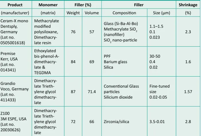
Four restoraive materials (three nano-composites and a mi-cro-hybrid composite) in combinaion with the manufacturers’ recommended adhesive systems were used. The combinaions between resin composites and adhesive systems were: (Group 1) Ceram-X mono (nano-ceramic)/Prime & Bond NT; (Group 2) Premise (nano-illed)/OpiBond Solo Plus; (Group 3) Grandio (nano-hybrid)/Admira Bond; and (Group 4) Z100 (micro-hybrid)/ Adper Scotchbond Muli-Purpose (MP). Shade A3 was used for all materials as described in Tables 1 and 2.
Each group consisted of 40 restoraions, 20 with a gingival mar-gin in enamel and 20 with a mar-gingival marmar-gin in denine/cementum. The restoraive materials and their adhesive systems were applied according to the manufacturers’ instrucions. The caviies were re-stored in three increments of approximately 2mm each and each increment of composite was light-cured for 40 seconds using a standard halogen light-curing unit (output intensity 600mW/cm2).
Ater seven days storage in disilled water at 37°C, the restoraions were inished and polished with aluminium oxide-coated lexible disc according to the manufacturer’s instrucions.
The root apices were sealed with a resin modiied glass-ionomer cement and the teeth were coated with two layers of nail-varnish except for an area approximately 1-2mm around the margins of the Class II restoraions. All the teeth were thermocycled according to the Internaional Organizaion for Standardizaion (ISO) TR11405 standard of 500 cycles, at 5° and 55°C, with a 15 second dwell ime in a bufered (pH 7) 0.5% methylene blue soluion dye.
Ater thermocycling, the specimens were cleaned and embed-ded in a slow-seing epoxy resin and allowed to set overnight. All the teeth were cut bucco-lingually in the centre and each res-toraion was then secioned mesiodistally with a 0.35mm thick blade in a diamond disk cuter. Three secions per restoraion of approximately 0.5mm thickness provided six surfaces for evalu-aion. Dye penetraion was evaluated at the gingival margin by two previously calibrated examiners (as observed at 100x mag-niicaion under a stereomicroscope). Both surfaces of the three secions of each restoraion were scored for dye penetraion by both examiners independently. Any discrepancies between the two examiners were re-evaluated by both unil a consensus score was reached. As a measure of agreement Cohen’s Kappa staisic was used. The severity of dye penetraion was analysed according to a 0-3 scale system (Figure 2):
0 = no dye penetraion
1 = Dye penetraion up to but less than half the cavity depth 2 = Dye penetraion up to and more than half the cavity depth,
but not extending to the axial wall
3 = Dye penetraion up to the axial wall and beyond.
Figure 1: Restoraion Template.
Figure 2: A cut secion of the tooth showing the scoring of
dye penetraion.
Table 1: Restoraive materials used in the study (data supplied by the manufacturer)
Product Monomer Filler (%) Filler Shrinkage
(manufacturer) (matrix) Weight Volume Composiion Size (µm) (%)
Ceram-X mono SiO2 nano-paricle
1.1–1.5
84 69 PPFBarium glass Silica
87 71.4 Convenional Glass paricles Silicium dioxide
72 66 Zirconia/silica 3.5-0.01 2.8
The data was collected and staisically analysed using a non-parametric one-way ANOVA (Kruskal-Wallis) at the 5% signii-cance level. The Mann-Whitney U test was then used for pair-wise (muliple) comparison between the groups (p <0.05).
RESULTS
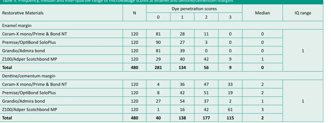
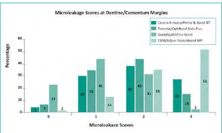
The observed frequency, median and inter-quarile range of mi-croleakage scores at the enamel and denine/cementum margins are depicted in Table 3. Figure 3 shows the percentage of microle-akage scores at enamel margins and Figure 4 depicts the percent-age of microleakpercent-age scores at denine/cementum margins for all the restoraive material groups. The Kruskal-Wallis test showed
a staisically signiicant diference in microleakage amongst the material groups at both enamel and denine/cementum margins (p<0.05). The Mann-Whitney U test showed the following results for both the enamel and denine/cementum margins:
Gingival margin in enamel
Z100/Adper Scotchbond MP showed signiicantly higher leakage scores compared to other groups tested (p<0.05). There was no staisically signiicant diference between Ceram-X mono/Prime & Bond NT, Premise/OpiBond Solo Plus and Grandio/Admira Bond as regards to leakage scores in enamel.
Gingival margin in denine/cementum Table 2: Adhesive systems, composiions, and manufacturers’ instrucions
Product/manufacturer Composiion Manufacturer instrucions
Prime & Bond® NT, Dentsply Germany,
Di- & trimethacrylate resins −
Cetylamine hydroluoride −
Acetone −
Acid etching for 15 seconds 1.
Rinse for 15 seconds 2.
Blot dry (wet bond technique) 3.
applicaion of two adhesive coats (20 seconds) 4.
Air dry for 5 seconds 5.
Light acivaion for 10 seconds 6.
Acid etching for 15 seconds 1.
Rinse for 15 seconds 2.
Dry for 5 seconds 3.
Adhesive applicaion 15 seconds 4.
Gently air dry 3 seconds 5.
Light acivaion 20 seconds 6.
Acid etching of enamel for 20 seconds and den-1.
ine 15 seconds Rinse for 20 seconds 2.
Dry for 5 seconds 3.
Adhesive applicaion 30 seconds 4.
Gently air dry 5 seconds 5.
Light acivaion 20 seconds 6.
AdperTM , ScotchbondTM 3M ESPE, USA
Acid etching for 15 seconds 1.
Rinse for 15 seconds 2.
Dry for 5 seconds 3.
Primer applicaion 4.
Dry gently for 5 seconds 5.
Adhesive applicaion 6.
Light cure 10 seconds 7.
HEMA = Hydroxyethyl methacrylate; BIS-GMA = Dimethacrylate; PENTA = Phosphonate penta-acrylate ester; GPDM = Glycerol Phosphate Dimethacrylate
Table 3: Frequency, median and inter-quarile range of microleakage scores at enamel and denine/cementum margins
Restoraive Materials N Dye penetraion scores Median IQ range
0 1 2 3
Enamel margin
Ceram-X mono/Prime & Bond NT 120 81 28 11 0 0
1
Premise/OpiBond SoloPlus 120 90 27 3 0 0
Grandio/Admira bond 120 81 39 0 0 0
Z100/Adper Scotchbond MP 120 29 40 42 9 1
Total 480 281 134 56 9 0
Denine/cementum margin
Ceram-X mono/Prime & Bond NT 120 4 36 47 33 2
1
Premise/OpiBond SoloPlus 120 8 42 51 19 2
Grandio/Admira bond 120 27 54 37 2 1
Z100/Adper Scotchbond MP 120 1 16 42 61 3
Grandio/Admira bond showed signiicantly lower microleakage scores compared to the other materials tested (p<0.05), while Z100/ Adper Scotchbond MP showed the highest microleakage scores that were signiicantly diferent from all the others groups (p<0.05). There was no staisically signiicant diference between Ceram-X mono/Prime & Bond NT and Premise/OpiBond Solo Plus.
When comparing the leakage between enamel and denine/ce-mentum margins within each material group, the leakage at the denine/cementum margins for all materials tested was greater than the leakage at the enamel margins.
DISCUSSION
In this study all restoraive material groups showed lower leakage scores at the enamel margins compared to the denine/cementum margins in class II restoraions. The efeciveness of bonding to enamel in reducing microleakage is demonstrated by the results of this study and other studies with resin composites.6,25-29
On the other hand all material groups showed greater leakage at the denine/cementum margins. These results are in agree-ment with those of other microleakage studies in class II restora-ions with denine/cementum margins.30-32 Bonding to denine is
more diicult and less predictable compared to bonding to enamel
because denine is a more complex sub-strate.9 It is about 75% inorganic in nature
and consists of apaite iller crystallites in a collagen matrix with luid illed tubular structures connecing the pulp to the den-ine-enamel juncion.1,9 Cementum is also
a complex substance that does not provide microretenion for bonding agents even ater acid-etching because of the hypomin-eralized and hyperorganic outer layer.33 The
diference in composiion and structure of enamel and denine/cementum may have resulted in more leakage at the denine/ce-mentum margins.
However, Group 3 (Grandio/Admira Bond) showed the lowest microleakage scores (Table 1) at the denine/cementum margins and Z100/Adper Scotchbond MP showed the highest leakage scores. Ceram-X mono/ Prime & Bond NT and Premise/OpiBond Solo Plus showed intermediate leakage at denine/cementum margins. The applica-ion of denine bonding agents is highly technique sensiive and requires meicu-lous atenion to detail to achieve the best results.5 Following acid etching,
mainte-nance of moist deninal surface is of great importance in denine bonding. Moisture prevents the collapse of collagen ibrils in demineralised denine, thus allowing the penetraion of adhesive monomers into the denine to form a hybrid layer.34 The
technique is commonly known as the wet bonding technique. However, it is problem-aic to quanify the amount of moisture to be let ater acid etching. Moreover, speciic solvents that present in the adhesive sys-tems (e.g. acetone and ethanol vs water) may require diferent amounts of moisture; overweing and overdrying can lead to the formaion of an altered hybrid layer knows as hybridoid layer.35
The lexibility and elasicity of the bonding layer provides a gra-dient of elasicity between the resin-bonding areas that may ab-sorb the stresses induced during polymerizaion.36 Prime & Bond
NT, Admira Bond and OpiBond Solo Plus contain nano-illers while Adper Scotchbond MP contains unilled resin. In adhesive systems that form a thick layer or contain a illed low-viscosity resin, the elasic modulus is much pronounced and might relive the stresses produced during polymerizaion.37
The diferences may not only be related to diferent bonding agents or methodology diferences, the polymerizaion shrink-age of resin composites also plays a major role in debonding of the adhesive interface thus increasing the microleakage.27,38 It
is also considered to be one of the major problems that sill im-poses limitaions in the applicaion of direct aestheic restoraive techniques.9,14 The polymerizaion shrinkage values of the tested
materials imply that this may be one of the factors responsible for the diferent behaviour evident in the study. According to the manufacturers’ Z100 and Ceram-X mono have volumetric polym-erizaion shrinkage values of 2.8% and 2.3% respecively, while
487
Figure 3: Percentage of microleakage scores at enamel margins for each material group
Premise and Grandio have shrinkage values of 1.6% and 1.57%. Premise (84% by weight) and Grandio (87% by weight) are highly illed resin composites with a nominal resin matrix content that results in low shrinkage values and contracion stresses. The high-er leakage scores of Z100 and Chigh-eram-X mono may be the result of the higher polymerizaion shrinkage values.
The degree of microleakage at gingival margins located in den-ine/cementum margins was more in each group than that of enamel margins. The indings are in agreement with those of mi-croleakage studies in Class II caviies with nano-composites,21,22,24
however no study used similar combinaion of composite/adhe-sive systems. Ozel et al reported that the locaion of the gingival
margin afected the microleakage of nano-composites and the gingival margin 1mm above CEJ provided signiicant reducion in cervical microleakage of nano-composite restoraions.22 In
con-trast, no signiicant diference between microleakage at gingival margins in denine/cementum of Class II restoraions with hybrid composite and nano-composite materials had been reported.22,24
The nano-composites used in this study were recently intro-duced commercially. Independent microleakage studies with these materials are scarce. Since this was an in vitro study, long-term clinical trials are needed to assess the performance of these materials clinically.
CONCLUSIONS In this
1. in vitro study, microleakage in Class II restoraions was
detected at the gingival margins placed in enamel and den-ine/cementum.
Ceram-X mono, Premise and Grandio with their respecive ad-2.
hesive systems performed equally well at margins in enamel while Z100/Adper Scotchbond MP showed signiicantly higher microleakage scores.
Grandio/Admira Bond showed the least microleakage and 3.
Z100/Adper Scotchbond MP showed the highest microleak-age at margins in denine/cementum, while Ceram-X mono and Premise performed intermediately.
In general, Grandio/Admira Bond performed the best at both 4.
enamel and denine/cementum margins.
Declaraion: No conlict of interest was declared
REFERENCES
Loguercio AD, de Oliveira Bauer RJ, Reis A, Miranda Grande RH. In vitro mi-1.
croleakage of packable composites in Class II restoraions. Quintessence Int
2004;35:29-34.
Türkün LS, Aktener BO, Ateş M. Clinical evalutaion of diferent posterior res-2.
in composite materials: A 7-year report. Quintessence Int 2003;34:418-426.
Herrero AA, Yaman P, Dennison JB. Polymerizaion shrinkage and depth of 3.
cure of packable composites. Quintessence Int 2005;36:25-31.
Cenci MS, Demarco FF, de Carvalho RM. Class II composite resin restoraions 4.
with two polymerizaion techniques: relaionship between microtensile bond strength and marginal leakage. J Dent 2005;33:603-610.
Hilton TJ, Schwartz RS, and Ferracane JL. Microleakage of four class II resin 5.
composite inserion techniques at intraoral temperature. Quintessence Int
1997;28:135-145.
Neiva IF, de Andrada MAC, Baraieri LN, Monteiro Junior S, Riter AV. An in 6.
vitro study of the efect of restoraive technique on marginal leakage in pos-terior composites. Oper Dent 1998;23:282–289.
Trushkowsky RD, Gwinnet AJ. Microleakage of class V composite, resin 7.
sandwich, and resin-modiied glass ionomers. Am J Dent 1996;9:96-99.
Ferdianakis K. Microleakage reducion from newer estheic restoraive ma-8.
terials in permanent molars. J Clin Pediatr Dent 1998;22:221-229.
Yazici RA, Celik C, Ozgunaltay G. Microleakage of diferent resin composite 9.
types. Quintessence Int 2004;23:790-794.
Franco EB, Lopes LG, Lia Mondelli RF, Da Silva E Souza MH, Lauris JRP. Efect 10.
of the cavity coniguraion factor on the marginal microleakage of estheic restoraive materials. Am J Dent 2003;1:211-214.
Alani AH, Toh CG. Detecion of microleakage around dental restoraions: 11.
a review. Oper Dent 1997;22:173-185.
Versluis A, Douglas WH, Sakaguchi RL. Thermal expansion coeicient of den-12.
tal composites measured with Strain Gauges. Dent Mater 1996;12:290-294.
Bayne SC, Thompson JY, Swit Jr EJ, Stamaiades P, Wilkerson M. A characterizaion 13.
of irst-generaion lowable composites. J Am Dent Assoc 1998;129:567-577.
Yap AUJ, Lim CC, Neo JCL. Marginal sealing ability of three cervical restor-14.
aive systems. Quintessence Int 1995;26:817-820.
Déjou J, Sindres V, Camps J. Inluence of criteria on the results of in vitro 15.
evaluaion of microleakage. Dent Mater 1996;12:342-349.
Mitra SB, Dong WU, Holmes BN. An applicaion of nanotechnology in ad-16.
vanced dental materials. J Am Dent Assoc 2003;134:1382-1390.
Ho C. Composite aristry using the Premise – the new breed of nanoiller. 17.
Dent Pract 2004:138-142.
Dresch W, Volpato S, Gomes JC, Ribeiro NR, Reis A, Loguercio AD. Clinical 18.
evaluaion of a nanoilled composite in posterior teeth: 12-month results.
Oper Dent 2006;31:409-417.
Milnar FJ. Selecing nanotechnology-based composites using colorimetric 19.
and visual analysis for the restoraion of anterior deniion: A case report.
J Esthet Restor Dent 2004;16:89-101.
Kournetas N, Chakmakchi M, Kakaboura A, Rahiois C, and Geis-Gerstorfer J. Mar-20.
ginal and internal adaptaion of Class II ormocer and hybrid resin composite res-toraions before and ater load cycling. Clin Oral Invest 2004;8:123–129.
Korkmaz Y, Ozel E, Atar N. Efect of lowable composite lining on microle-21.
akage and internal voids in Class II composite restoraions. J Adhes Dent
2007;9:189-194.
Ozel E, Korkmaz Y, Atar N. Inluence of locaion of the gingival margin on 22.
the microleakage and internal voids nanocomposites. J Contemp Dent Pract