Microleakage of a Microhybrid Composite Resin Using Three
Different Adhesive Placement Techniques
Article in The journal of adhesive dentistry · February 2004 Source: PubMed
CITATIONS
15
READS
90
5 authors, including:
Simone Deliperi
Tufts University
24PUBLICATIONS 380CITATIONS
SEE PROFILE
Aikaterini Papathanasiou
Tufts University
7PUBLICATIONS 223CITATIONS
SEE PROFILE
All content following this page was uploaded by Simone Deliperi on 04 January 2017.
Microleakage of a Microhybrid Composite Resin Using
Three Different Adhesive Placement Techniques
Simone Deliperi
a/David N. Bardwell
b/Aikaterini Papathanasiou
c/Samer Kastali
d/
Franklin García-Godoy
ea Visiting Instructor and Research Associate, Tufts University School of Dental Medicine, Boston, MA, USA, and Department of Restorative Dentistry, Univer-sity of Cagliari, Italy
b Associate Clinical Professor of Restorative Dentistry and Director of Postgra-duate Esthetic Dentistry, Tufts University School of Dental Medicine, Boston, MA, USA.
c Assistant Professor and Clinic Coordinator of Postgraduate Esthetic Den-tistry, Department of Restorative DenDen-tistry, Tufts University School of Dental Medicine, Boston, MA, USA.
d Assistant Professor, Director of Advanced Clinical Dentistry Center, Assistant Director Post Graduate Esthetic Dentistry, Tufts University School of Dental Medicine, Boston, MA, USA.
e Assistant Dean for Research, Professor, Department of Restorative Dentistry and Department of Pediatric Dentistry, Nova Southeastern University, Ft. Lauderdale, FL, USA.
Purpose: To evaluate the efficacy of two adhesive systems in reducing microleakage when applied with three
different adhesive placement techniques.
Materials and Methods: Sixty freshly extracted caries-free human premolars and molars were used. MO/DO Class
II standardized preparations were performed with the gingival margin placed 1 mm above the CEJ. Teeth were randomly divided into 2 groups (group I: Prime& Bond NT, Dentsply/Caulk; group II: Single Bond, 3M Espe). Each group was divided into 3 subgroups: (A) application of 2 coats and one cure: IA-IIA; (B) 2 coats and 2 cures of each adhesive system: IB-IIB; and (C) one coat of each adhesive along with the manufacturers’ B1 flowable resin (0.5-mm thick layer) cured together at once: IC-IIC. Each coat was cured for 20 s at 800 mW/cm2 using a
quartz-tungsten halogen light (Elipar Trilight, 3M ESPE). Teeth were then restored using 2-mm increments of an A2 microhybrid composite (Esthet-X, Dentsply/Caulk). All teeth were stored in distilled water at 37°C for 24 h, ther-mocycled (500x, 5° to 55°C, 30 s dwell) and then placed in a 0.5% methylene blue dye solution for 24 h at 37°C. Samples were sectioned longitudinally and evaluated for microleakage at the gingival margin under a stereomicro-scope at 20X magnification. Dye penetration was scored using an ordinal scoring system, where 0: no penetration; 1: enamel penetration; 2: gingival dentin penetration; 3: axial dentin penetration. Kruskal-Wallis and Mann-Whitney tests were used.
Results: A Mann-Whitney U-Test revealed no statistically significant difference between subgroups. Although not
statistically significant, P&B NT (two coats and one cure) revealed the lowest microleakage scores.
Conclusion: In the experimental model adopted for this study, microleakage was not affected either by the adhesive
or its placement technique.
Key words: adhesive system, Class II restoration, composite resin, microleakage.
J Adhes Dent 2004; 6: xx–xx. Submitted for publication: 06.05.03; accepted for publication: 28.07.03.
Reprint requests: Dr. Simone Deliperi, Via G. Baccelli,10/b, 09126 Cagliari, Italy. Tel:+39-347-953-3259, Fax: +39-178-601-5920. e-mail: simone.deliperi@ tufts.edu
he use of composite resin may be associated with marginal discoloration, recurrent decay, and postop-erative sensitivity. This is the result of composite resin polymerization shrinkage, which may be responsible for the formation of a gap between composite resin and the cavity walls. This gap may vary from 1.67% to 5.68% of the total volume of the restoration, and it may be filled with oral fluids and bacteria.2
Various studies3,7,8 pointed out that the marginal
ad-aptation of composite resin is not related only to enamel and dentin bond strength, but is influenced by several oth-er factors. At present, clinicians are aware that cavity con-figuration or C-factor,7 the modulus of elasticity of
res-in,14,15,31 the restorative technique,5,17 and the rate of
polymerization4,10,13 can play important roles in the
clini-cal performance of composite resin restorations.
Deliperi et al
Presently, the use of low-viscosity composite resins and filled adhesives as liner materials is increasing in popularity.6,16,26 Even though these new materials have
been introduced onto the market, the problem of ade-quate composite resin marginal adaptation – and mi-croleakage elimination – is far from being solved. An im-portant role in this area may be played by enamel-dentin adhesive systems and application technique. In the last five years, fourth generation three-step total-etch adhe-sive systems have been progresadhe-sively replaced by fifth generation two-step total-etch adhesive systems. In spite of a simplified application technique, fifth generation adhesive systems are more technique sensitive than their precursors.30,32 This phenomenon may be related to
a higher solvent-to-monomer ratio, which may be respon-sible for the formation of an uneven hybrid layer.
Platt et al24 reported improved dentin bond strength
for 2 coats vs one coat of Prime & Bond NT. Van Meer-beek et al33 recommended “brushing thinning rather than
air thinning” to avoid reducing the adhesive film thickness to an extent that the air-inhibited layer corresponds to a great part of the resin layer, reducing the bond effective-ness. According to Rueggeberg and Margeson,25 the top
15 µm of the adhesive resin cannot polymerize because of oxygen inhibition. Any layer thinner than this cannot be polymerized, thereby preventing establishment of a bond. To overcome this problem, Unterbrink and Lieben-berg29 proposed the use of flowable composites as filled
adhesives; they combined the use of a single component adhesive as a dentin primer and a thin layer of flowable composite as a filled adhesive, which were cured together to avoid oxygen inhibition of very thin adhesive layers. Conversely, the importance of precuring direct composite restorations was previously reported.18
The purpose of this study was to evaluate the ability of two different fifth generation adhesive systems to reduce microleakage when applied with three different place-ment techniques: (A) two coats and one cure, (B) two coats and two cures, and (C) one coat of each adhesive along with the manufacturers’ B1 flowable resin cured
to-gether at once.
The null hypotheses tested were: (1) the application of two coats and one cure and two coats and two cures of fifth generation adhesive systems will result in a similar mean microleakage score; (2) the application of one coat along with flowable resin will result in increased mi-croleakage scores because of incomplete resin polymer-ization.
MATERIALS AND METHODS
Sixty freshly extracted caries-free human premolars and molars were kept in distilled water at 4°C for 24 h. The dimensions of the preparation were standardized: 4 mm long, 3 mm wide, and 5 mm deep. MO/DO Class II cavity preparations were prepared with the gingival margin 1 mm above the CEJ using a 245 carbide bur (Brasseler, Savannah, GA, USA) with a high-speed handpiece and copious amounts of water. The teeth were divided into two groups, which were subdivided into three subgroups.
Each prepared tooth was etched with 34% H3PO4
(Tooth Conditioner Gel, Dentsply/Caulk Mildford, DE, USA) for 15 s, rinsed for 20 s, and then gently blown to remove excess water, being careful to maintain a moist surface. A nanofilled acetone-based adhesive system (Prime& Bond NT, Dentsply/Caulk) was used in group I, and an unfilled ethanol/water-based adhesive system (Single Bond, 3M Espe, St. Paul, MN, USA) was used in group II.
Each group was divided into 3 subgroups: (A) applica-tion of two coats of each adhesive system with the two coats cured together for 20 s at 800 mW/cm2; (B) two
coats of each adhesive system and two cures with each coat cured for 20 s at 800 mW/cm2; and (C) one coat of
each adhesive system along with the manufacturers’ B1 flowable resin (even, 0.5-mm thick layer) cured together at once. Dyract flowable compomer resin (Dentsply/ Caulk) was used with Prime& Bond NT and Filtek flowable composite resin (3M Espe) was used with Single Bond (Table 1). Each coat of adhesive and flowable resin was cured for 20 s at 800 mW/cm2 using a quartz-tungsten
halogen light (Elipar Trilight, 3M ESPE). The light intensity of the curing light was evaluated using a light meter (cur-ing radiometer, Kerr/Demetron, Orange, CA, USA) before starting each restoration.
A 0.0015-inch (0.038 mm) Toffelmire metal matrix band was used to reconstruct the proximal surface and simulate clinical conditions. The restorations were com-pletely filled with an A2 microhybrid composite (Esthet-X, Dentsply/Caulk) using an apico-occlusal vertical layering technique with each layer not exceeding 2 mm. A conven-tional continuous mode of polymerization (800 mw/cm2
continuous energy output for 20 s) was used to cure each composite resin layer. Restorations were finished using carbide burs, and polished using Enhance (Dentsply/ Caulk) points and cups and Prisma Gloss (Dentsply/ Caulk) polishing paste. All teeth were stored in distilled water at 37°C for 24 h.
The restored teeth were thermocycled 500x in a 5° – 55°C water bath with a dwell time of 30 s in each bath. The samples were then blotted dry with a paper towel. A clear nail polish was applied on the entire tooth surface with the exception of the restoration and the area sur-rounding the cavosurface margins and then air dried. All specimens were then immersed in 0.5 methylene blue dye solution for 24 h. The dye solution was buffered to pH 7 in order to avoid demineralization of the tooth structure, which would have given false readings. Teeth were rinsed
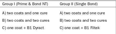
Table 1 Groups according to adhesive and procedure
Group I (Prime & Bond NT) Group II (Single Bond)
A) two coats and one cure A) two coats and one cure B) two coats and two cures B) two coats and two cures
in running water, blotted dry, and embedded in an acrylic resin block (Orthodontic Resin, Dentsply/Caulk). The teeth were sectioned longitudinally from mesial to distal with a water-cooled diamond-wheel saw (Isomet-Buehler, Lake Bluff, IL, USA). Dye penetration at the gingival mar-gin was examined using a stereomicroscope at 20X by two independent evaluators precalibrated at 85% reliabil-ity. If any disagreements in score between the two evalu-ators were reported, the higher score was taken. Sam-ples were scored utilizing an ordinal scoring system, where 0: no penetration; 1: enamel penetration; 2: gingi-val dentin penetration; 3: axial dentin penetration. Statis-tical analysis was performed using Kruskal-Wallis Anova at p < 0.05 and the Mann-Whitney U-Test.
RESULTS
None of the adhesive placement techniques tested in this study completely eliminated microleakage. The data showing the extent of leakage in groups I and II are sum-marized in Tables 2 and 3. In group I, subgroup C exhibit-ed a higher microleakage score with a prevalence of axial dentin penetration; dentin penetration was reduced for subgroups B and especially A. The latter had a higher number of samples with no gap formation. Dentin pene-tration of the dye solution was also higher in the three subgroups of group II; subgroup C showed a lower level of axial dentin penetration in this group.
The Kruskal-Wallis ANOVA test revealed no statistically significant difference between the two groups (p = 0.1378). Further analysis by the Mann-Whitney U-test was undertaken to compare the six different subgroups; no statistically significant difference was found between these subgroups.
DISCUSSION
Maintaining a moist dentinal surface is of crucial impor-tance in enhancing bond strength in vitro.12,27 Moisture
prevents collagen fibrils from collapsing into deminera-lized dentinal surfaces after acid etching, thus allowing
the adhesive monomers to penetrate into dentin, forming a hybrid layer.19 This technique is commonly known as the
wet bonding technique and has been considered a para-mount mechanism in adhesive dentistry. Quantifying the amount of moisture that should be left after acid etching is, however, problematic. Moreover, differing amounts of moisture may be required with specific solvents con-tained in each adhesive system (eg, acetone and ethanol vs water); overwetting and overdrying phenomena may occur and lead to the formation of an altered hybrid layer known as a hybridoid layer.28,33
In this study, particular attention was paid to preserv-ing a moist dentin surface prior to the start of bondpreserv-ing procedures, both with Prime & Bond NT and Single Bond. The excess water was removed using an air syringe; this method is particularly technique sensitive. The formation of totally or partially collapsed collagen layers may be responsible for differences in microleakage scores within groups IA and IB. The use of damp cotton pellets to remove excess water may help to create an ideal moist dentinal surface without the risk of dentin desiccation.23
Perdigão et al21 reported a dramatic decrease in bond
strength in vitro after air drying dentin for 15 s. This was a common result with acetone, ethanol, and ethanol/wa-ter-based adhesive systems, but not with a waethanol/wa-ter-based adhesive system, which demonstrated an intrinsic rewet-ting capability. A rewetrewet-ting time twice as long as the time spent on drying was required to restore bond strength in water-free adhesive systems. In this context, one could expect better performance of Single Bond, which contains water. Conversely, there is a trend for Single Bond to show higher microleakage scores. This was particularly rele-vant for groups IIA and IIB, in which the adhesive system was applied in multiple coats avoiding the formation of a too-thin hybrid layer and assuring its complete polymeriza-tion. Although Single Bond is an unfilled adhesive sys-tem, it contains polyalkenoic acid, contributing to the thick resin layer even when using a single coat.
Moreover, doubling the number of coats recommended by the manufacturer could affect the performance of this adhesive system. Conversely, a more uniform hybrid and resin layer can result from doubling the number of coats of Prime & Bond NT; this procedure can promote the
for-Table 2 Group I microleakage scores
Subgroup Scores Samples
Kruskal-Wallis Anova revealed no statistically significant difference between IA vs IB vs IC (p = 0.104)
Table 3 Group II microleakage scores
Subgroup Scores Samples
Deliperi et al
mation of a stress-absorbing resin layer which is able to reduce microleakage.9,11,31 The use of multiple coats
along with a flowable resin on the uncured adhesive sys-tem were atsys-tempts at excluding the formation of a too-thin unpolymerized adhesive resin layer, created by oxy-gen inhibition. In both cases, no improvement in mi-croleakage score was observed. The amount of dentin moisture may have played a more important role than the effect of the oxygen inhibited layer over the hybrid layer. Notable is the fact that this study contradicts the theory by Unterbrink and Liebenberg29 that the use of flowable
composites as filled adhesives reduces leakage. Labora-tory or clinical data have never been presented in support of this theory. This is the first study to scientifically ana-lyze this technique in vitro.
The level of moisture may be less important clinically than under laboratory conditions. A recent study reported similar results for Prime & Bond NT and Single Bond applied either on moist or dried dentin after six months of clinical service.22 The discrepancy between laboratory
and clinical studies may be related to the different level of dentin’s intrinsic moisture in vitro and in vivo. Vital den-tin and its fluid filled tubules may be a critical factor. It can be hypothesized that dentin possesses a self-rewetting capacity in vivo; this means that when slightly dried, den-tin can recover some degree of moisture, leading to a reduction in technique sensitivity.
A recent in vitro study6 conducted in our laboratory by
the same clinician reported a lower microleakage score for Prime & Bond NT when used with the two coats/two cure technique. In the previous study, the packable com-posite resin Pyramid (Bisco, Schaumburg, IL, USA) was utilized instead of the microhybrid composite resin Es-thet-X. We hypothesized that the different composite re-sin was not the only factor responsible for such a differ-ence; indeed, the light intensity could have played a very important role (600 vs 800 mW/cm2). The higher light
in-tensity used in the current study may be responsible for a faster polymerization reaction of the adhesive system and microhybrid composite resin, thus leading to the de-velopment of higher interfacial stress and increased gap formation. The use of self-cured and dual-cured bonding agents should be encouraged. They can contribute to a reduction in microleakage. The polymerization reaction progresses at a slower rate than in light-cured adhesive systems; additionally, the porosities incorporated during mixing may be responsible for a stress relaxation mecha-nism.1,20,34
CONCLUSION
The results of this study confirmed the high degree of technique sensitivity associated with the use of fifth gen-eration adhesive systems and composite resin restora-tions. The null hypotheses were only confirmed in part. The attempt to establish a thick resin layer on the top of the hybrid layer through the application of multiple coats resulted in reduced microleakage scores for the
nano-filled acetone-based adhesive system Prime & Bond NT, but not for the unfilled ethanol/water-based adhesive sys-tem Single Bond. However, this was not statistically sig-nificant. The association of Prime & Bond NT with Dyract flowable compomer resin and Single Bond with Filtek flow-able composite resin did not prevent microleakage. Fur-ther research should be conducted to provide clinicians simpler and faster adhesive systems that can guarantee a long-term clinical seal at the cavosurface margins.
ACKNOWLEDGMENTS
The authors would like to express their gratitude to Dentsply/Caulk and 3M ESPE North America for providing materials.
REFERENCES
1. Alster D, Feilzer AJ, De Gee AJ, Mol A, Davidson CL. The dependence of shrinkage stress reduction on porosity concentration in thin resin layers. J Dent Res 1992;71:1619-1622.
2. Davidson CL, De Gee AJ, Feilzer AJ. The competition between the composite-dentin bond strength and the polymerization contraction stress. J Dent Res 1984;63:1369-1399.
3. Davidson CL, De Gee AJ. Relaxation of polymerization contraction stresses by flow in dental composites. J Dent Res 1984;63:146-148.
4. Deliperi S, Bardwell DN, Papathanasiou A. In vitro evaluation of composite microleakage using different methods of polymerization. Am J Dent 2003; 16:73A-76A.
5. Deliperi S, Bardwell DN. An alternative method to reduce polymerization shrinkage in direct posterior composite restorations. J Amer Dent Assoc 2002;133:1387-1398
6. Deliperi S, Bardwell DN, Papathanasiou A, Perry R. Microleakage of resin liners and packable composites using filled and unfilled adhesives. Am J Dent 2003;16:351-355.
7. Feilzer AJ, De Gee AJ, Davidson CL. Setting stress in composite resin in relation to configuration of the restoration. J Dent Res 1987;66: 1636-1639.
8. Feilzer AJ, De Gee AJ, Davidson CL. Curing contraction of composite re-storatives and glass ionomer cements. J Prosthet Dent 1988;59:297-300. 9. Fortin D, Swift EJ Jr, Denehy GE, Reinhardt JW. Bond strength and microleakage of current dentin adhesives. Dent Mater 1994;10:253-258. 10. Goracci G, Mori G, Casa de Martinis L. Curing light intensity and marginal leakage of resin composite restorations. Quintessence Int 1996;27: 355-362.
11. Inoue S, Vargas M, Abe Y, Yoshida Y, Lambrechts P, Vanherle G, Sano H, Van Meerbeek B. Microtensile bond strength of eleven contemporary adhesives to dentin. J Adhes Dent 2001;3:237-245.
12. Kanca III J. Resin bonding to wet substrate. I: bonding to dentin. Quintes-sence Int 1992;23 39-41.
13. Kanca J, Suh BI. Pulse activation: reducing resin-based composite con-traction stresses at the cavosurface margins. Am J Dent 1999;2:107-112. 14. Kemp-Scholte CM, Davidson CL. Complete marginal seal of class V resin composite restorations effected by increased flexibility. J Dent Res 1990; 69:1240-1243.
15. Kemp-Scholte CM, Davidson CL. Marginal integrity related to bond strength and strain capacity of composite resin restorative systems. J Prosthet Dent 1990;64:658-664.
16. Labella R, Lambrechts P, Van Meerbeek B, Vanherle G. Polymerization shrinkage and elasticity of flowable composites and filled adhesives. Dent Mater 1999;15:128-137.
17. Leibenberg WH. Successive cusp build-up: An improved placement tech-nique for posterior direct resin restorations. J Canad Dent Assoc 1996;62: 501-507.
18. McCabe JF, Rusby S. Dentine bonding – the effect of precuring the bonding resin. British Dent J 1994;176:333-336.
19. Nakabayashi N, Kojima K, Masuhara E. The promotion of adhesion by the infiltration of monomers into tooth substrates. J Biomedical Mater Res 1982;16:265-273.
Vol 6, No 2, 2004 5
21. Perdigão J, Frankenberger R. Effect of solvent and rewetting time on dentin adhesion. Quintessence Int 2001;32:385-390.
22. Perdigão J, Carmo ARP, Geraldeli S, Dutra HR, Masuda MS. Six-month clinical evaluation of two dentin adhesives applied on dry vs moist dentin. J Adhes Dent 2001;3:343-352.
23. Perdigão J. Dentin bonding as a function of dentin structure. Dent Clin N Am 2002;46:277-301.
24. Platt JA, Almeida J, Gonzales-Cabezas C, Rhodes B, Moore BK. The effect of double adhesive application on the shear bond strength to dentin of compomers using three one bottle adhesive systems. Oper Dent 2001;26: 313-317.
25. Rueggeberg FA, Margeson DH. The effect of oxygen inhibition on an unfilled/filled composite system. J Dent Res 1990;69:1652-1658. 26. Swift EJ Jr, Triolo P Jr, Barkmeier WW, Bird JL, Bounds SJ. Effect of low
viscosity resins on the performance of dental adhesives. Am J Dent 1996; 9:100-104.
27. Tay FR, Gwinnett JA, Wei SHY. Micromorphological spectrum from over-drying to overwetting acid-conditioned dentin in water-free, acetone-based, single bottle primer/adhesives. Dent Mater 1996;12:236-244. 28. Tay FR, Gwinnett JA, Wei SHY. Relation between water content in acetone/
alcohol-based primer and interfacial ultrastructure. J Dent 1998;26: 147-156.
29. Unterbrink GL, Liebeberg WH. Flowable composites as “filled adhesive”: literature review and clinical recommendations. Quintessence Int 1999; 30:249-257.
30. van Dijken JWV. Clinical evaluation of three adhesive systems in Class V non carious lesions. Dent Mater 2000;16:285-291.
31. Van Meerbeek B, Willems G, Celis JP, Roos JR, Braem M, Lambrechts P, Vanherle G. Assessment by nanoindentation of the hardness and elasticity of the resin dentin bonding area. J Dent Res1993;72:1434-1442. 32. Van Meerbeek B, Yoshida Y, Snauwaert J, Hellemans L, Lambrechts P,
Vanherle G, Wakasa K, Pashley DH. Hybridization effectiveness of a two step versus a three step smear layer removing adhesive system examined correlatively by TEM and AFM. J Adhes Dent 1999;1:7-23.
33. Van Meerbeek B, Vargas M, Inoue S, Yoshida Y, Peumans M, Lambrechts P, Vanherle G. Adhesives and cements to promote preservation dentistry. Oper Dent 2001;(suppl 6):119-144.
34. Yoshikawa T, Burrow MF, Tagami J. The effects of bonding system and light curing method on reducing stress of different C-factor cavities. J Adhes Dent 2001;3:177-183.
Clinical relevance: Composite resin microleakage