136
©Turk J Pharm Sci, Published by Galenos Publishing House.
*Correspondence: E-mail: [email protected], Phone: +62315033710 ORCID-ID: orcid.org/0000-0002-3391-1877 Received: 04.02.2017, Accepted: 01.06.2017
ÖZ
Amaç: Bu çalışmanın amacı, andrografolid-karboksimetil kitosan nanopartiküllerinin oluşumunun andrografolidin fiziksel özellikleri, in vitro salım
profili ve in vivo antimalaryal aktivitesi üzerine etkisini araştırmaktır.
Gereç ve Yöntemler: Nanopartiküller iyonik jelasyon yöntemi-püskürterek kurutma ile çapraz bağlayıcı olarak CaCI2 kullanılarak etken madde: polimer: CaCl2=40: 250: 100 bileşimi ile hazırlandı. Elde edilen partiküller büyüklükleri ve morfolojisi, fiziksel durumu, etken madde içeriği, in vitro
etken madde salımı ve Plasmodium berghei ile enfekte sıçanlarda in vivo antimalaryal aktivite açısından değerlendirildi.
Bulgular: DTA ve XRD sonuçları, nanopartikül sistemlerinin daha düşük bir erime noktasına ve daha düşük kristallik derecesine sahip olduğunu gösterdi. Nanopartiküllerden çözünen etken madde 6.5 kat artmıştır ve in vivo antimalaryal etkinlik andrografolid ile karşılaştırıldığında 1.65 kat daha
fazladır.
Sonuç: Andrografolid-karboksimetil kitosan nanopartiküllerinin oluşumu andrografolidin fiziksel özelliklerini etkiledi. Andrografolidin kristalinitesinin azalması, andrografolidin erime noktasının daha düşük olmasına neden olmuştur. Bu değişiklikler, etken maddenin çözünmesine ve daha sonra etkinliğine olumlu bir etki sağlamıştır.
Anahtar kelimeler: Andrografolid, karboksimetil kitosan, in vitro salım, in vivo antimalaryal, iyonik jelleşme, püskürterek kurutma
Objectives: The purpose of this study was to investigate the effect of andrographolide-carboxymethyl chitosan nanoparticles formation on the physical characteristics, in vitro release profile and in vivo antimalarial activity of andrographolide.
Materials and Methods: Nanoparticles were prepared by ionic gelation method-spray drying using CaCl2 as the crosslinker with a composition of drug: polymer: CaCl2=40: 250: 100. The obtained particles were evaluated for its size and morphology; physical state, drug content, in vitro drug
release and in vivo antimalarial activity on Plasmodium berghei infected mice.
Results: The results of DTA and XRD showed that nanoparticle systems had a lower melting point and lower crystallinity degree. The drug dissolved from the nanoparticles was increased up to 6.5 times and the in vivo antimalarial activity was 1.65 times higher compared to andrographolide.
Conclusion: The formation andrographolide-carboxymethyl chitosan nanoparticles affected the physical characteristics of andrographolide. The decrease crystallinity of andrographolide resulted in a lower melting point of andrographolide. Such changes provided a positive impact to the drug dissolution and then its activity.
Key words: Andrographolide, carboxymethyl chitosan, in vitro release, in vivo antimalarial, ionic gelation, spray drying
ABSTRACT
1Airlangga University, Faculty of Pharmacy, Department of Pharmaceutics, Surabaya, Indonesia
2Airlangga University, Faculty of Pharmacy, Department of Pharmacognosy and Phytochemistry, Surabaya, Indonesia
Retno SARI1*, Aty WIDYAWARUYANTI2, Franciscus B. Tedy ANINDITA1, Sinta Kusuma ASTUTI1, Dwi SETYAWAN1
Andrografolid Karboksimetil Kitosan Nanopartiküllerinin Geliştirilmesi:
Karakterizasyon,
in vitro
Salım ve
in vivo
Antimalaryal Aktivite Çalışması
Development of Andrographolide-Carboxymethyl
Chitosan Nanoparticles: Characterization,
in vitro
INTRODUCTION
According to the World Health Organization report 2014, 17.1% of all essential medicines are classified as BCS II (high permeability and low solubility) and 10.6% of them are classified as BCS IV (low permeability and low solubility). A drug can be absorbed by the body and provide a pharmacologic effect for the body if the drug in a dissolved state.1 Thus, a low solubility
drug results in low bioavailability. Andrographolide, a diterpene lactone obtained from Andrographis paniculata, has extensive
pharmacologic effects such as inflammatory, anti-diarrhea, anti-HIV, anti-malarial, hepatoprotective, anticancer, antioxidant, and antihyperglycemic properties. Andrographolide application is restricted due to its low water solubility, short half-life time (2 hours) and low permeability. Pharmacokinetic studies showed that andrographolide was quickly absorbed and metabolized in rats and humans.2,3 Several methods have been
used to improve the low solubility of andrographolide. These methods are chemical modification, solid dispersion, liposomes, and nanoparticles. By improving the solubility, it is expected to increase the release rate and bioavailability.2,4 Formation
of nanoparticles andrographolide-eudragit® EPO increased
bioavailability 2.2 times compared with pure andrographolide in oral administration.5 Nanoparticles are a dispersion of
solid particles with a diameter ranging between 10-1000 nm consisting of drug which is dispersed, trapped or enveloped in a matrix of nanoparticles. Nanoparticles can be used as a drug carrier in drug therapy or vaccine adjuvant, and because of their small size, they can increase the absorption of the drug into the biologic membrane by facilitated diffusion.6-8
Carboxymethyl chitosan is a derivative of the water-soluble chitosan. Having amine and carboxyl groups in the molecule enables it to be used as a carrier in drug delivery systems because these materials are biocompatible, biodegradable, and non-toxic.9 Carboxymethyl chitosan nanoparticles can
be prepared by emulsification, gelation ionic, coacervation, spray drying, sonication, emulsion droplet-coalescence, reverse micellar, and sieving. The mechanism of formation of carboxymethyl chitosan nanoparticles with an ionic gelation method is based on the electrostatic interaction between carboxyl groups of carboxymethyl chitosan and a positive charge of the crosslinker, CaCl2. The addition of CaCl2 is intended to form a bond between the divalent cations Ca2+ ions
of CaCl2 with -COO- of carboxymethyl chitosan.9,10
Malaria is an infectious disease caused by the parasite Plasmodium, which is characterized by fever, anemia, and splenomegaly.11 There were 198 million cases of malaria and
584 thousand deaths (78% children under 5 years of age) due to malaria worldwide by 2013. Resistance to antimalarial drugs such as chloroquine and sulfadoxine-pyrimethamine is a problem that results in increased morbidity and mortality.12
Therefore, the development of antimalarial drug delivery systems still needs to be achieved. In the present study, carboxymethyl chitosan nanoparticles of andrographolide were made by ionic gelation–spray drying to improve the physical properties and antimalarial activity of andrographolide. The particles obtained were evaluated for their morphology, physical state, in vitro
release, and in vivo antimalarial activity on Plasmodiumberghei
-infected mice.
EXPERIMENTAL
Material
Andrographolide (RD Health Ingredients Co., Ltd.); Carboxymethyl chitosan (degree of substitution 81.9%, 96.5% degree of deacetylation, viscosity 1% 22 mPa.s, China Eastar Group Co., Ltd.); Calcium chloride CaCl2.2H2O pro analysis (Merck); methanol; 96% ethanol pro analysis; distilled water;
Plasmodium berghei ANKA strain obtained from the Eijkman
Institute for Molecular Biology, Jakarta and maintained SATREPS ITD (Institute of Tropical Disease); Alceivers medium; Giemsa dye in phosphate buffer; absolute methanol.
Preparation of andrographolide-carboxymethyl chitosan nanoparticles
The andrographolide-carboxymethyl chitosan nanoparticles were prepared by ionic gelation using CaCl2 as a crosslinker then spray dried. Two hundred fifty milligrams of carboxymethyl chitosan were dissolved in 100 mL of distilled water. Carboxymethyl chitosan solution was then poured into andrographolide solution (40 mg in 5 mL methanol) and stirred briefly at 500 rpm. The andrographolide-carboxymethyl chitosan solution was added into CaCl2 solution (100 mg in 40 mL ethanol-water=1:9) and the mixture solution was stirred constantly for 4 hours at 500 rpm. Non-crosslinked particles were also prepared through simple mixing of the polymer solution and andrographolide solution. Dry particles were obtained by spray drying using SD Basic, LabPlant with 1.0 mm nozzle diameter at inlet temperature 100°C, flow rate 5 mL/min
and pressure 2 mBar.
Particle morphology and size evaluation
The size, shape, and surface morphology of the particles were observed using scanning electron microscopy. Particles were embedded in a holder made of aluminum and coated with gold palladium prior to analysis. Pictures were taken at various magnifications at 20.00 kV.
Fourier transform infrared (FT-IR)
Particles were made as a pellet by mixing with KBr powder then pressed with a hydraulic pump to form a transparent pellet. Sample observation was conducted at wavelength 4000-450 cm-1 (Jasco FT-IR 5300, Easton MD, USA).
Differential thermal analysis (DTA)
Thermal analysis of the sample was conducted using a differential thermal analyzer (DTA FP-65 P-900 Thermal, Mettler Toledo, USA). About 5 mg of particles were placed in a crucible pan, sealed and observed for a thermogram. The thermogram was recorded at temperatures of 50 to 250°C with
a heating rate 10°C/min.
X-ray diffractometry
The light source employed was a Kα Cu Ni. The voltage and
the current were set at 40 kV and 40 mA. Samples were analyzed at 2Ø and angle between 5-40º. The diffractogram
of the andrographolide-carboxymethyl chitosan particles was compared with the diffractogram of base andrographolide.
Drug content
The andrographolide content in nanoparticle was determined using high-performance liquid chromatography (HPLC) Agilent 1100 Series using reverse phase with a mobile phase consisting of methanol-orthophosphoric acid (50:50, pH 3) at a wavelength of 228 nm. Five milligrams of accurately weighed particles were dissolved in 10.0 mL of methanol, filtered through a
0.2-µm membrane filter, and then the 5 mL sample was injected
into the HPLC column. The assay is performed in triplicate. The drug content was calculated using the equation below: % Drug content= drug amount x 100%
particle weight
In vitro release study
The in vitro release study was performed in 50 mL 0.1% w/v
sodium lauryl sulfate (SLS) media at 37±0.5°C, 120 rpm using
a water bath shaker. SLS was used to improve the dissolution of andrographolide.13 Andrographolide-carboxymethyl chitosan
particles equivalent to 2 mg of andrographolide were weighed accurately and put into the media. A 1.0-mL sample was taken at a predetermined time for 3 hours and the same volume of media was added after sampling. Andrographolide was also evaluated as control. The samples were analyzed using HPLC as described above. This evaluation was performed triplicate.
In vivo antimalarial activity test
The in vivo antimalarial activity test was conducted according
to the method of Peter. The 4-day suppressive test of blood schizonticidal action.14 Male mice aged 1.5-2 months of the
strain Balb/C weighing 20-30 grams were supplied by the Department of Parasitology, Faculty of Medicine, University of Brawijaya, Malang. All mice were acclimatized in the animal house and were fed with standard diet and water ad libitum. The
use of animals in this study was approved by the Animal Care and Use Committee (ACUC) of the Veterinary Faculty, Airlangga University (591-KE). All mice were injected intraperitoneally with 200 mL Plasmodium berghei infected mice’s blood, which
contained approximately 105 parasitized erythrocytes. Sixteen
mice were used and were divided into four groups. Group 1 and group 2 were orally treated twice daily for four days with andrographolide suspended in carboxymethyl chitosan solution, and andrographolide-carboxymethyl chitosan particles suspended in water, respectively, with doses equivalent to 12.5 mg andrographolide/kg. Group 3 received carboxymethyl chitosan solution and group 4, as the untreated/control group, received water. Over a period of 6 days, tail blood was withdrawn and parasitemia was monitored by examining Giemsa-stained thin blood smears using an optical microscope. Parasitemia was calculated using the following equation:
% parasitemia= number of parasitized erythrocytes x 100% number of erythrocytes
Percentage of parasitemia inhibition was calculated on day 5 using equation below.
Figure 1 shows the morphology of andrographolide-carboxymethyl chitosan nanoparticles and non-cross-linked andrographolide-carboxymethyl chitosan. Cross-linked andrographolide-carboxymethyl chitosan was seen as non-spherical with a hollow shape in a micrograph, whereas the non-cross-linked andrographolide-carboxymethyl chitosan produced more spherical and smooth particles. The andrographolide crystal was observed in cross-linked particles. It indicated that andrographolide was solidified thus hindered the formation of spherical and smooth particles shape. Cross-linked andrographolide-carboxymethyl chitosan nanoparticles had a range of sizes from 600 nm to 3000 nm, whereas the non-cross-linked andrographolide-carboxymethyl chitosan particle sizes were within the range of 500 nm to 2500 nm.
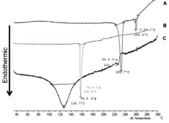
DTA
From the DTA thermogram in Figure 2, it was identified that andrographolide had a melting range 231.6°C with a sharp
Figure 1. SSEM of crosslinked andrographolide-chitosan particles (A) and non crosslinked andrographolide- chitosan particles (B) (magnification 20000x)
endothermic peak, and non-cross-linked andrographolide-carboxymethyl chitosan particles had a sharp endothermic peak with melting point of 162.9°C. The thermogram of
andrographolide-carboxymethyl chitosan nanoparticles shifted to the lower melting point of 125.7°C with a wider endothermic
peak compared with andrographolide and the non-cross-linked andrographolide-carboxymethyl chitosan particles. It indicated that the formation of cross-linked particles resulted in lower ordered structural molecule patterns compared with the non-cross-linked particles and base andrographolide.
X-ray diffractrometry
As shown in Figure 3, the diffractogram of andrographolide indicated crystalline peaks with high intensity on 2Ø 9.83°,
14.81°, 15.69°, 15.85°. CaCl2 itself had a diffraction peak at 14.74°
2Ø. In the diffractogram of andrographolide-carboxymethyl chitosan physical mixture, several crystalline peaks with low intensity at 12.14°, 15.77° and 18.55° were detected.
The diffractogram of andrographolide-carboxymethyl chitosan nanoparticles showed that diffraction peaks of andrographolide, carboxymethyl chitosan, and CaCl2 disappeared, but new crystalline peaks at 31.63° 2Ø appeared. These results showed that the formation of cross-linked particles of andrographolide-carboxymethyl chitosan prepared by ionic gelation-spray drying had lower crystallinity compared with andrographolide itself.
Drug content
From the drug content evaluation using HPLC, the andrographolide content in the particles was found as 12.09±0.26%. The result was further used to calculate the
amount of andrographolide-carboxymethyl chitosan for the in vitro release test and in vivo antimalarial activity test.
In vitro release study
The in vitro release test was performed in 0.1% SLS to facilitate
drug dissolution. The result demonstrated that the amount of andrographolide dissolved in 15 min from the nanoparticle systems (69.06%) was greater compared with andrographolide. After 30 min, the amount of andrographolide dissolved from the nanoparticles was up to 10 times higher than with base andrographolide (Figure 4).
The slope that indicated the release rate of andrographolide from carboxymethyl particles and base andrographolide were 12.7158±0.8054% dissolved/min1/2 and 2.0221±0.2702%
dissolved/min1/2, respectively. The result indicated that the
formation of andrographolide-carboxymethyl chitosan could increase the release rate of andrographolide 6.3 times compared with andrographolide itself. This was due to the formation of particles prepared by ionic gelation-spray drying lead to changes in the crystallinity of the drug as well as has being shown by the results of the DTA thermogram and X-ray diffractogram in Figure 2 and Figure 3. The entrapment of andrographolide into the cross-linked carboxymethyl chitosan continued with fast solidification of the particles during the spray drying process, which caused inhibition of crystal growth, could result in an amorphous form and crystal size reduction of andrographolide. The changes of crystallinity structure into the amorphous form and reduction of particle size of poorly soluble drug would be advantageous because it would enhance the solubility and then its bioavailability.15,16
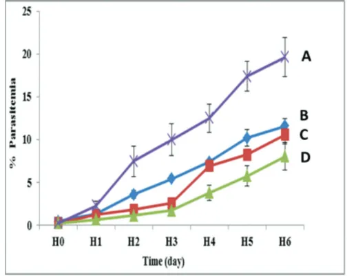
In vivo antimalarial activity test
Figure 5 shows that parasitemia growth occurred during the evaluation in all groups. The treated groups led to a slow growth of parasitemia because the untreated/control group
Figure 4. In vitro release profile of andrographolide-carboxymethyl chitosan particles and andrographolide in 0.1% SLS media at 37±0.5°C (n=3) Figure 3. X-ray diffractogram of (A) carboxymethyl chitosan, (B) CaCl2, (C) andrographolide (D) crosslinked andrographolide-chitosan particles, dan (E) physical mixture of andrographolide-chitosan
had rapid growth parasitemia. The antimalarial activity test results presented in Figure 5 and Figure 6 revealed that the increasing number of Plasmodium berghei infected erythrocytes
(parasitaemia) were lower than the control/untreated group in all treated groups.
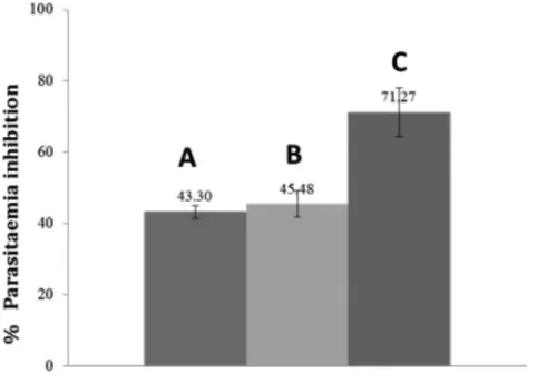
The growth inhibition of parasitemia on day five of andrographolide system-carboxymethyl chitosan nanoparticles-treated group was 71.27±6.83%, which was higher compared with the andrographolide-treated group (43.30±1.83%) and carboxymethyl chitosan-treated group (45.48±3.71%). The formation of andrographolide nanoparticles with carboxymethyl chitosan changed the physical state of andrographolide, which lowered its melting point and degree of crystallinity, as shown in Figure 2 and Figure 3. These results will improve the dissolution of andrographolide and further, will provide a favorable effect on its activity. The parasitemia growth inhibition of the andrographolide-carboxymethyl chitosan particles increased 1.65 times compared with andrographolide and was statistically significantly different (Table 1).
CONCLUSION
The formation system of andrographolide-carboxymethyl chitosan nanoparticles affected the physical characteristics of andrographolide. The crystallinity decrease of andrographolide resulted in a lower melting point of andrographolide. Such changes had a positive impact on the drug dissolution and then its activity. The release rate of andrographolide from carboxymethyl chitosan nanoparticles increased up to 6.3 times and in vivo antimalarial activity in Plasmodium berghei-infected
mice was significantly enhanced up to 1.65 times compared with base andrographolide.
ACKNOWLEDGEMENTS
This research was financially supported by Research Grant of Faculty of Pharmacy, Airlangga University, 2016.
Conflict of Interest: No conflict of interest was declared by the authors.
REFERENCES
1. Wais U, Jackson AW, Zuo Y, Xiang Y, He T, Zhang H. Drug Nanoparticles by Emulsion-Freeze-Drying viathe Employment of Branched Block Copolymer Nanoparticles. J Control Release. 2015;222:141-150.
2. Roy P, Das S, Bera T, Mondol S, Mukherjee A. Andrographolide nanoparticles in leishmaniasis: Characterization and invitro evaluations. Int J Nanomedicine. 2010;5:1113-1121.
3. Roy P, Das S, Auddy RG, Mukherjee A. Engineered andrographolide nanosystems for smart recovery in hepatotoxic conditions. Int J Nanomedicine. 2014;9:4723-4735.
4. Du H, Yang X, Li H, Han L, Li X, Dong X, Zhu Q, Ye M, Feng Q, Niu X. Preparation and evaluation of andrographolide-loaded microemulsion. J Microencapsul. 2012;29:657-665.
5. Chellampillai B, Pawar AP. Improved Bioavailability of Orally Administered Andrographolide from pH-sensitive Nanoparticles. Eur J Drug Metab Pharmacokinet. 2010;35:123-129.
6. Park K, Yeo Y. Microencapsulation Technology. In: Swarbrick J, ed. Encyclopedia of Pharmaceutical Technology, 3rd ed. New York; Informa
Healthcare; 2007:2315-2325.
Table 1. One-way ANOVA (p=0.05) to determine the effect of andrographolide, carboxymethyl chitosan and andrographolide-carboxymethyl chitosan particles on parasitaemia inhibition of 0 infected mice
Group n % Parasitaemia inhibition ± SD ANOVA
Result Conclusion
Andrographolide treated group 4 43.30±1.83a
F=11.373 p=0.002
Significantly different
Carboxymethyl chitosan treated group 4 45.48±3.71a
Andrographolide-carboxymethyl chitosan particles treated group 4 71.27±6.83b
Note: a,b Signs refer to no difference between the groups. n: Sample number
Figure 6. Histogram of parasitaemia inhibition percentage of
7. Rao JP, Geckler KE. Polymer nanoparticles: Preparation techniques and size-control parameters. Progress in Polymer Science. 2011;36:887-913. 8. Yuan Z, Ye Y, Gao F, Yuan H, Lan M, Lou K, Wang W. Chitosan-graft-ß-cyclodextrin nanoparticles as a carrier for controlled drug release. Int J Pharm. 2013;446:191-198.
9. Upadhyaya L, Singh J, Agarwal V, Tewari RP. The implications of recent advances in carboxymethyl chitosan based targeted drug delivery and tissue engineering applications. J Controlled Release. 2014;186:54-87. 10. Mourya VK, Inamdar NN, Tiwari A. Carboxymethyl chitosan and its
applications. Adv Mat Lett. 2010;1:11-33.
11. Menteri Kesehatan Republik Indonesia. Peraturan Menteri Kesehatan Republik Indonesia Nomer 5 Tahun 2013 tentang Pedoman Tata Laksana Malaria. Jakarta: Menteri Kesehatan Republik Indonesia; 2013:7-11. 12. WHO, World Malaria Report 2014, WHO Press, (Switzerland, 2014).
Available from: http://www.who.int/malaria/publications/world_ malaria_report_2014/report/en/
13. Ghosh VK, Bhope SG, Kuber VV, Gaikwad PS, Patil MJ. Development and Validation of Dissolution Test Method for Andrographolide from Film Coated Polyherbal Tablet Formulation. Int J Pharm Pharm Sci. 2012;4:307-311.
14. Bantie L, Assefa S, Teklehaimanot T, Engidawork E. Invivo antimalarial activity of the crude leaf extract and solvent fractions of Croton macrostachyus Hocsht. (Euphorbiaceae) against Plasmodium berghei in mice. BMC Complement Altern Med. 2014;14:79.
15. Buckton, G., Solid state properties. In: Aulton, M,E., editor. Pharmaceutics: The Science of Dosage Form Design, 2nd edition, Toronto: Churcill Livingstone; 2002:145-148.