How to cite:
Liana A, Purnomo, Sumardi I, Daryono BS (2017)The classifica-tion of Bambusa spp. from Celebes Based on the Micromorpho-logical Characters of Leaf Epidermis. J. Trop. Life. Science 7 (3): 197 – 203.
*Corresponding author: Budi Setiadi Daryono
Laboratory of Genetics and Breeding, Faculty of Biology Universitas Gadjah Mada
Jalan Teknika Selatan, Yogyakarta, Indonesia 55281 E-mail: [email protected]
VOL. 7, NO. 3, pp. 197 – 203, September, 2017 Submitted February 2017; Revised May 2017; Accepted June 2017
The Classification of
Bambusa
spp. from Celebes Based on
the Micromorphological Characters of Leaf Epidermis
Alin Liana 1, Purnomo 2, Issirep Sumardi 3, Budi Setiadi Daryono 4* 1 Faculty of Biology, Universitas Gadjah Mada, Yogyakarta, Indonesia
2 Laboratory of Plant Systematics, Faculty of Biology, Universitas Gadjah Mada, Yogyakarta, Indonesia 3 Laboratory of Plant Structure and Development, Faculty of Biology, Universitas Gadjah Mada, Yogyakarta, Indonesia
4 Laboratory of Genetics and Breeding, Faculty of Biology, Universitas Gadjah Mada, Yogyakarta, Indonesia
ABSTRACT
Species of Bambusa had widespread in Celebes, especially for Bambusa striata and Bambusa vulgaris. As an effect of the lacking of flowering, species identification mainly depends on leaf epidermal micromorphology, and vegetative features have proven to be useful in bamboo taxonomy. The objective of this research was to describe the classifica-tion of Bambusa from Celebes based on the micromorphological characters of leaf epidermis. The specimens were collected from the wild population. The samples of leaf were collected from five members of Bambusa i.e.: Bambusa
blumeana, Bambusa maculata, B. striata, B. vulgaris and Bambusa sp. Micromorphological characters were identified
using Scanning Electron Microscope (SEM). Leaf epidermis characters separated B. blumeana from other species of
Bambusa. Furthermore, B. striata were closely related to B. maculata in a variation of bulliform cells. As an invention,
we release peltate hair as a new type of trichome in Bambusa. The presence of numerous prickles, trichomes, and bulliform cells may be especially useful in delimiting species.
Keywords: Micromorphology, scanning electron microscope (SEM), leaf epidermis, Bambusa, Celebes INTRODUCTION
Famously, bamboos can take between 7 and 120 years to flower. Because of this rarity of flowering, the identification of bamboo generally using vegetative char-acters [1]. However, vegetative charchar-acters are often in-fluenced by the ecological factors [2]. Therefore, the an-atomical technique was undertaken to establish taxo-nomic studies.
The function of the bamboo epidermis anatomical characters in classification has been known for a long time. Bamboos identification based on leaf anatomical characters were done since 1907 [2]. The nature of the anatomical considered very important in the study of taxonomists at various levels taxa [3, 4, 5].
Furthermore, the researchers offer a variety of leaf epidermal structures as diagnostic characters in the iden-
tification of bamboo. The composition of epidermal fea- tures along internodal structures can be used to differ-entiate between species [6]. The leaf epidermis charac-ters of Asian Bamboo was compared with an emphasis on papillae character [7]. The pattern of papillae on the leaf epidermis in Arundinarieae approaches the phylo-genetic taxonomy [8]. Micromorphological study of ep-idermal structures such as silica bodies, bulliform cells and various types of hair can provide valuable infor-mation on the anatomical properties of plants and also used in taxonomic studies [9].
This research analyzed the leaf epidermis of
Bam-busa blumeana, Bambusa maculata, Bambusa striata,
Bambusa vulgaris and Bambusa sp. to contribute a
and Bambusa sp.
For the SEM analysis, leaf pieces were submerged in xylene for at least 4 hours in order to remove the waxy covering from the leaf epidermis. Afterward, materials were dried using hairdryer and then were mounted on stubs coated with gold. After gold sputtering, the speci-mens were observed in a JEOL (JSM-6510LA). The ter-minology of epidermis appendages follows Wu (1962), Gomes and Neves (2009) and Zhang et al. (2011) [2, 8, 10].
The data were analysis using Multivariate Statistical Program (MVSP) version 3.1 for identification of cluster analysis and principal component analysis (PCA) of
Bambusa.
RESULTS AND DISCUSSION
Both the upper and the lower epidermis are con-structed of epidermal cells and modified epidermal cells. There are two types of epidermal cells namely long cells and short cells. Some epidermal cells are highly modified and become quite different in appearance from the orig-inal cells. They are important in classification and de-scribed as hair (macrohair, microhair and prickles), sil-ica bodies, stomata and bulliform cells [2].
Poaceae leaves have traditionally been observed with a light microscope, but some researchers have demonstrated the use of Scanning Electron Microscope (SEM) on the study of the leaf epidermis surface to pro-
aspects such as papillae, prickles, macrohair, microhair, silica bodies, bulliform cells and stomata apparatus [11]. To confirm the type, the frequency of occurrence, com-position, and present or absent of some structures is cru-cial for taxonomic purposes.
Here is presented characters used to differentiate species in the genus Bambusa (Table 1). The characters include trichomes, silica cells, bulliform cell, and sto-mata structure. Analysis of leaf epidermis on Bambusa
was done based on the structure of the epidermis on both surfaces, a variety of shapes and sizes.
The analysis of leaf epidermis in Bambusa species revealed that epidermal elements on both surfaces vary in structures. Leaf epidermis cells on the upper epider-mis showing silica bodies, bulliform cells and trichomes. Silica bodies present in B. maculata, B. striata, B.
vul-garis and Bambusa sp., but absent in B. blumeana.
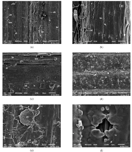
Bul-liform cells are clearly present in the intercostal zones of all spesies Bambusa were tested. Stomatal apparatuses, papillae, and various trichomes observed on the lower epidermis. The leaf surface characteristics of all the ex-amined species are shown in Figure 1 and 2.
There are three types of trichomes observed in Bam-busa leaf epidermis namely prickles, microhair and pel-tate hair. Prickles have two types, the first type is nearly elliptical at the base and cuspidate at the apex, are found on both surface of leaves over the costal zone; the second type is rounded at the base and acute at the apex, are
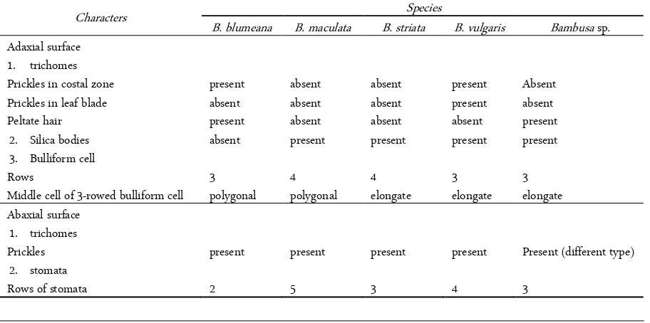
Table 1. Comparison of leaf epidermis characters among species of Bambusa
Characters Species
B. blumeana B. maculata B. striata B. vulgaris Bambusa sp. Adaxial surface
1. trichomes
Prickles in costal zone present absent absent present Absent
Prickles in leaf blade absent absent absent present absent
Peltate hair present absent absent absent present
2. Silica bodies absent present present present present
3. Bulliform cell
Rows 3 4 4 3 3
Middle cell of 3-rowed bulliform cell polygonal polygonal elongate elongate elongate Abaxial surface
1. trichomes
Prickles present present present present Present (different type)
2. stomata
found in the lower epidermis over the intercostal zone. Microhairs are bicellular. The apical cell has a very thin wall, which may be separated during plant material preparation [10]. Microhair is distributed both of upper and lower epidermis of all species Bambusa observed. Peltate hairs are multicellular. Peltate hairs consist of a short stalk and a flat disk. Each partition on the disk is a single cell. This type generally saved secretory fluids to
protect the plant from external factors [15]. Peltate hairs are new trichome observed in this study. This trichome type has not been reported found on Bambusa. Peltate hairs found on the upper epidermis of B. blumeana and
Bambusa sp.
Silica bodies are derivatives of epidermal cells. There is a short cell consists of two types of cells, the silica cells, and cork cells. Silica cells and cork cells are often
(a) (b)
(c) (d)
(e) (f)
successively formed in pairs along the leaf. The cells are developed from isotropic silica mass and usually con-taining microscopic granules in the middle of structures. Silica cells are thought to have protective mechanisms of plants from pests and environmental stresses [15]. The silica cells on the upper epidermis are dumbbell of all species Bambusa were studied. These results are con-sistent to Wu (1962) that Bambusa has a dumbbell shape
silica cells in the costal zone of upper epidermis [2]. Thus the silica cells on the upper epidermis cannot be used to differentiate species of Bambusa. Silica cells in the costal zone of lower epidermis are rectangular found in all species tested, while silica cells on intercostal zone of lower epidermis found in all Bambusa species, except
in Bambusa sp.
Some types of leaves may curl in dry air or others
(a) (b)
(c) (d)
(e) (f)
environmental conditions. This mechanism supported by bulliform cells. The shape, size, and disposition of these cells can be used for classification and identifica-tion purpose [16]. Bambusa species has a wide variety of bulliform cells types, the number of rows constituting the band varies three or four, with a polygonal or an elongated of middle cell. Hence the bulliform cells struc-ture are useful for classification.
Stomata in Poaceae lined up on the leaf blade and Bamboo generally on lower epidermis [15]. Stomata are very difficult to observe because of the growth of papillae that cover the surface [10]. In many studies, papillae character is considered appropriate to be a differentiator,
taxonomic representative. However, the character of pa-pillae not appropriate to distinguish members of Bam-boo tropical Asia [7]. In this study showed that Bambusa
only has four finger-like protuberances with the same structure.
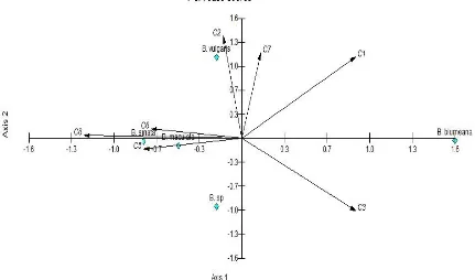
B. blumeana is a member of Bambusa with the
low-est coefficient of similarity to another species Bambusa, namely 0.587 (Figure 3). Accoding to morphological classification, B. striata is a variety of B. vulgaris. How-ever on micromorphological data B. striata to be closely related to B. maculata. B.maculata has been considered as part of B.vulgaris for a long time, but because of dif-ferences in the vegetative and generative characters, it is
Figure 3. Dendrogram of Bambusa Celebes based on micromorphological characters
ure 4). These diagram is showing direction and role of each character, which showing as vectors with different length. Based on eigenvalues is known to form a group of species consisting of Bambusa species, except B.
blumeana that split its own. B. blumeana were split due
to prickles character and present of peltate hair. This re-sult is supported by field observation that among mem-bers of Bambusa, B. blumeana have different morpho-logical characters since main branches elongated and vinelike, spreading horizontally and coiled around culm. Key to Bambusa species based on leaf epidermis characters:
1. No peltate hairs on upper epidermis . . . 2 1. A few peltate hairs on upper epidermis . . . 3 2. Silica cells absent on the upper epidermis, middle cells polygonal in a 3-rowed bulliform band . . . . . . . . B. blumeana
2. Silica cells present on the upper epidermis, middle cells elongate in a 3-rowed bulliform band . . .
Bambusa sp.
3. Prickles present on upper epidermis over costal zone . . . B. vulgaris
3. Prickles absent on upper epidermis over costal zone . . . . . 4
4. Three rows of stomata on each side of costal on lower epidermis . . . B. striata
4. Five rows of stomata on each side of costal on lower epidermis . . . . B. maculata
CONCLUSION
From above explanation, we conclude that leaf epi-dermis characters (i.e., prickles, trichomes, and bulli-form cells) were useful for distinguishing between Bam-busa species. Therefore, the use of anatomical characters to complete the morphological data on species identifi-cation may be suggested.
ACKNOWLEDGMENT
We would like to thank Dr. Harini, Integrated Re-search and Testing Laboratory, Universitas Gadjah Mada Yogyakarta for her skillful technical assistance in scanning electron microscope operation.
REFERENCES
1. Kelchner SA, Bamboo Phylogeny Group (2013) Higher level phylogenetic relationship within the bamboos (Poaceae:
leaf anatomy. Botanical Bulletin of Academia Sinica 3: 83 – 114.
3. de Villiers BJ, Tilney PM, van Wyk EB (2010) The taxo-nomic significance of leaf anatomical characters in Cussonia and related genera (Araliaceae). Botanical Journal of the Lin-nean Society 164 (3): 246 – 263. doi: 10.1111/j.1095-8339.2010.01085.x.
4. Zarrei M, Wilkim P, Ingrouille MJ et al. (2010) The system-atic importance of anatomical data in Gagea (Liliaceae) from the Flora Iranica area. Botanical Journal of the Linnean So-ciety 164 (2): 155 – 177. doi: 10.1111/j.1095-8339.2010.01081.x.
5. Liu W, Zhu XY (2011) Leaf epidermis characters and taxo-nomic revision of Schizophragma and Pileostegia (Hydran-heaceae). Botanical Journal of the Linnean Society 165 (3): 285 – 314. doi: 10.1111/j.1095-8339.2010.01101.x. 6. Pattanath PG, Rao KR (1969) Epidermal and internodal
structure of the culm as aid to identification and classifica-tion of bamboo. In: recent advices in the Anatomy of Trop-ical seed plants eds. New Delhi, Hindustan Publishing Cor-poration, pp 79 – 196.
7. Yang H, Wang H, Li D (2008) Comparative morphology of the foliage leaf epidermis, with emphasis on papillae charac-ters, in key taxa of woody bamboos of the Asian tropics (Po-aceae: Bambusoideae). Botanical Journal of the Linnean So-ciety 156 (3): 411 – 423. doi: 10.1111/j.1095-8339.2007.00736.x.
8. Zhang HY, Yang YM, Liu XZ (2011) Bamboo species rela-tions revealed by random amplified polymorphism chloro-plast DNA. African Journal of Agricultural Research 6 (5): 1241 – 1245. doi: 10.5897/AJAR10.794.
9. Pole M (2010) Cuticle morphology of Australasian Sapin-daceae. Botanical Journal of the Linnean Society. 164 (3): 264 – 292. doi: 10.1111/j.1095-8339.2010.01086.x. 10. Gomes DMS, Neves LJ (2009) Scanning electron microscopy
of the leaf epidermis of Merostachys Spreng. (Poaceae: Bam-busoideae). Acta Botanica Brasilica 23 (2): 516 – 525. doi: 10.1590/S0102-33062009000200023.
11. Palmer PG, Tucker AE (1981) A scanning electron micro-scope survey of the epidermis of East African grasses I. Washington, Smithsonian Institution Press.
12. Palmer PG, Tucker AE (1983) A scanning electron micro-scope survey of the epidermis of East African grasses II. Washington, Smithsonian Contributions to Botany. doi: 10.5479/si.0081024X.53.
electron microscope survey of the epidermis of East African grasses III. Washington, Smithsonian Contributions to Botany. doi: 10.5479/si.0081024X.55.
14. Ascensão L, Marques N, Pais MS (1995) Glandular tri-chomes on vegetative and reproductive organs of Leonotis leonulus (Lamiaceae). Annals of Botany 75 (6): 619 – 626. doi: 10.1006/anbo.1995.1067.
15. Judziewicz EJ, Clark LG, Londono X, Stern MJ (1999) Amer-ican bamboos. Washington, DC, Smithsonian Institution Press.
16. Cutler DF, Botha T, Stevenson DW (1939) Plant anatomy: An applied approach. New Jersey, Blackwell Publishing. 17. Widjaja EA (1997) New taxa in Indonesian bamboos.