In vivo and in vitro evidence for the glycoxidation of low density
lipoprotein in human atherosclerotic plaques
Yoshinobu Imanaga
a, Noriyuki Sakata
a,*, Shigeo Takebayashi
a, Akira Matsunaga
b,
Jun Sasaki
b, Kikuo Arakawa
b, Ryoji Nagai
c, Seikoh Horiuchi
c, Hiroyuki Itabe
d,
Tatsuya Takano
daSecond Department of Pathology,School of Medicine,Fukuoka Uni
6ersity,45-1,7-chome Nanakuma,Jonan-ku,Fukuoka814-0180,Japan bSecond Department of Medicine,School of Medicine,Fukuoka Uni
6ersity,45-1,7-chome Nanakuma,Fukuoka814-0180,Japan cDepartment of Biochemistry,Kumamoto Uni6ersity School of Medicine,2-2-1,Honjo,Kumamoto860-0811,Japan dDepartment of Microbiology and Molecular Pathology,Faculty of Pharmaceutical Sciences,Teikyo Uni6ersity,1091-1Suarashi,
Sagamiko-machi,Kanagawa199-0195,Japan
Received 25 January 1999; received in revised form 6 September 1999; accepted 15 September 1999
Abstract
Although there have been suggestions that the glycation and oxidation of low density lipoprotein (LDL) might increase its atherogenic potential, little is known about the presence of glycoxidative LDL in human atherosclerotic lesions. We developed specific antibodies against different immunological epitopes of AGE structures, includingNo-(carboxymethyl)lysine-protein adduct
(CML), a glycoxidation product, and structure(s) other than CML (nonCML), and a monoclonal antibody against oxidized phosphatidylcholine (oxPC), as an epitope of oxidized LDL. Immunohistochemical analysis demonstrated that the CML- and oxPC-epitopes were accumulated mainly in macrophage-derived foam cells in atherosclerotic lesions, including fatty streaks and atherosclerotic plaques. On the other hand, the nonCML-epitope and apolipoprotein B were localized mainly in extracellular matrices of atherosclerotic lesions. The CML- and oxPC-epitopes were characterized by a model antigen-generating system using the copper ion-induced peroxidation and/or glucose-induced glycation of LDL. The glycoxidation of LDL caused the formation of CML-epitope with increasing concentrations of copper ion and glucose. It was also formed to some extent in LDL incubated with high concentrations (500 mM) of glucose. However, no CML-epitope was observed in oxidized LDL induced by copper ion alone. On the other hand, the formation of oxPC-epitope in LDL was dependent on copper ion-induced peroxidation, but independent of glucose-induced glycation. The addition of chelators, ethylenediaminetetraacetic acid and diethylenetriaminepen-taacetic acid, reduced the increase in electrophoretic mobility and TBARS caused by the peroxidation and glycoxidation of LDL, but had no effects on the formation of fructosamine caused by the glycation and glycoxidation of LDL. Chelators as well as aminoguanidine protected the formation of CML-epitope in glycated or glycoxidative LDL. Although the formation of oxPC-epitope was completely inhibited by the addition of chelators, it was partially protected by aminoguanidine. These in vitro results suggest that the glycoxidative modification of LDL may occur in the arterial intima, and may contribute to the development of human atherosclerotic lesions. © 2000 Elsevier Science Ireland Ltd. All rights reserved.
Keywords: Atherogenesis; Glycoxidation of LDL; Oxidized phosphatidylcholine; No-(carboxymethyl)lysine (CML)-protein adduct; ELISA; Immunohistochemistry
www.elsevier.com/locate/atherosclerosis
1. Introduction
Although oxidized low density lipoprotein (LDL) reportedly plays an important role in atherogenesis, it cannot sufficiently account for the increased
susceptibil-ity of patients with diabetes mellitus to atherosclerosis. Advanced glycation end products (AGEs) are formed from a series of non-enzymatic chemical reactions be-tween reducing sugars, proteins, lipids and nucleic acid [1]. The final step of these reactions gives rise to AGEs which consist of heterogeneous chemical compounds. We [2] previously reported that AGEs accumulate in human atherosclerotic lesions and their location de-* Corresponding author. Tel.: +81-92-8011011, ext 3285; fax:
+81-92-8638383.
pends on the different epitopes of AGEs. Palinski et al. [3] demonstrated the colocalization of oxidation-specific adducts and AGE-specific epitopes within intimal le-sions of euglycemic WHHL rabbits. Thus, it is possible that in vivo lipid peroxidation and glycation reactions may be closely linked and enhance atherosclerotic lesions.
No-(carboxymethyl)lysine-protein adduct (CML) and pentosidine, major products of glycoxidation, have been suggested to be general markers of the oxidative modification of glycated proteins in atherosclerosis and diabetes [4 – 6]. A close link between glycoxidation and lipid peroxidation has been shown to exist and to play a role in tissue damage in atherogenesis, diabetes and aging [6,7]. Glycated LDL has been shown to be more susceptible to oxidative modification of its lipids and protein than native LDL, suggesting that glycoxidation contributes to the formation of oxidized LDL [8,9]. Superoxides are likely to play an important role in the mechanism by which glycoxidation enhances the oxi-dized modification of LDL, because Amadori com-pounds and intermediates of the browning reaction are known to be a source of superoxides [10]. CML is formed by the oxidative cleavage of Amadori com-pounds [11]. The formation of CML has been recently shown to be mediated by hydroxy radical which is generated by a Fenton reaction between Fe2+
and Amadori compounds [12]. An immunohistochemical analysis with monoclonal anti-CML antibody (6D12) showed that CML accumulated in macrophage-derived foam cells and extracellular matrix in atherosclerotic lesions [13,14]. On the other hand, oxidized phos-phatidylcholine (oxPC), which is recognized by the monoclonal antibody FOH1a/DLH3, has been shown to be formed by the copper ion-induced oxidation of LDL and to exist in human atherosclerotic plaques and plasma [15,16]. Thus, it is possible that glycoxidative modification may be involved in the formation of both CML- and oxPC-epitopes in LDL and may play a role in atherogenesis.
This study was undertaken to investigate the possibil-ity that the glycoxidative modification of LDL occurs in human aorta, and contributes to the progression of atherosclerosis. We examined the localization of CML-and oxPC-epitopes in atherosclerotic lesions of human aorta and the mechanism of CML and oxPC formation in LDL in vitro.
2. Materials and methods
2.1. Chemicals
O-Phenylenediamine dihydrochloride and naphthol-AS-BI-phosphoric acid were purchased from Sigma (St. Louis, MO). Crystallized bovine serum albumin (BSA)
was purchased from the Seikagaku Kogyo Co. (Tokyo, Japan). Horseradish peroxidase (HRP)-conjugated rab-bit anti-mouse IgG antibody and goat anti-rabrab-bit IgG antibody were purchased from CAPPEL (Durham, NC). Monoclonal anti-smooth muscle actin antibody, monoclonal human macrophage (HAM56) anti-body, biotinylated rabbit anti-mouse IgG antianti-body, biotinylated swine anti-rabbit antibody and alkaline phosphatase-labeled streptavidin were obtained from DAKO (Carpinteria, CA). Biotinylated goat anti-mouse IgM antibody was obtained from VECTOR (Burlingame, CA). Monoclonal anti-human apolipo-protein B (Apo-B) antibody was obtained from MONOSAN (Netherlands). The monoclonal antibody against oxidized LDL, FOH1a/DLH3, was prepared by Itabe et al. and has been shown to recognize oxidized phosphatidylcholine (oxPC) as a marker of oxidized LDL [15]. The monoclonal anti-CML (6D12) and poly-clonal anti-nonCML antibodies were prepared as previ-ously described [2,17,18]. Non-immune, chromato-graphically purified rabbit and mouse IgG were ob-tained from ZYMED (San Francisco, CA). A 96-well microtitration plate was purchased from the Greiner Co. (Frickenhausen, Germany). All other chemicals were of the best grade available from commercial sources.
2.2. Tissue samples
For the immunohistochemical analysis, tissue sam-ples of the aorta were obtained at autopsy from 20 nondiabetic patients (ten males and ten females; mean age, 53.8 years; range, 1 – 75 years). The autopsy was carried out within 2 – 6 h postmortem. The causes of death are listed in Table 1. All of the subjects were free both clinically and pathologically of diabetes mellitus. The samples consisted of grossly normal regions (dif-fuse intimal thickening), fatty streaks, atherosclerotic plaques and complicated lesions. The tissue specimens were washed with PBS, and embedded in OCT com-pound. The embedded samples were rapidly frozen in dry ice-acetone. Serial frozen sections were made by a cryostat and stored at−80°C until use. The sections were stained with hematoxylin and eosin or oil red O to evaluate lipid deposition in the lesions.
2.3. Immunohistochemical methods
antibodies used in this study included polyclonal anti-nonCML, monoclonal anti-CML (6D12), anti-oxPC (FOH1a/DLH3) and anti-Apo-B antibodies, and mon-oclonal anti-a smooth muscle actin and anti-human
macrophage (HAM 56) antibodies. After additional washes (in PBS, three times), biotinylated second anti-body (1:200) was added and the samples were allowed to sit for 30 min at room temperature. The slides were then rinsed with PBS and incubated for 30 min with alkaline phosphatase-labeled streptavidin (1:100) at room temperature. The alkaline phosphatase coloring reaction was carried out by incubating the slides with naphthol-AS-BI-phosphoric acid as a substrate and hexazotized new fuchsin as a coupler in 0.2 M Tris – HCl buffer (pH 8.2). The reaction mixture contained levamisole (24 mg/ml) to block endogenous alkaline phosphatase activity. After washing with PBS, the sec-tions were counterstained with Meyer hematoxylin. Control tests for the specificity of immunostaining in-cluded the substitution of non-immune sera or PBS for the primary antibodies. To further verify the specificity of the staining of monoclonal anti-CML and polyclonal anti-nonCML antibodies, controls were performed by the preadsorption of anti-AGEs and anti-CML from these primary antisera with purified AGE-BSA and CML-BSA, respectively. Three atherosclerotic lesions, i.e. diffuse intimal thickening (DIT), fatty streak (FS) and atherosclerotic plaque (AP), were examined in each case under a light microscope. Complicated lesions with
calcification and ulceration were excluded from this study. The degree of extracellular staining of antigens was evaluated by calculating the number of cases with positively stained extracellular matrix as a percentage of the total number of cases examined. Regarding the degree of staining of antigens in macrophage/foam cells, we counted the number of positively stained cells as a percentage of the total number of cells within the defined field, and then classified the values into three degrees, including no stained cells (−), and less (+) or more (+ +) than 50% under low-power light microscopy.
2.4. Preparation of low density lipoprotein
LDL was isolated from freshly drawn plasma samples (250 ml) from different human donors. Native LDL (nLDL) was prepared in the presence of 1 mM ethylenediaminetetraacetic acid (EDTA) by a potassium bromide stepwise density gradient ultra-centrifugation [19]. After ultra-centrifugation, the fractions with a density of 1.019 – 1.063 were obtained as LDL and then dialyzed at 4°C against excessive 0.15 M NaCl and 1 mM EDTA (pH7.4). nLDL was sterilized by Millipore filtration (0.45 mm) and stored in the dark at
4°C.
In vitro oxidation, glycation and glycoxidation of low density lipoprotein. All solutions were sterilized by filtration through 0.45-mm pore membranes into sterile
conical tubes, and the modifications were carried out in the absence of EDTA at 37°C for 2 weeks under sterile conditions. The oxidative modification of LDL was performed by incubating freshly prepared LDL with 5 or 10 mM CuSO4. Glycated LDL was prepared by
incubating freshly prepared LDL with 25 or 500 mM glucose under a stream of N2 gas. The glycoxidative
modification of LDL was performed by incubating freshly prepared LDL with both 5 – 10 mM CuSO4 and
25 – 500 mM glucose. The modification was stopped by extensive dialysis of the reaction mixtures against 0.15 M NaCl and 1 mM EDTA at 4°C. Furthermore, LDL was incubated in PBS without glucose and copper ion at 37°C for 2 weeks, which was referred to as nongly-cated/nonoxidized LDL. Native or nonglycated/ nonox-idized LDL was used as control.
2.5. Analysis of TBARS, electrophoretic mobility and early glycation products
The extent of oxidation in modified LDL was esti-mated by the formation of thiobarbituric acid reactive substrate (TBARS) and by changes in the elec-trophoretic mobility on agarose gel [20]. The formation of early glycation products in modified LDL was as-Table 1
Age, sex and diagnosis of subjects examined
Sex Diagnosis at autopsy 4 28 M Acute myelocyticleukemia
M 31
5 Dilatated cardiomyopathy 47
6 M Gastric adenocarcinoma. 7 50 F Cervical carcinoma of uterus
Carcinoma of gall bladder 8 60 F
60
9 F Pulmonary tuberculosis 61 F
10 Mucinous adenocarcinoma of the ovary Cerebellar thromboembolism, myocardial F
11 63
infarction
Acute gastric ulcer with arterial rupture 66 15 69 M Prostaticadenocarcinoma, gastric
adenocarcinoma M
69 Mitral valve regurgitation 16
F
17 71 Angiotropic lymphoma, breast cancer 72
18 M Lung cancer, renal cell carcinoma Lung cancer
M 73 19
75
sessed by determining fructosamine using the Fruc-tosamine Test (Hoffman-La Roche, Switzerland), as previously described [21].
2.6. E6aluation of the formation of CML- and oxPC -epitopes by noncompetiti6e ELISA
The formation of CML and oxPC was estimated by noncompetitive ELISA with anti-CML and anti-oxPC antibodies, respectively. All of the procedures were carried out at room temperature. The wells of microt-iter plates were coated with various concentrations of samples in 0.1 ml of carbonate buffer for 60 min. The wells were washed three times with washing buffer (PBS containing 0.05% Tween 20), and then incubated with 0.5% gelatin for 60 min to block nonspecific binding. After the wells were washed with washing buffer, 100ml
of anti-CML or anti-oxPC antibody in PBS containing 0.1% BSA and 0.05% Tween 20 was added to each well and incubated for 60 min. After further washings, the wells were incubated with 100 ml of
peroxidase-conju-gated second antibody for 60 min. The reactivity of peroxidase was determined by incubation with o -phenylenediamine dihydrochloride for appropriate in-tervals, and the absorbance at 492 nm was measured on an ELISA plate reader.
2.7. Effects of chelators and aminoguanidine on oxidation, glycation and the formation of CML- and oxPC-epitopes
Incubation of LDL with copper ion and/or glucose was carried out at 37°C for 2 weeks in the presence of metal chelators, 1 mM diethylenetriaminepentaacetic acid (DTPA) and 1 mM EDTA, or 100 mM amino-guanidine. After extensive dialysis against PBS, elec-trophoretic mobility, TBARS, fructosamine contents and the formation of CML- and oxPC-epitopes were determined as described above.
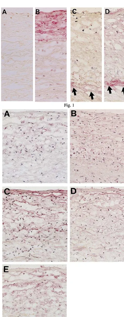
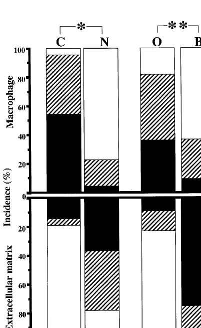
Fig. 1. Immunohistochemical localization of nonCML-epitope and apolipoprotein B in diffuse intimal thickening (DIT) of aorta. The red alkaline phosphatase reaction product is localized at antigenic sites. Although nonCML was not formed in DIT of aorta from a 21-year-old subject (A); apolipoprotein B was observed in the intima (B). An extra-cellular deposit of nonCML was seen in collagen fibers in DIT from a 68-year-old subject (C, arrows). As shown in D, apolipo-protein B was observed not only in nonCML-positively stained collagen (arrows), but also in other extracellular spaces. Original magnification is ×350 for all micrographs.
2.8. Statistical analysis
Numerical data are expressed as the mean9SE. Statistical analyses were performed using Mann – Whit-ney U-test, Chi-square test and unpaired Student’st-test. P-values of less than 0.05 were considered significant.
3. Results
3.1. Immunohistochemical localization of CML- and oxPC-epitopes in atherosclerotic lesions
AGEs, including CML- and nonCML-epitopes, showed an age-dependent staining pattern in diffuse intimal thickening (DIT). As shown in Fig. 1A and B, neither CML- nor nonCML-epitope was formed in DIT
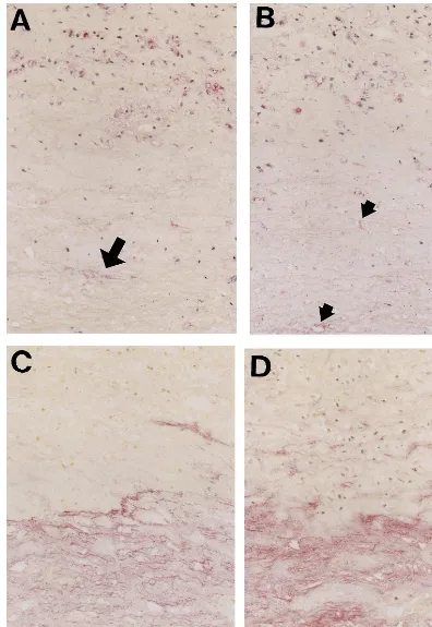
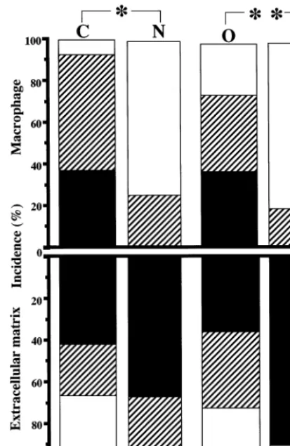
of subjects younger than middle age, but apolipoprotein B (Apo-B) was detected in their intima. Scattered staining of CML- and nonCML-epitopes was found in the collagen fibers of intima in old subjects (Fig. 1C). Apo-B was observed not only in AGE-positive collagen, but also in other extracellular matrix of DIT (Fig. 1D). However, DIT was negative for oxidized phosphatidylcholine (oxPC), an epitope of oxidized LDL, in all of the subjects. In fatty streaks, all of the subjects showed numerous macrophage-derived foam cells that accumulated both CML- and oxPC- epitope in their cytoplasm (Fig. 2A, B). On the other hand, nonCML-epitope and Apo-B were detected mainly in extracellular matrices of intima (Fig. 2C, D). Apo-B was also observed within a few macrophage/foam cells in intima. As shown in Fig. 3, most subjects exhibited a marked deposition of AGEs and oxidized LDL in atherosclerotic plaques. CML-epi-tope showed costaining with oxPC-epiCML-epi-tope in the cyto-plasm of most macrophage-derived foam cells (Fig. 3A, B). Positive staining of CML- and oxPC-epitopes was also found in the extracellular matrix and atheroma. On the other hand, nonCML-epitope and Apo-B were observed exclusively in acellular connective tissues and amorphous atheroma (Fig. 3C, D). The positivity of various antigens in the extracellular matrix or macrophage/foam cells in fatty streaks and atheroscle-rotic plaques is shown in Figs. 4 and 5, respectively. In fatty streaks, macrophage-derived foam cells showed significantly higher positivity for CML- and oxPC-epi-topes than for nonCML-epitope and Apo-B, respec-tively. In contrast, in the extracellular matrix, the positivity for nonCML-epitope and Apo-B was signifi-cantly higher than that for CML- and oxPC-epitopes, respectively. Similarly, in atherosclerotic plaques, the number of CML- and oxPC-positive macrophage/foam cells was significantly greater than that of nonCML- and Apo-B-positive macrophage/foam cells. Although the positivity for Apo-B was significantly higher than that for oxPC-epitope in the extracellular matrix, no difference was observed between the positivity for CML- and nonCML-epitopes.
3.2. In 6itro oxidation, glycation and glycoxidation of low density lipoprotein
As shown in Table 2, the electrophoretic mobility, TBARS and fructosamine contents of modified LDL were compared to those of native and nonglycated/ nonoxidized LDLs. All modifications of LDL, includ-ing oxidation, glycation and glycoxidation, caused a significant increase in electrophoretic mobility, TBARS and fructosamine contents in a dose-dependent manner with either copper ion or glucose. While copper ion-induced oxidation had a much greater effect on elec-trophoretic mobility than glucose-induced glycation, the latter had a much greater effect on the formation of fructosamine. The addition of 1 mM EDTA and 1 mM Fig. 4. Positivity for the CML-, nonCML-, and oxPC-epitopes or
Fig. 5. Positivity for the CML-, nonCML-, and oxPC-epitopes or apolipoprotein B in macrophage/foam cells (upper row) and extracel-lular matrix (lower row) in atherosclerotic plaques. C, CML-epitope; N, nonCML-epitope; O, oxPC-epitope; B, apolipoprotein B. Solid bars: incidence of cases which show more than 50% staining of antigens in macrophage/foam cells or extracellular matrix. Diagonal bars: incidence of cases which show less than 50% staining of antigens in macrophage/foam cells or extracellular matrix. Open bars: inci-dence of cases which show no staining of antigens in macrophage/ foam cells or extracellular matrix. *PB0.0001, **PB0.001, ***PB0.005 by the Mann – Whitney U-test.
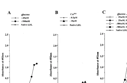
concentration (25 mM) of glucose, but was generated to some extent by incubation with a high concentration (500 mM) of glucose (Fig. 6A). As shown in Fig. 7A, incubation of LDL with glucose alone did not generate the oxPC-epitope. On the other hand, the oxidative and glycoxidative modification of LDL caused the forma-tion of oxPC-epitope, which was dependent on copper ion, but independent of glucose (Fig. 7B, C).
3.4. Effects of chelators and aminoguanidine on the formation of CML- and oxPC-epitopes in low density lipoprotein
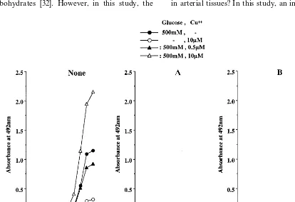
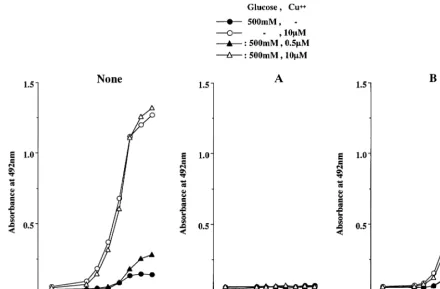
As shown in Fig. 8A, the addition of chelators completely blocked the formation of CML in LDL incubated with copper ion and/or a high concentration (500 mM) of glucose. Aminoguanidine decreased the formation of CML-epitope in glycated and glycoxida-tive LDL by approximately 70 – 98% (Fig. 8B). Fig. 9 shows the effects of chelators and aminoguanidine on the formation of oxPC-epitope. The chelators com-pletely blocked the formation of oxPC in glycoxidative and oxidized LDL (Fig. 9A). On the other hand, the addition of aminoguanidine reduced the formation of oxPC-epitope by approximately 50% in oxidized and glycoxidative LDL incubated with 10mM copper ion or
both 500 mM glucose and 10mM copper ion (Fig. 9B).
4. Discussion
The present study demonstrates for the first time indirect evidence for the presence of glycoxidative LDL, which may contribute to the progression of human atherogenesis.
Advanced glycation end products (AGEs) consist of heterogeneous chemical compounds, including No -(car-boxymethyl)lysine (CML) [11], pentosidine [22] and pyrraline [23]. Although the in vivo localization of AGEs has been extensively studied in various tissues, such as artery, skin and kidney, some controversy still exists about its location in atherosclerotic lesions. We [2] previously demonstrated that CML-epitope was lo-cated within macrophage-derived foam cells while non-CML-epitope was located mainly in the extracellular space in atherosclerotic lesions. The formation of CML and pentosidine has been shown to depend on the oxidation of glycated protein [24]. On the other hand, although the chemical structure of nonCML-epitope has not yet been identified, we showed that nonCML-epitope was formed by oxidation-independent glycation [25]. Thus, glycoxidative products may be accumulated in macrophage-derived foam cells, and oxidation-inde-pendent glycated proteins may be formed in the extra-cellular matrices in atherosclerotic lesions.
DTPA inhibited the increased electrophoretic mobility and TBARS in oxidative, glycated and glycoxidative LDL, but did not have any effect on the enhanced formation of fructosamine in glycated and glycoxida-tive LDL.
3.3. In 6itro formation of CML- and oxPC-epitopes in low density lipoprotein by oxidation, glycation and glycoxidation
Table 2
Electrophoretic mobility, TBARS and fructosamine of modified low density lipoprotein
Electrophoretic
Administration TBARS (nmol
LDL Fructosamine
(mmol/l) MDA/mg)
mobility Cu++(mM) EDTA (mM) DTPA (mM)
Glucose (mM)
– 1 1.5990.10
– – 6.7990.67
– Native LDL
–
Nonglycated/ – – – 1.34 1.5990.20 13.390.63
nonoxidized LDL
– 2.19
25 – 6.1990.50* 22.590.84*
Glycated LDL –
500 – – – 2.72 4.1390.45 47.492.10*
– – – 2.75 5.3890.37* 18.690.53*
Oxidised LDL 0.5
– 3.50 7.2590.26*
– 18.490.88*
10 –
Glycoxidative 25 0.5 – – 2.63 6.2390.98 21.091.00*
LDL
– 3.19 5.0690.13*
10 – 24.190.80*
25
500 0.5 – – 2.94 4.6490.22* 52.691.40*
–
500 10 – 3.13 3.2390.24 56.290.89*
1.0 1.0 1.25 1.1590.61
– 43.091.31*
Effect of 500 chelators
1.0 1.10
– 10 1.0 0.6990.08 8.6791.20
1.0 1.21 1.1490.29
1.0 48.291.36*
0.5 500
1.0
500 10 1.0 1.21 0.9090.09 57.891.60*
*PB0.0001, compared with native LDL,n=4.
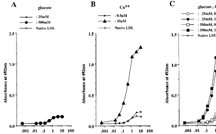
Fig. 7. oxPC-epitope formed by incubating LDL with glucose (A), copper ion (B) or glucose and copper ion (C). The formation of oxPC-epitope was estimated by non- competitive ELISA with anti-oxPC antibody (FOH1a/DLH3). LDL incubated with copper ion alone or both glucose and copper ion showed increased immunoreactivity to anti-oxPC antibody, which depended on the concentration of copper ion, but not on that of glucose. Incubation with glucose alone did not increase the immunoreactivity of LDL to anti-oxPC antibody. The experiments were repeated three times. The data represent the mean values for triplicate determinations.
Several studies have shown that the oxidation of low density lipoprotein (LDL) is implicated in atherogene-sis. Foam cells, which were found in atherosclerotic lesions, were formed from macrophages by taking up oxidized LDL through several receptors, and con-tributed to the progression of the lesions [26,27]. Im-munochemical and immunohistochemical analyses have demonstrated the presence of oxidized LDL in human plasma and atherosclerotic lesions [15,28]. A ‘model’ compound of AGEs, 2-furoyl-4[5]-(2-furanyl)-1H-imi-dazole, has been shown to colocalize with epitopes generated during the oxidation of LDL, e.g. MDA-lysine and 4-hydroxynoneal-MDA-lysine, in the atheroscle-rotic lesions of euglycemic rabbits [3]. We [14] previously showed the colocalization of epitopes of AGEs, CML and nonCML, and apolipoprotein B in intimal lesions of human aorta. Therefore, it is possible that the nonenzymatic glycation of proteins, including LDL and extracellular matrix components, may be closely linked to lipid peroxidation in the arterial in-tima. The present study demonstrated that CML-epi-tope, a major product of the oxidative modification of glycated proteins, and oxPC, an epitope of oxidized LDL, existed mainly in the cytoplasm of macrophage-derived foam cells in atherosclerotic lesions. On the other hand, nonCML, an oxidation-independent epi-tope, and apolipoprotein B were located mainly in the
extracellular matrix of intimal lesions. These results suggest that glycoxidation may be involved in the oxi-dative modification of LDL in atherosclerotic lesion. AGEs have been shown to increase with age in collagen of various tissues, including arteries and skin [7,29]. Our previous studies [2,14] showed that AGE-modified collagen significantly increased with age in DIT of human aorta. In this study, DIT of the subjects younger than middle age was positive for apolipo-protein B, but negative for AGEs, including CML and nonCML. In DIT of elderly persons, apolipoprotein B was observed not only in nonCML- positive collagen, but also in other extracellular matrices. However, DIT was negative for oxPC, an epitope of oxidized LDL, in all of the subjects. LDL has been shown to be bound specifically by AGE of collagen [30]. Thus, AGEs ob-served in collagen may contribute to increased LDL trapping in DIT of aorta.
and copper ion, LDL dramatically increased its im-munoreactivity to anti-CML antibody, and this de-pended on the concentration of either glucose or copper ion. Moreover, the formation of CML-epitope in gly-coxidative LDL was inhibited by chelators and aminoguanidine. On the other hand, CML was not formed by incubating LDL with copper ion alone or with 25 mM of glucose. These results suggest that the CML-epitope may be formed by the glycoxidative mod-ification of LDL. Although CML was not detected in LDL incubated with 25 mM of glucose alone, the incubation with a high concentration (500 mM) of glucose led to the formation of CML in LDL to some extent. AGEs generate in a manner dose-dependent on glucose, and its formation requires a period of several weeks. The time-course study showed that a period of over 3 weeks was required to detect CML in BSA incubated with glucose alone [18]. Therefore the incu-bation time may be too short to achieve adequate CML levels in LDL incubated with 25 mM of glucose. On the other hand, the formation of CML in LDL incubated with a high concentration of glucose might be caused by the autoxidation of glucose [31].
Recently, CML has been shown to be formed by the copper-ion induced oxidation of LDL, which may be derived from products of lipid peroxidation rather than from carbohydrates [32]. However, in this study, the
CML-epitope was not detected in LDL incubated with copper ion alone. This discrepancy may be due to the different assay systems for CML. On the other hand, the incubation of LDL with copper ion in the presence or absence of glucose generated the oxPC-epitope. The formation of oxPC-epitope in LDL was dependent on copper ion-catalyzed oxidation, but independent of in-cubation with glucose. These results suggest that the oxPC-epitope may be formed only by the copper ion-induced peroxidation of LDL.
Although the mechanism of CML formation is not fully understood, CML has been proposed to be formed in vitro through three pathways, i.e. via the oxidative fragmentation of Amadori products [11], from a Schiff base [33], and through modification with glyoxal generated directly by the autoxidation of glu-cose [31]. In vivo, CML is thought to be primarily formed by the oxidative cleavage of Amadori products [34]. In this study, the glycoxidative modification of LDL generated CML as well as fructosamine, an early glycation product. The formation of CML was inhib-ited not only by chelators, but also by aminoguanidine. Thus, CML might be generated mainly by the copper ion-catalyzed oxidation of Amadori products in this in vitro model of glycoxidation of LDL.
Where does LDL undergo glycoxidative modification in arterial tissues? In this study, an
Fig. 9. Effects of chelators (A) and aminoguanidine (B) on the formation of oxPC-epitope in LDL incubated with 500 mM glucose and/or copper ion. The addition of chelators, including 1 mM EDTA and 1 mM DTPA, completely blocked the formation of oxPC-epitope in the copper ion-induced oxidation or glycoxidation of LDL. Aminoguanidine inhibited the formation of oxPC-epitope in the glycoxidative modification of LDL to some extent, but had no effect on that in oxidized LDL. The experiments were repeated three times. The data represent the mean values for triplicate determinations.
cal analysis showed that macrophage-derived foam cells were strongly stained by both anti-CML and anti-oxPC antibodies. However, CML- and oxPC-epitopes were also observed in extracellular matrices including colla-gen fibers and atheroma. The nonCML epitope and Apo-B were localized mainly in extracellular spaces in all intimal lesions. Recently, malondialdehyde, a glyca-tion-independent lipid peroxidation product, was de-tected in diabetic nodular lesions with the accumulation of CML and pentosidine [35]. Macrophages are known to mediate the endocytic uptake of oxidized LDL and AGEs through scavenger receptors as well as AGE receptor [26,27,36,37]. Thus, at least in advanced lesions, LDL can undergo glycoxidative modification in extra-cellular spaces and be taken up by macrophages, which may lead to the intracellular accumulation of glycoxida-tive LDL recognized by both anti-CML and anti-oxPC antibodies. However, this study could not rule out the possibility that modified LDL may undergo endocytosis by macrophages, followed by glycoxidative modifica-tion.
Diabetes is one of the main risk factors of atheroscle-rosis. The metabolic events responsible for its
the presence or absence of glucose. These results sug-gest that LDL undergoes glycoxidative modification, which may contribute to the development of human athero-sclerotic lesions.
Acknowledgements
The authors are grateful to Yuko Aoyagi for her excellent technical assistance. This work was funded by a Grant-in-Aid from the Ministry of Education, Science and Culture of Japan (No. 10670183).
References
[1] Vlassara H, Bucala R, Striker L. Pathogenic effects of advanced glycosylation: biochemical, biologic, and clinical implications for diabetes and aging. Lab Invest 1994;70:138 – 51.
[2] Sakata N, Imanaga Y, Meng J, Tachikawa Y, Takebayashi S, Nagai R, et al. Immunohistochemical localization of different epitopes of advanced glycation end products in human atherosclerotic lesions. Atherosclerosis 1998;141:61 – 75. [3] Palinski W, Koschinsky T, Butler SW, Miller E, Vlassara H,
Cerami A, et al. Immunological evidence for the presence of advanced glycosylation end products in atherosclerotic lesions of euglycemic rabbits. Arterioscler Thromb Vasc Biol 1995;15:571 – 82.
[4] Baynes JW. Role of oxidative stress in development of complica-tions in diabetes. Diabetes 1991;40:405 – 12.
[5] Schleicher ED, Wagner E, Nerlich AG. Increased accumulation of the glycoxidation productNo-(carboxymethyl)lysine in human tissues in diabetes and aging. J Clin Invest 1997;99:457 – 68. [6] Meng J, Sakata N, Takebayashi S, Asano T, Futata T, Nagai R,
et al. Glycoxidation in aortic collagen from STZ-induced dia-betic rats and its relevance to vascular damage. Atherosclerosis 1998;136:355 – 65.
[7] Odetti P, Pronzato MA, noberasco G, Cosso L, Traverso N, Cottalasso D, et al. Relationships between glycation and oxida-tion-related fluorescences in rat collagen during aging. Lab In-vest 1994;70:61 – 7.
[8] Sakurai T, Kimura S, Nakano M, Kimura H. Oxidative modifi-cation of glycated low density lipoprotein in the presence of iron. Biochem Biophys Res Comm 1991;177:433 – 9.
[9] Kawamura M, Heinecke JW, Chait A. Pathophysiological con-centrations of glucose promote oxidative modification of low density lipoprotein by a superoxide-dependent pathway. J Clin Invest 1995;94:771 – 8.
[10] Sakurai T, Tsuchiya S. Superoxide production from nonenzy-matically glycated protein. FEBS Lett 1988;236:406 – 10. [11] Ahmed MU, Thorpe SR, Baynes JW. Identification ofNo
-(car-boxymethyl)lysine as a degradation product of fructoselysine in glycated protein. J Biol Chem 1986;261:4889 – 94.
[12] Nagai R, Ikeda K, Higashi T, Sano H, Jinnouchi Y, Araki T, et al. Hydroxyl radical mediates No-(carboxymethyl)lysine forma-tion from Amadori product. Biochem Biophys Res Comm 1997;234:167 – 72.
[13] Kume S, Takeya M, Mori T, Araki N, Suzuki H, Horiuchi S, et al. Immunohistochemical and ultrastructural detection of ad-vanced glycation end products in atherosclerotic lesions of hu-man aorta with a novel specific monoclonal antibody. Am J Pathol 1995;147:654 – 67.
[14] Sakata N, Imanaga Y, Meng J, Tachikawa Y, Takebayashi S, Nagai R, et al. Increased advanced glycation end products in
atherosclerotic lesions of patients with end-stage renal disease. Atherosclerosis 1999;142:67 – 77.
[15] Itabe H, Takeshima E, Iwasaki H, Kimura J, Yoshida Y, Imanaka T, et al. A monoclonal antibody against oxidized lipoprotein recognizes foam cells in atherosclerotic lesions. J Biol Chem 1994;269:15274 – 9.
[16] Itabe H, Yamamoto H, Imanaka T, Shimamura K, Uchiyama H, Kimura J, et al. Sensitive detection of oxidatively modified low density lipoprotein using a monoclonal antibody. J Lipid Res 1996;37:45 – 53.
[17] Horiuchi S, Araki N, Morino Y. Immunochemical approach to characterize advanced glycation end products of the Maillard reaction: evidence for the presence of a common structure. J Biol Chem 1991;266:7329 – 32.
[18] Ikeda K, Higashi T, Sano H, Jinnouchi Y, Yoshida M, Araki T, et al. No-(carboxymethyl)lysine protein adduct is a major im-munological epitope in proteins modified with advanced glyca-tion end products of the Maillard reacglyca-tion. Biochemistry 1996;35:8075 – 83.
[19] Radgrave TG, Rogert DCK, West CE. Separation of plasma lipoproteins by density gradient ultracentrifugation. Anal Biochem 1975;65:42 – 9.
[20] Ohkawa H, Ohishi N, Yagi K. Assay for lipid peroxides in animal tissues by thiobarbituric acid reaction. Anal Biochem 1979;95:351 – 8.
[21] Johnson RN, Metcalf PA, Baker JR. Fructosamine: a new approach to the estimation of serum glycosylation. An index of diabetic control. Clin Chim Acta 1982;127:87 – 95.
[22] Sell DR, Monnier VM. Structure elucidation of a senescence cross-link from human extracellular matrix: implication of pen-toses in aging process. J Biol Chem 1989;264:21597 – 602. [23] Hayase F, Nagaraj RH, Miyata S, Njoroge FG, Monnier VM.
Aging of proteins: immunological detection of a glucose-derived pyrrole formed during Maillard reaction in vivo. J Biol Chem 1989;264:3758 – 64.
[24] Dyer DG, Blackledge JA, Thorpe SR, Baynes JW. Formation of pentosidine during nonenzymatic browning of protein by glu-cose: identification of glucose and other carbohydrates as possi-ble precursors of pentosidine in vivo. J Biol Chem 1991;266:11654 – 60.
[25] Ikeda K, Nagai R, Sakamoto T, Sano H, Araki T, Sakata N, et al. Immunochemical approaches to AGE-structures: characteri-zation of anti-AGE antibodies. J Immunol Methods 1998;215:95 – 104.
[26] Endemann G, Stanton LW, Maddem KS, Bryant CM, White RT, Protter AA. CD36 is a receptor for oxidized low density lipoprotein. J Biol Chem 1993;269:11811 – 6.
[27] Freeman MY, Ekkel Y, Rohrer L, Penmen M, Freeman NJ, Chisolm GM, et al. Expression of type I and type II bovine scavenger receptors in Chinese hamster ovary cells: lipid proplet accumulation and nonreciprocal cross competition by acetylated and oxidized low density lipoprotein. Proc Natl Acad Sci USA 1991;88:4931 – 5.
[28] Palinski W, Rosenfeld ME, Yla-Herittuala S, Gurtner GC, Socher SS, Butler SW, et al. Low density lipoprotein undergoes oxidative modification in vivo. Proc Natl Acad Sci USA 1989;86:1372 – 6.
[29] Reiser KM. Nonenzymatic glycation of collagen in aging and diabetes: minireview. Proc Soc Exp Biol Med 1991;196:17 – 29.
[30] Brownlee M, Vlassara H, Cerami A. Nonenzymatic glycosyla-tion products on collagen covalently trap low-density lipo-protein. Diabetes 1985;34:938 – 41.
[32] Requena JR, Fu MX, Ahmed MU, Jenkins AJ, Lyons TJ, Thorpe SR. Lipoxidation products as biomarkers of oxidative damage to proteins during lipid peroxidation reactions. Nephrol Dial Transplant 1996;11(Suppl 5):48 – 53.
[33] Glomb MA, Monnier VM. Mechanism of protein modification by glyoxal and glycoaldehyde, reactive intermediates of the Maillard reaction. J Biol Chem 1995;270:10017 – 26.
[34] Wells-Knecht MC, Thorpe SR, Baynes JW. Pathways of forma-tion of glycoxidaforma-tion products during glycaforma-tion of collagen. Biochemistry 1995;34:15134 – 41.
[35] Horie K, Miyata T, Maeda K, Miyata S, Sugiyama S, Sakai H, et al. Immunohistochemical colocalization of glycoxida-tion products and lipid peroxidation products in
dia-betic renal glomerular lesions. J Clin Invest 1997;100:2995 – 3004.
[36] Yui S, Sasaki T, Araki N, Horiuchi S, Yamasaki M. Induction of macrophage growth by advanced glycation end products of the Maillard reaction. J Immunol 1994;152:1943 – 9.
[37] Goldstein JL, Ho YK, Basu SK, Brown MS. Binding site on macrophages that mediates uptake and degradation of acetylated low density lipoprotein, producing massive cholesterol deposi-tion. Proc Natl Acad Sci USA 1979;76:333 – 7.
[38] Sell DR, Lapolla A, Odetti P, Fogarty J, Monnier VM. Pento-sidine formation in skin correlates with severity of complication in individuals with long-standing IDDM. Diabetes 1992;41: 1286 – 92.
.