Scientific Research www.textroa d.com
Production of Polyclonal Antibodies against Sucrose Transporter (SUT1) Protein
Expressed in
Escherichia coli
BL21 and Application for Immunodiagnosis
Popy Hartatie Hardjo1), Nurul Holifah2), Tri Handoyo3), Win Darmanto4) and Bambang Sugiharto2)
1
The Faculty of Biotechnology, University of Surabaya, Surabaya 2
Biology Department, Faculty of Mathematics and Science, University of Jember, Jember 3
Faculty of Agrotechnology, University of Jember, Jember 4
Biology Department, Faculty of Science and Technology, Airlangga University, Surabaya
Received: September 16, 2014 Accepted: Ja nua ry 10, 2015 ABSTRACT
Sucrose transporter (SUT1) protein plays important roles in sucrose translocation from leaves to other organs in plants, therefore it is interested to study the existence of SUT1 protein in plants. Detection of SUT1 protein in plants can be done by using specific antibodies for that protein. This research was done to prepare SUT1 polyclonal antibodies by using SUT1 recombinant protein produced in Escher ichia coli strain BL21. The production of SUT1 recombinant protein was done using fragment cDNA-SoSUT1 of sugarcane plants inserted inside plasmid pET28a, and expressed as a fusion protein containing N-terminal hexa-histidine tags. Expressed SUT1 was purified under denaturing conditions by affinity chromatography. Recombinant protein was purely used as antigen and was injected subcutanly in the back of female New Zealand White rabbits. Wester n Blot
analysis using SUT1 polyclonal antibodies could detect the existence of SUT1 recombinant protein until the concentration 1.0 ng. Those antibodies could detect the existence of SUT1 protein at sugarcane plants.
KEYWORDS: polyclonal antibodies, SUT1 recombinant protein, cDNA –SoSUT1.
INTRODUCTION
Sucrose is one of the most common and abundant carbon forms in plants. Most plants synthesize sucrose as a major photosynthetic product and use it for long distance carbon transport. Therefore sucrose transport in plants probably is highly regulated and sucrose transporters have indispensable roles in the regulation.
The sucrose translocation process from photosynthesis tissue to storage tissue in the plants was done with the help of sucrose transporter protein (SUT) as the intermediary. This protein was known as the indicator of t he amount of sucrose which could be accumulated in plants [1]. Considering the importance of protein role in sucrose translocation, some researchers succesfully isolated cDNA-SUT from different kinds of plants, such as potatoes and tomatoes [2], tobacco [1], Arabidopsis [3], rice [4] and sugarcane plants [5,6].
To study sucrose translocation process on plants, SUT1 protein analysis needed to be done. Plants protein detection could be done with several methods, such as double diffusion [7], Western blot and immunohistochemistry [8] using specific antibodies. Direct isolation and purification of SUT1 protein from plants was difficult because the amount of SUT1 protein in plants was very low and it was located in cell membrane.
cDNA-SoSUT1 in sugarcane plants [6] could be used to produce SUT1 recombinant protein through transformation and the expresion in E.coli. Moreover, SUT1 recombinant protein would be used to produce SUT1 polyclonal antibodies in rabbits.
Recently, pET28a vector could be used for protein expression in E.coli efficiently. In this plasmid, there were hexa-histidine tags at the edge of N-terminal to make the protein purification easily. On the other hand, this plasmid was occupied with efficient promoter, so DNA inserted can be transcripted and translated easily. By inserting cDNA-SoSUT1 in pET28a, SUT1 protein was expected to be expressed, so it could be isolated for the purpose of antibodies production.
The availability of SUT1 antibodies would be used to determine the amount of SUT1 prote in using Western blot method, so sucrose translocation process in plants could be studied. The knowledge of this sucrose translocation process was the important discovery to increase the translocation and the amount of sucrose in plants.
MATERIALS AND METHODS
Expression of recombinant protein
The construction ofpET28a-SoSUT1 was transformed into E. coli BL21and transformants were screened by antibiotic medium, restriction enzyme analysis and PCR using SoSUT1 primers forward
consisted of one cycle at 94 oC for 2 min, 30 cycles at 94 oC for 30 s, 55 oC for 60 s and 72 oC for 2 min, and a final extension step at 72 oC for 7 min. The amplified product was analyzed by 1% agarose gel electrophoresis. Recombinant plasmids were extracted from two clones using the Roche High Pur e Pla smid Isola tion kit and sequenced using the same primers for colony screening in order to confirm the integrity of the ORF. A single clone, containing pET28a-SoSUT1 was selected for expression studies. A single clone of E.coli BL21 contained with the construction of pET28a-SoSUT1 plasmid [9] were grown in 4 mL of liquid LB medium contained with 35 mg/L chloramphericol and 50 mg/L kanamycin in shaker incubator (37oC 150 rpm) overnight and it was used as starter. 4 mL starter culture was inoculated to 200 mL of liquid LB medium contained with 35 mg/L resuspended in NPI-10 buffer pH 8 (50mM NaH2PO4, 300 mM NaCl and 10 mM imidazole) and 100 μg/mL lysozyme was added. The pellets was sonicated for 3 minutes and centrifuged (12000 rpm) at 4 oC for 20 minutes. Supernatant (soluble fraction) was taken to examine the existence of SUT1 recombinant protein. Pellet, which contained cell debris (insoluble fraction), was resuspended with NPI -10 buffer and centrifuged at 12000 rpm, at 4oC for 20 minutes. Supernatant was thrown away and pellet resuspended with DNPI-10 buffer pH 8.0 (50 mM NaH2PO4, 300 mM NaCl and 10 mM imidazole and 8 M urea) and sonicated for 3 minutes to diffuse the membrane protein. Suspension was centrifuged at 12000 rpm, 20 oC for 20 minutes and supernatant obtained (insoluble fraction) were analyzed. Soluble and insoluble fraction were analyzed for SDS-PAGE to find the location of the expression of SUT1 recombinant protein.
Purification of recombinant SUT1 protein
The SDS – PAGE analysis was done with the concentration of 15% akrilamid for sepa r a ting gel which contained 30% akrilamid, Tris – HCl pH 8.8, SDS 10%, 50 μL ammonium persulfate (APS) and 5 μL N, N, N', N'-tetramethylethylenediamine (TEMED) and the concentration of 4.5 % akrilamid for stacking gel 30% akrilamid, Tris – HCL pH 6.8, SDS 10% and 4.5 μL TEMED [10]. After the location of SUT1 recombinant protein expression being discovered at insoluble fraction, supernatant purification (insoluble fraction) was done with the column of Ni – NTA resin affinity chromatography. The resin which was ready to use was placed in a chromatography column and equilibrated with DNPI – 10 buffer. Purification result of SUT1 recombinant protein was analyzed by SDS – PAGE to check the protein purity, and if it was still found contaminant protein, the cutting of the gel and the protein electroelussion would be done. Urea was removed by dialyzing buffer of
phosphate buffer saline (PBS) pH 7.4 contained with 8 g NaCl, 0.2 g KCl, 1.44 g Na2HPO4, 0.24 g KH2PO4 in 1 L buffer for 12 hours with constant stirring at 4 oC.. The following steps, after 12 hours, the buffer was replaced with a new same buffer. The concentration of refolded protein was measured by the Lowry method [11] using bovine serum albumin (BSA) as a standard. The protein was aliquoted before lyophilization [12]and stored at -20 oC.
Production and evaluation of polyclonal antibody raised against recombinant SUT1 protein
The production of SUT1 polyclonal antibodies was done by injecting SUT1 recombinant protein (antigene) in female New Zealand white rabbits. A week before injection, pre-immune blood serum was taken from veins
of the rabbits’ ears. Injection was done by mixing SUT1 recombinant protein (0.5 mg) with Freund’s Complete
Adjuva nt (FCA) ( 1 : 1 ) until it was homogeneous, and then it would injected subcutaneously at the rabbits’ back. After 2 weeks, booster injected was implemented by mixing the antigen of SUT1 recombinant protein (0.1 mg) with Freund’s Incomplete Adjuvant (FIA) (1 : 1) until it was homogeneous. It was done continuously once a week until 9 weeks [13].
Ouchterlony analysis
Ouchterlony analysis was done by dissolving a ga r ose 1 % with a ga r ose solutionbuffer which contained 0.5 M Tris – HCl, 0.1 M EDTA, NaCl and 0.1 M NaN3. The solution was heated in micr owa ve oven until it was homogeneous, then it was poured in gla ss pla te evenly. The solution was left to be cold and frozen, then the well was made with diameter of 2 – 3 mm and the distance between each well was 0.5 cm. Antigen and antibodies solution were put inside well side by side and they were incubatedfor 2 days and were observed. The precipitin line formed between well of antibodies and antigen were stained with Cooma ssie Br iliant Blue (CBB) 0.1 %.
The samples of SUT1 recombinant protein with the concentration of 1 ng, 10 ng, 100 ng and 1000 ng were analyzed using SDS – PAGE. The separated proteins were electroblotted onto nitrocellulose membrane using Semi-dry Trans-Blot at 180 mA for 2.5 hours. The membrane was washed 3 times using Tris Buffer Saline (TBS, 25 mM Tris-Cl pH 7.5, 150 mM NaCl, 3 mM KCl) for 5 minutes each. After being washed, protein in the membrane was blocked by submerging it in 2 % non-fat powdered milk in TBS for 30 minutes. Then, membrane was submerged in TBS contained with 2 % non-fat powdered milk and primary antibodies was given (SUT1 polyclonal antibodies) with the dilution of 2000x, incubated overnight with the gentle shaking. Membrane was rewashed 3 times with TBS for 5 minutes each, then it was given secondary antibodies, goat anti-rabbit IgG alkaline phosphatase (AP)-conjugate in TBS non-fat powdered milk 2 % and incubated for an hour at room temperature. The bands of interest were visualized by reaction with freshly prepared substrate: 5 -bromo-4-chloro 3-indolyl-phosphate (BCIP, 168_g/mL) and nitroblue tetrazolium (NBT, 332_g/mL), in developing buffer (0.1M Tris buffer, pH 9.5, containing 0.1M NaCl and 5 mM MgCl2). The protein which could be detected in certain concentration showed the sensitivity of antibodies titer.
The detection of SUT1 protein in plants
Crude leaf extracts were prepared by grinding the leaf tissue in 3 mL extraction buffer (50 mM MOPS - NaOH pH 7.5, 10 mM MgCl2.6H2O, 1 mM EDTA, 2.5 mM dithiothreitol (DTT), 10 µM phenyl methyl sulfonyl fluor ide (PMSF)), and 10% polyvinylpyr rolidone (PVP). Crude leaf extracts was centrifugated 12000 rpm at 4oC for 10 minutes. The pellet was resuspended with the same buffer for 3 times, then the pellets were resuspended with 150 µL extraction buffer (50 mM Tris-base pH 8.5, 1 mM EDTA, 2% SDS, 30% sucrose, 5 mM DTT), and was centrifugated 12000 rpm for 10 minutes at 4oC. Supernatant was stored in -80 oC to be analyzed with SDS-PAGE and Wester n blot.
RESULTS AND DISCUSSION
The existence of pE28a -SoSUT1 in E.coli BL21was confirmed by restriction enzyme analysis and PCR.
E.coli grown in solid LB medium and contained 35 mg/L chloramphenicol antibiotic and 50 mg/L kanamisin
proved that those E.coli contained pET28a-SoSUT1 plasmid. Restriction enzyme analysis which used Xba l and
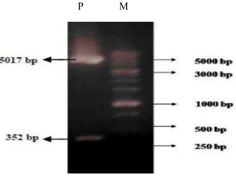
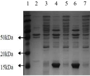
Xhol was intentionally used to prove that pET28a -SoSUT1 had contained cDNA-SoSUT1 fragment (Fig.1) that 2 DNA band in the size of 5017 bp and 352 bp were obtained. PCR analysis used a couple of F/R SUT1 primer to prove that cDNA-SoSUT1 fragment was inserted at pET28a-SoSUT1 as shown in Fig. 2. It was obtained DNA band with the size of 255 bp which was cDNA-SoSUT1 fragment. One of E.coli clones which contained pET28a-SoSUT1 construction, was then used for the production SUT1 recombinant protein. In Fig. 3, new protein with the size of 15 kDa was emerged at E.Coli insoluble fraction and it contained pET281-SoSUT1, however, that protein was not found in soluble fraction of pET28a and pET28a-SoSUT1, and also control insoluble fraction of pET28a.
Fig. 1. Nucleotide fragment as the result of pET28a-SoSUT1 restriction plasmid. pET28a-SoSUT1 which had been cut using Xhol and Xba l produce 2 nucleotide fragments with the size of 5017 bp and 352 bp.
P: pET28a-SoSUT1, M: protein marker
Fig. 2. Product of PCR E.coli which contained pET28a-SoSUT1 construction used a couple of SUT1 primer. Lane 1, 2, 3, 4: E.coli clones which contained pET28a-SoSUT1 construction. Lane 5: E.coli negative
control contained pET28a. M: DNA marker.
Fig. 3. SDS-PAGE analysis expressed fraction protein of soluble and insoluble at E.coli. Lane 1: protein marker, lane 2: protein of insoluble fraction pET28a E.coli, lane 3: protein of soluble fraction pET28a
E.coli, lane 4 and 6: protein of insoluble fraction pET28a-SoSUT1E.coli, lane 5 and 7: protein of soluble fraction pET28a-SoSUT1E.coli.
The purification result showed that SUT1 recombinant protein, which had been fused with hexa-histidine tag had been successfully purified by using Ni-NTA resin. Basically, Ni-NTA resin which contained Nickel ion (Ni2+) would be connected to protein which had fussion protein hexa-histidine tag. That bond could be eluted by imidazole with the high concentration of 100 – 250 mM. This was intended to release protein which contained hexa-histidine tag which was attached to Ni2+ ion. The contamination of other protein (Fig. 4A) was probably caused by washingbefore elution, which was not done maximum enough, therefore there was still non target protein engaged in elution. Furthermore, protein electroelution was done towards the contaminated one in order to obtain the pure protein (Fig. 4B). Protein electroelution was an easy method to isolate protein from polyacrylamide gel using electricity [14]. The purification result using this method was specific in which there was only one protein band of SUT1 target with the size of 15 kDa. The purification result was specific in electroelution because only target protein band was cut and removed using electricity.
Fig. 4. SDS PAGE analysis of SUT1 recombinant protein which was washed (A) and SUT1 recombinant protein after electroelution (B). A(lane1-2) SUT1 protein which was still mixed with other protein at
the beginning of the washing. A (lane 3-6) SUT1 protein washed in resin and separated from other protein. B SUTl: SUT1 recombinant protein after electroelution. M: protein marker.
Based on ouchter lony test (Fig. 5), serum taken for the first time (Ab1) at the 4th week after the first injection or the 1st week after booster, SUT1 antibodies was still not detected, it could be seen because there was not precipitinline. SUT1 antibodies inside rabbit had actually been formed because the first injection of antigen would stimulate B cells and formed antibodies and memory cells called primary response. Antibodies formed in primary response was still little and would increase in the next antigen injection, called secondary response [15]. SUT1 antibodies were detected in serum secondly taken at the 5th week after the first injection and at the 2nd week after booster. However, serum formed still had low titer, it could be seen from the thin line of precipitin. On the 6th week until the 7th week, antibodies titer increased, while on the 8th week until the 9th week, it was most likely to decrease. Precipitin line formed in Ouchter lony analysis showed there was match in the bond of antigen and antibodies and it was migrated one another inside gel. This precipitin reaction was specific because only the match antibodies and antigen which could be bond. This method was often used to check the specificity of polyclonal antibodies [16].
Ag
Fig. 5. Ouchterlony analysis of SUT1 polyclonal antibodies and SUT1 recombinant protein antigen. Pre : pre- immunization serum; Ab1 – Ab 6 : weekly serum (the 4th week until the 9th week after the first injection). Ab: antibody, Ag: antigen (SUT1 recombinant protein)
Based on Western blot analysis (Fig. 6), SUT1 polyclonal antibodies of Ab4 serum could detect the existence of SUT1 antigen until the concentration 1 ng with antibodies dilution 1 : 2000. As seen in the result, it could be concluded that SUT1 polyclonal antibodies had high titer because it could detect the existence of antigen with the very low concentration, such as 1 ng.
Fig. 6. Sensitivity test of the capability of SUT1 polyclonal antibodies using Western blot analysis with several concentrations of SUT1 recombinant protein antigen. A: 1000 ng, B: 100 ng, C: 10 ng, D:1 ng
SUT 1 protein detection on plants was done with Ouchterlony and Western blot analysis using sugar - cane protein as antigen. The result of Ouchterlony analysis showed the precipitine line formed between antibodies and insoluble fraction. SUT1 polyclonal antibodies could bind SUT1 protein at insoluble fraction (Fig.7) and it indicated that the protein existed on cell membrane . In soluble fraction, antibodies – antigen bond reaction was not happened, so precipitin line was not formed. This was suitable with the report of [17] that sucrose transporter which was isolated from sugar beet leaves existed on cell membrane.
Fig. 7. Ouchterlony analysis of SUT1 protein on sugarcane plants using serum of the 4th SUT1
polyclonal antibodies. A: insoluble fraction. B: soluble fraction ; precipitin line formed at insoluble fraction. Ab: antibody
precipitin line 4th 5th 6th 7th 8th 9th week
The SUT1 polyclonal antibodies has been used succesfully for the detection of SUT1 protein on sugarcane leaves by Western blot analysis (Fig. 8), and specific SUT1 protein band is detected with the size of 65 kDa.
Fig. 8. Western blot analysis of SUT1 protein on sugarcane leaves with SUT1 polyclonal antibodies. Lane 1 and 2: SUT1 protein, lane 3: protein marker.
CONCLUSION
SUT1 recombinant protein, which was the result of cDNA-SoSUT1 fragment overexpression on E.coli
BL 21cells, could be used to produce SUT1 polyclonal antibodies in rabbits. The result of polyclonal antibo dies against SUT1 recombinant protein can be applied successfully for SUT1 efficient detection in plants.
ACKNOWLEDGEMENT
This work was supported by Grant Fundamental Research, Dikti, Indonesia. 2011 (on the behalf of Prof. Bambang Sugiharto, Ph.D.).
REFERENCES
1. Lemoine, R., B. Lukas, B. Laurence, S. Soulaiman, K. Christina, R. Matthieu, G. Cecile, D. Serge, and B.F. Wol, 1999. Identification of a pollen-spesific transporter-like protein NtSUT3 from tobacco. FEBS Letters, 454:325-330.
2. Riesmeier, J.W., B. Hirner, W.B. Frommer, 1993. Potato sucrose transporter expression in minor veins indicates a role in phloem loading. The Plant Cell, 5:1591-1598.
3. Weise, A., Barker, L., Kuhn, C., Lalonde, S., Buschmann, H., Frommer, W.B., and J.M. Ward, 2000. A new subfamily of sucrose transporters, SUT4, with long affinity/high capacity localized in enucleate sieve elements of plants. Plant Cell, 12:1345 – 1355.
4. Matsukura, C.A., T. Saitoh, T. Hirose, R. Obsugi, P. Perata, J. Yamaguchi, 2000. Sugar uptake and transport in rice embryo: expression of companion cell-specific sucrose transporter (OsSUT1) induced by sugar and light. Plant Physiology, 124: 85-93.
5. Rae, AL., JM. Perroux, CPL. Grof, 2005. Sucrose partitioning between vascular bundles and storage parenchyma in the sugarcane stem. A potensial role for the ShSUT1 sucrose transporter. Planta, 220:817-825.
6. Sugiharto, B., 2009. Isolasi full lengthSoSUT1, Laporan Penelitian Hibah Kompetensi (no published).
7. Cheung, H., K. Chan, and C. Cheng, 2002. Production of polyclonal antibody against recombinant goldfish prolactin and demonstration of itsusefulness in a non-competitive antigen-capture ELISA. Comp. Biochem. Physiol. Biochem. Mol.Biol., 131: 37-46.
8. Hackell, N., N. Schauer, F. Carrari, A.R. Fernie, B. Grimm, and C. Kuhn, 2006. Sucrose transporter
LeSUT1 and LeSUT2 inhibition affects tomato fruit development in different ways. Plant Journal,45: 180– 192.
9. Sugiharto, B., M. Hazmi, Slameto, dan P. Dewanti, 2010. Peningkatan produksi gula melalui overekspresi gen sucr ose phospha te synthase dan sucr ose tr ansporter pada tanaman tebu (Sa ccha r um spp. hybrids). Laporan Penelitian Hibah Kompetensi (no published).
11. Pohl, T., 1990. Concentration of Protein and Removal of Solutes. InM.P. Deutscher (Ed.) Guide to Protein Purification. Methods in Enzymology, 182: 68-83.
12. Dunbar, BS. and E.D. Schwoebel, 1990. Preparation of Polyclonal Antibodies. InM.P. Deutscher (Ed.) Guide to Protein Purification. Methods in Enzymology, 182:663-669.
13. Harrington, M.G., 1990. Elution of protein from gel. InM.P. Deutscher (Ed.) Guide to Protein Purification. Methods in Enzymology, 182: 448-495.
14. Abbas, A. K., A.H. Lichtman, and S. Pillai, 1994. Cellular and molecular immunology; UpdatedEdition. Elsevier Inc. p. 107-158.
15. Kunkel, J.G., 1988. Immunological techniques in insect biology. Gilbert & Miller Eds. p. 1-49.


