44 Cornain Med J Indones
The
SignifÏcance
of HlA-Antigens
in
Dermatology
SantosoCornain
Abstrak
"Hunan leukocyte antigens group A" (H L4) telah diketahui nenpunyai hubungan pada derajat tertentu dengan berbagai penyakit
lailit.
Risiko relatif derajat rendah sanpai sedang yang dihasillan pada penelitian-penelitian terdahulu perlu dijelaslan dengan penelitian-penelitian serupa pada populasi-populasi lain. Upaya semaca,il itu telah diselenggaraknn pada 'International Histocom-ptttibility Workshop and Conference". Makalah ini nenbahas kenaknaanHIÀ
dalan dernatologi, baik hubungan antaraHlÀ
dengan berbagai penyakit kulit maupun beberapa aspek lain dari ekspresi HLt4 pada konponen-kotnponen kulit. Penelitian pendahuluanluni
pada psoriasis dengan analisa "linkage disequilibriutn', nentberikan kesan hubungan antara HlÀ-851 dan HLt4-Cw7 dengan psoriasis. Telah dilaporkan bahwa ekspresi HLA-DR pada keratinosit telah ditenukan pada berbagai derntatosis. Ekspresi antigen tersebut pada sel dendritik danlinfosit
T telah dikaitkan dengan perilaku biologiknya, aktivasinya dan patogenesis pada penyakit kulit tertentu. HLA-DR diekspresikan pada Iintfona jenis selT,yaitu Mycosisfungoides dan sindroua Se7ary. Pada sebagian tuilrcr kulit antigen HLA berkurang ataunihil
dan antigenHlÀ
tertentu tnungkin bersifat protektif. Walaupun denikian, hubungan se,,nca,nitu
uasih kontroversial.Abstract
Hutnan leukocyte antigens group A (HLA) have been indicated to have sone degree ofassociationwithvarious skin diseases. Low to ,troderate relative risk revealed in previous studies needs to be clarified by sitnilar studies in other populations. Such an atterilpt has been organiTed
in
the International Histoconpatibility Workshop and Conference. The paper discussed the significance of HLA in dennatology, both the association ofHIÀ
and various skin diseases and several other ospects ofHlÀ
expression on skin conponents. Our prelitninary studyin
psoriasis v,ith linkage disequilibriwn analysis, saggested the association berween HLA-851 and HIÀ-Cw7 with psoriasis. It has been reported that HlÀ-DR expression on keratinocl,tes has been observed in various dennatoses. This particular anliBenic expression on dendritic cells andT lynphocytes has been related to their biological behavior, activation and pathogenesis in certain skin diseases. Hl,4-DRwas etpressed in T cell ll,tttphotttos, natnely Mycosisfungoides and Sezary's syndrone. Sone skin tuntors showed. reduction or absence ofHlÀ
antigetut and certainHIÀ
antigennight
be protective. However, such relationship was still controversial.Keywords:
HIA
antigens, psoriasis, dennatoses, T lynphotnas, mycosisfungoides, Sezary's syndrotne.INTRODUCTION
Human leucocyte
antigensgroup
A
(HLA)
have beenstudied
to have
certain relationship
with the risk
or
susceptibility
to
various
diseases.f,2During the
lasttwo
decadesthe studies
in skin
diseasesor
systemic
diseaseswith
certain
cutaneous manifestations
haverevealed some
degreesof association between
HLA
and
various skin
diseases. Some have shownsufficient
evidence
with low to moderate
relative risks, while
some remain to be studied
further.
Such an attempt has beenconsistently
carried out bothglobally until
the lasteleventh.
International Histocompatibility
Workshop
and
Conference
and regionally
until
the last fourth
Asia-Oceania
Histocompatibility
Workshop
andCon-ference. Since
participation by
most
of the countries
would help to complete the analysis,
we therefore
started
to
join the regional workshop
with
the
In-donesian
population.'
In addition
to
the
population
study we alsoinitiated
the diseasestudy.
For thelatter,
Vol 4, No 1, January-March 1995
inter
alia
we have
made
apreliminary study on
the
association
betweenHLA and psoriasis.
So far, reports on
both
the extensionof
studiesof
the association between
HLA
and various skin diseasesand
other
related aspects have been accumulating.4-24In
this
paper,
we would
like to
discuss
briefly
about
the significance
of HLA in
dermatology,
con-cerning
both the association betweenHLA
andvarious
skin diseases and
several other aspectsofHLA
expres-sion
onskin
components.HLA
AND ITS
SIGNIFICANCE
AND
ASSOCJA-TION
WITII
SKIN DISEASES
Since the
discovery
of
the
first histocompatibility
an-tigen
in
man
by
Daussetin
1958, anumber
of
inves-tigations
have
beenconsistently performed
to collect
sufficient
evidence and to make betterdefinition
of
themajor histocompatibility
system
of
man,
which
wasfurther
designated asHLA
(human leukocyte antigen
group
A).
TheHLA
complex
is located in the short armof
hunnanchromosome number
6, andcontains genes
encoding
the HLAantigenic
specificities of
theHLA-A,
HLA-B, HLA-C
(ClassI)
andHLA-D,
-DRR,
-Dp,
-DQ
(ClassII).
Besides that there are complem ent (C2,C4,
C3dreceptor) encoding genes
(ClassIII),
effector
stimulating
genesfor lympholysis,
genes of the Rogers andChido red
cell
groups, Immune
response(Ir)
andimmune
associated
('Ia-'like)
genesencoding
B
cell
alloantigens,
geneof
phosphoglucomutase-3
(pGM
3) and PG5, etc.The
purpose
of
the
HLA
antigen determination
(HLA
typing) in
organ transplantation
has beenwell
documented. Theinvestigations
have beencarried
out toextend the knowledge
of
thegenetic predisposition
in
various diseasesthrough studying
the associationof
the HLAantigen
andits
gene,both
in
the population
and
in
thefamily, with
various diseases.
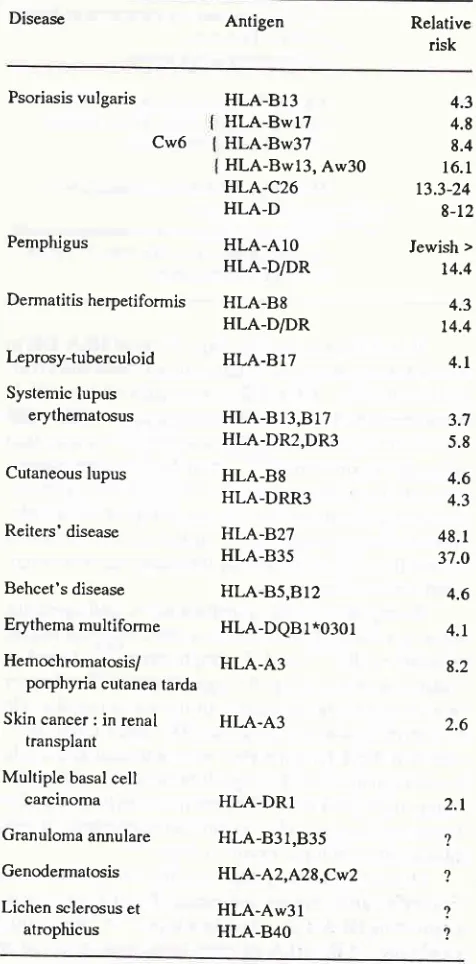
l'2The association between
HLA and
skin
diseases has beenshown by calculating
therelative risks.
The current
status
of
the
associationt,2'4-22ur.
indicated
in Table
1.Both
theprincipal
skin
diseases and thesystemic
diseases with
some cutaneous manifestations are
in-cluded.
While
the association
appeared
to
be
still
varied
for
certain
diseases,
further confirming
or
clarifying
investigations
are encouraged.Our
prelimi-nary study
on
19Indonesian psoriasis patients, using
linkage
disequilibrium
analysis, suggested theassocia-tion
between
the
HLA-B5l
and
HLA-Cw7
with
psoriasis.
HU
Antigen
45OTEER ASPECTS
OF
ELA
EXPRESSION ON
SKIN COMPONENTS
The
HLA
studiescould
alsohelp
in
understanding
thepathogenesis, histogenesis
(determination
of
the
cell
origin)
of certainskin diseases,
biological
behaviorsof
skin
components andlymphocytic
infiltrates.23-36S""
[image:2.595.301.539.262.744.2]Table 2.
Table l. The association between HLA and skin diseases
Disease Antigen Relative
risk
Psoriasis vulgaris
Cw6
Pemphigus
Dermatitis herpetiformis
Leprosy-tuberculoid
Systemic lupus erythematosus
Cutaneous lupus
Reiters'disease
Behcet's disease
Erythema multiforme
Hemochromatosis/ porphyria cutanea tarda
Skin cancer : in renal transplant
Multiple basal cell carcinoma
Granuloma arnulare
Genodermatosis
Lichen sclerosus et atrophicus
HLA-813
4.3HLA-8w17
4.8HLA-8w37
8.4HLA-Bwl3,
Aw30
16.1HLA-C26
13.3-24HLA-D
8-12Iewish >
t4.4
4.3
t4.4
4.t
3.7 5.8
4.6
4.3
48.
I
37.O HLA-AIOHLA-D/DR
HLA-B8 HLA-D/DR
HLA-B17
HLA-BI3,B17
HLA-DR2,DR3
HLA-B8
HLA-DRR3
HLA-B27 HLA-835
HLA-B5,BI2
4.6HLA-DQB1*0301
4.1HLA-A3
8.2HLA-A3
HLA-DRI
HLA-831,835
HLA-A2,A28,CW2
HLA-Aw3l
HLA-B4O2.6
2.t
?
?
2.
46 Cornain
Table2. HLA and pathogenesis
Situation
IILA
expression related to pathological changesHLA-DR expression on keratinocytes in skin diseases (dermatosis):
lichen planus, mycosis fungoides, cutaneous BJymphoma, peudolymphoma, lupus erythematosus, para-psoriasis en plaque, bullous pemphigoid, drug reaction, contact dermatitis, actinic keratosis, pityria-sis rosea, vitiligo, verrucous carcinoma, etc.
tr{LA-DR expression on dendritic cells Â
deposition of immune complexes at dermo-epidermal junction :
infl ammatory skin diseases
HLA-DR expression on activated T cells :
sarcoidosis, granuloma, lichen planus, discoid lupus erythematosus
HLA-AIl,
HLA-DR4 expression were reduced or absence :Skin cancers (factors: immunosuppression/ renal transplant, lack of cytotoxic T cells, human papilloma virus)
It
isof interest
that the expressionof
HLA-DR in
normal
stateonly occur in Langerhans'
cells andsyrin-geal
epithelium.
HLA-DR
is normally
undetected in
keratinocytes.
However,
the expression
of
HLA-DR
on
keratinocytes have
beenobserved
in
various skin
diseases (dermatosis),23-27
incltding:
lichen
planus,mycosis fungoides,
cutaneousB
lymphoma,
pseudo-lymphoma,
lupus erythematosus, parapsoriasis enpla-que,
bullous
pemphigoid,
drug
reaction,
contact
dermatitis,
actinic
keratosis,
pityriasis
rosea,vitiligo,
verrucous carcinoma,
etc.In
regard
with
the
significance in
pathogenesis,the expression
of
HLA-DR
has been encountered ondendritic
cells28'2eand
T
lymphocytes3o-32found in
relation with
certain
pathologic
changes.The
former
was related to the
deposition
of
immune complexes
in
the dermo-epidermal
junction. The latter might
indi-cate that the
T
lymphocytes were activated
andmight
be
consistent
to
the
biological
behaviours
of
the skin
components
andthe
pathogenesisof
certain
skin
dis-eases, such as sarcoidosis granuloma,^psoriasis,
lichen
planus,
discoid
lupus erythematosus.rr
It
is of
interest,
that
mycosis fungoides
andSezary's syndrome
âre cutaneousT
cell
lymphomas,
expressing
HLA-DR
or
Ia-like
antigen.ra'3)Reduction
or absence
of
theHLA
antigens have been observedin
Med J Indones
some
skin
tumors.22'36'37HLA-Atl
has been
con-sidered to haveprotective effect
againstskin
cancer,2lwhich
together
with cytotoxic T cells might
be
inter-active
with
extraneousfactor
such as humanpapilloma
virus. However,
such
relationship might
bestill
con-troversial
asit
wasnot
observedin
other
studies.20SUMMARY AND CONCLUSION
HLA
complex has been studied extensively,
both
globally
andregionally.
The association
of
HLA
andskin
diseaseshas
indicated a well define
associationwith
certain degree
of
relative
risks
in
some
and remains to be studiedfurther
in others. Ourpreliminary
result
in
psoriasis
suggested
the
association
of
theHLA-B5I
andHLA-Cw7 with
the disease.The
HLA-DR
expression
on
keratinocytes
hasbeen observed
in
various dermatosis.
Such anexpres-sion on dendritic cells
and
T
lymphocytes has
beenrelated
to
their
biological behavior, activation
andpathogenesis
in
certain
skin
diseases.Mycosis
fun-goides and Sezary's syndrorme
areT
cell
lymphoma
which
express
HLA-DR.
Some
skin tumors
showedreduction
or absence ofHLA
antigens and certainHLA
antigen
might
beprotective. However,
such
relation-ship
wasstill
controversial.
REFERENCES
1. Hors J. HLA et maladies. In: Dausset
I,
Marika PLA (Eds).HLA
complexe majeur d'histocompatibilite de l'homme. Paris. Flammarion Medecine-Sciences, 1985; 227 -56. 2. CrumptonMf
(Ed). HLAin
Medicine. Br MedBull
1987;43: t-245.
3. Cornain S, Zain
MM,
GandhaI.
HlA-antigens of normal individuals in Indonesia. In: AizawaM
(Ed). HLA in Asia-Oceania. Sapporo: Hokkaido University Press, 1986;258-61.4. Chan SH. HLA and Skin disease in the Chinese. Ann Acad Med Singapore 1983; 12: 3-5.
5. Suzuki
T,
Matsushita S, NakamizoY,
et al.
HLA
and psoriasisin
Asia populations:Ioint
report.In:
AizawaM
(Ed). HLA in Asia- Oceania. Sapporo: Hokkaido University Press, 1986:349-53.
6. Chiewsilp
I,
SukumaranKD,
SujirachatoK,
OngKf,
Mongkolsuk T, Pecharanond Y.
HLA
antigens in leprosy:Ioint
report.In:
AizawaM
(Ed).HLA in
Asia-Oceania Sapporo: Hokkaido University Press, 357-63.7.
Arnett FC.
HLA
and genetic predispositionto
lupus erythematosus and other dermatologic disorders.I
Acad Dermatol 1985; 13: 472- 81.8. Naito S, Kong FH, Hawkins BR, Mehra
NK,
Serjeantson SW, Hammond MG. Joint report:HLA
and disease: SLE.In: Aizawa
M (Ed).HLA
in Asia-Oceania. Sapporo: Hok-kaido University Press, 1986; 357-63.7
Vol 4, No
I,
January-March 19959. Rantapaa-Dahlqvist S, Strom H, Bjelle A, Moller E. Clinical symptoms and HLA antigens in a family with Reiter.s
dis-ease. Scand fRheumatol 1985; 14: 149-58.
trO. Wong RC, Ellis CN, Diaz LA. Behcet's disease. Int J
Der-matol 1984;23:25-32.
11. Mizoguchi
M,
MatsukiK,
MochizukiM,
et al.
Human leukocye antigen in Sweet's syndrome and its relationship to Behcet's disease. Arch Dermatol 1988; 124:1069-73-12. Friedman-Birnbaum Rr, Gideoni O, Bergman R, pollack S.
A study
ofHLA
antigen association in localized and general-ized granuloma annulare. Br J Dermatol 1986; I 15: 329-33. 13. Beaumont C, Fauchet R, Phung LN, De Verneuil H, GuegenM, Nordmann Y. Porphyria cutanea tarda and
HLAlinked
hemochromatosis. Evidence against a systematic
associa-tion. Gastroenterology 1987 ; 92: I 833-8.
14. Edwards CQ, Griffen LM, Kushner Jp. Increased frequency
of
HLA-A3
in
subjectswith
sporadic porphyria cutanea tarda. Tissue antigens 1988;31: 250-3.15. Amer
M, Afifi
N,
IskanderI,
DiabN.
Human leukocyte antigen in genodermatosis. IntI
Dermatol l9B4;232 53-5.16. Holt Pf, Darke C. HLA antigens and BF allotypes in lichen sclerosis et atrophicus. Tissue antigens 1983; 22: g9-91. 17. Purcell
KG,
SpencerLV,
SimpsompM,
Helman SW,Oldfather
IW,
Fowler JF
Ir.
HLA
antigensin
lichensclerosus et atrophicus. Arch Dermatol l9O;126:1043-5. 18. Hall RP, Otley C. Immunogenetics of dermatitis
herpetifor-mis. Semin Dermatol
l99l;
l0:
240-5.19. Khalil I, Lepage V, Douay C et al. HLA
DeBl
*0301 allele is involvedin
the susceptibility to erythema multifome.I
Invest Dermatoll99l;
97:697-700.20. Czarnecki D, Watkins F, Leahy S et. Skin cancers and HLA frequencies in renal transplant patients. Dermatology 1992;
185:9-ll.
21. Bouwes Bavinck JN, Kootte AM, Van der Woude FI, Van
denbroucke JP, Vermeer BJ, Class FH. On a possible protec_
tive effect
of
HLA-AIl
against skin cancer and keratotic skin lesions in renal transplant recipients. J Invest Dermatoll99l;91:
269-72.22. Czarnecl<i D, Lewis A, Nicholson I, Tait B. Multiple basal
cell carcinomas and HLA frequencies in southem Australia.
J Am Acad dermatol
l99l;24:559-61.
23.
Volc
PlatzerB, Majdi
O, KnappW,
et aal. Evidenceof
HLA-DR antigen biosynthesis by human keratinocytes in
disease. J Exp Med 1984; 159: ll.84-9.
24. Lamp,rt
IA.
Expressionof
HLA-DR (Ia like) antigen on epidermal keratinocytesin
human dermatoses.Clin
Exp Immunol 1984; 57 : 93- 100.25.
HLA-DR
antigen bearing keratinocytesin
various der-matologic disorders. Acta Derm Venereol 1985; 65: 9-13. 26.Aiba
S, TagamiH.
HLA-DR
antigen expression on thekeratino cyte surface
in
dermatoses characterized by lym-phocytic exocytosis (e.g. pityriasis rosea).Br
J Dermatol 1984;IIl:285-94.
27. Aubock I, Romani
N,
GrubauerG,
Fritschp.
I{LA-DRexpression on keratinocytes in a common feature ofdiseased skin. Br J Dermatol 1986;
l14:
465-72.28. Drijkoningen M, De-Vos R, De-Wolf-peeters C, Degreef H, Desmet
V.
Immunoelectron microscopic investigationof
HLA-DR positive dendritic cells at the dermo-epidermal junction in skin disorders associated with the depositionof
immune complexes. Ultrastruct Pathol 1986;
l0:
123-g.29. Baker BS, Lambert S, Powles AV, Valdimersson H, Fry L. Epidermal DRr+TG dendritic cells
in
inflammatory skindiseases. Acta Derm Venereol 1988;68: 2Og-17.
30. Willemze R, Vermeer BJ, Meijer CI. Immunohistochemical studies in lymphocytic infiltration of the skin (Jessner) and
discoid lupus erythematosus.
A
comparative study. J Am Acad Dermatol 1984;ll:
832-40.31. Martin AG, Kleinhenz ME, Elmets CA. Immunohistologic identification of antigen-presenting cells
in
cutaneous sar-coidosis. J Invest Dermatol 1986; 86: 625-9.32. Mishra BB, Poulter LW, Ianossy G, fames DG. The distribu-tion of lymphoid and macrophage like cell subsets of sarcoid and Kveim granulomata: possible mechanism
of
negative PPD reaction in sarcoidosis. Clin Exp Immunol l9g3; 54: ?05-15.33. Bjerke JR, Matre R. Demonstration of
IAlike
antigens on T lymphocytes in lesion ofpsoriasis, lichen planus and discoid lupus erythematosus, Acta Derm Venereol l9g3; 63: 103-7.34. Haynes BF, Hensley
LL,
Iegasothy BV. Differentiationof
human T lymphocytes: II. Phenotypic difference in skin and
blood malignant T-cells
in
cutaneousT<ell
lymphoma.f
Invest Dermatol 1982; 78:323-6.35. Tjerlund UM, Scheynius A, Kabelitz D, Klareskog L. Anti_
Ia-
reactive cellsin
mycosis fungoides:a
studyof
skin biopsies, single epidermal cells and circulatingT
lym_ phocytes. Acta Derm Venereoll98l; 6l:
2gl-301. 36. Natali PG, Viora M, Nicotra MR, Giacomini p, Bigotti A,Ferrone S. Antigenic heterogenicity of skin tumors of non_
melanocyte origin: analysis with monoclonal antibodies to tumor-associated antigens and
to
histocompatibility an_tigens. JNCI 1983;
7l:
439- 47.37. Holden CA, Sanderson AR, MacDonald DM. Absence
of
human leukocyte antigen molecules in skin tumors and some
cutaneous appendages: evidence using monoclonal an_
tibodies. J Am Acad Dermatol 1983;9: 967-7L.