, \ I.
,
INFECTION OF BABESIA BIGEMINA IN CATTLE
AND METHODS OF CONTROL
SCRIPT
By
JUANIS YAJUNI B 161100
FACULTY OF VETERINARY MEDICINE
BOGOR AGRICULTURAL UNIVERSITY
Babesia bigemina is the species which occurs mostly
through out the tropics and subtropical areas. It gains
access to the susceptible cattle through the bite of an· ig
fected tick and penetrates an erythrocyte. The
pathogene-sis of セN@ bigemina is considered to be as highly pathogenic and may cause heavy mortality in susceptible stock. Infeci
ed cattle rapidly become dull and listless and lose their
appetite, the erythrocytes are destroyed resulting anaemia,
icterus, emaciation, weakness, and a drop in milk
product-ion. Death and loss of productivity due to セN@ bigemina account for the economic loss.
Trypan blue was the first drug to be used in treating
セN@ bigemina but is now less frequently used because of its
discoloration effect of flesh. Acaprin at the rate of 1 mg
/ Kg given subcutaneously and Acridine derivates given
in-travenously
15
to 20 ml of5
%
solution or5
ml of a5
%
citra ted solution given intramuscular are most widely
used. Berenil is also very effective at the rate of 2 - 3
mg / Kg given by deep intramuscular injection.
Ticks are the only vector. Therefore babesiosis due
to セN@ bigemina is best controlled by eliminating the tick. Immunization of susceptible stock, treatment of infected
animals, and the control of stock movements are other
me-thods of controlling.
The author wishes to place on record his deepest sense
of gratitude to Dr. Gatut Ashadi as the adviser and Drh. Umi
Cahyaningsih as member for their help, encouragement and
guidance in the preparation of this script.
He also thankfully acknowledges his gratefulness to
the dean of the Faculty of Veterinary Medicine and all the
member of his staff for assisting and guiding him during
the course of the study at the university.
The author also wishes to express his sincere thanks
to the Chief Minister's Department in Sabah for awarding
the scholarship and other facilities while studying in
Bogor. To all librarians of the University libraries and
the Research Institute for Animal Disease Library in Bogor,
the author express his gratitude for their assistance in
various ways.
Last but not the least, the author records his
deep-est appreciation to his parents, brothers, sisters and
beloved wife and children for their prayers, encouragment
and help aimed at inspiring the author during the course
of his academic enterprise.
AND 1'lETHODS OF CONTROL
SCRIPT
By
JUANIS YAJUNI, Degree in Veterinary Medicine (
1984 )
B 161100
Drh.
sih
Dr Gatut Asha
A:dvistrr----'
AND METHODS OF CONTROL
SCRIPT
A Script
Presented to the Faculty of Veterinary Medicine
of Bogor Agricultural University
In Partial Fulfillment for the Degree of
Doctor of Veterinary Medicine
Adviser
Member
( Dokter Hewan )
By
JUANIS YAJUNI
Dr. Gatut Ashadi
Drh. Umi Cahyaningsih
FACULTY OF VETERINARY MEDICINE
BOGORAGRICULTURAL UNIVERSITY
I.
II.
I I I .
SUMMARY
• • • • • • • • • • • • " • • , • • • • • • . • ' . s" e_e·e_e·s . . . .e-• e-• e-• e-• e-• e-• e-• e-• e-• e-• e-• e-• e-• e-• e-• e-• e-• e-• e-• e-• e-• • e·' •
• • • • • • • • • • . • . • • • • • • • • •• e· e· • •
,.,
... .
ACKNOWLEDGEMENTS
LIST OF CONTENTS
LIST OF TABLE
LIST OF FIGURES
INTRODUCTION
LITERATURE REVIEW
• • • • • • • • • • • • • • • • • • • • • • • ••
e-• e-• e-• e-• e-• e-• e-• e-• e-• e-• e-• e-• e-• e-• e-• e-• e-• e-• e-• e-• e-• e-• • e-.
.
. .
. . . .
.
. .
. .
.
.
. .
.
.
. . .
.
.
.
A.
B.
C.D.
E.
F. G.H.
1.J .'
HISTORY AND OCCURRENCE
.. • • • • • • • • • e' e" • • • •CLASSIFICATION
.
...
..
'.
MORPHOLOGY
DIAGNOSIS
.
.
. .
.
.
.
. .
.
. . .
. . .
.
. .
.
. . .
".
.
.
• • • • • • •• e" • • • • -s . ' • • • • • • • • • • • • ' .
TRANSMISSION
PATHOGENESIS
CLINICAL SIGNS
• • • • • • •. . . e-.· ••
.
...
... .
.
. . .
.
.
.
. .
. . .
. . . .
.
.
. .
.
.
.
.
.
.
.
.
. . .
. . .
PATHOLOGY ANATOMY
ECONOMIC LOSS
eo • • • • • • • • • • • • • • • '"' • • • • • . ,TREATMENT AND CONTROL
• • • • • • • • • • " • • e·, •CONCLUSION
REFERENCES
e· • • • • • • • • • • • • • • • • • • • • • • • • • • • • • •
. . . • e''''
Table Page
1. Percentage of infection in: cattle
according to age • • • • • • • • • • • • • • • • • • • • • • • • • • • • • •
5
Figures Page
1. Erythrocytic development of Babesia bigemina
...
9 2. Diagrammatic representation of a trophozoiteof Babesia bigemina . . . • • . . . • . . . I - I • •
9
3.
Development of Babesia bigemina in Boophilusrnicroplus
...
...
124.
Postulated life cycle of Babesia bigemina inthe cow and tick • • • • • • • • • • • • • • • • • • • • • • • • • • • • • • • 12
The Babesia are intraerythrocytic, tick--transmitted
protozoan parasites. Babes (1888) in Romania was the
first to describe Babesia parasites ゥセ@ the blood of cattle and sheep. Various species of Babesia are known to
in-fect cattle, however, the most economically important
are those caused by Babesia bovis and Babesia bigemina.
Babesia bigemina was accurately described by Smith and
Kilborne ( Burner, 1973). These workers also made the
discovery of epochal importance because this was the
first protozoan disease shown to be so transmitted. In
1930, Babesia sp. had been described in all of the
domestic animals, the geographical distribution of the
parasite was world wide and ticks were the only vectors.
Babesiosis is presently considered as one of the
most important constraints in production of cattle in
A. HISTORY AND OCCURRENCE
Babesia bigemina was first discovered by Smith.
and Kilborne in. the United States ill. 1893. The cause
of an important disease of cattle called Tick fever,
red water fever, piroplasmosis and formally in North
America, Texas fever. Hosts principally the bovine,
also in zebus, water buffalo (Gibbons, 1963) and deer
(Soulsby, 1978).
Occurs through out the tropics and subtropical
areas including America, The West Indes, Australia,
Africa, tropical mainland Asia, Indonesia, The
Philip-pines (Hall, 1977 and Blood et al., 1979). According
to Gibbons (1963) Babesia bigemina infection in
gene-ral occurs in the warmer areas.
In the cattle body, セN@ bigemina is present in the red blood cells during the fibrile stage in the form
of round or pear-shaped bodies (Udall, 1954).
Accor-ding to Hall (1977) and The British Veterinary ABso.
siations (1976), the incidence of the infection of
セN@ bigemina can. be seasonally related to the occurrence and activity of the vectors. Movement from the free
areas into enzootic areas results in high morbidity
and mortality rates in cattle which have either never
contact with it for at least a year and have thereby,
partially or completely lost in their immunity.
Therefore it is a disease most commonly
encount-ered in cattle in transit to slaughter or to seasonal
or regional pasture, and rarely appears in cattle born.
and raised in. enzootic areas whose premmunition is
es-tablished gradually as the,natural resistance of young
animal subsides. When it does occur in cattle in
en-zootic areas its appearance is due to the stress of
intercurrent debilitating disease or to starvation or
parturition. It is also common to find that
inter-current infections with another micro-organisme like
Anaplasma, may cause break down of premunity and
initiation of babesiosis. (Hall, 1977 and Blood et al.,
1979)
European breeds are much more susceptible than
zebus, probably because of tick resistance of the lat-·
ter breed. Nevertheless, both are likely to become
infected if they are introduced into enzootic areas
without premunity. According to Blood et al.(1979),
heaviest losses occur in marginal areas where the tick
population is highly variable depending on the environ
mental conditions. The morbidity rate is such
circum-stances is often. 90% and the mortality may be of the
same order. With early, efective treatment the
According to Kreier
(1977),
it is of major eco-nomic importance in cattle because of the excessivehusbandry methods employed to raise this animal. In
enzootic areas cattle usually become infected while
relatively young, suffer only mild reactions, and a
subsequently resistant to severe clinical infection.
Previously uninfected adult cattles are highly
suscep-tible. The disease is mostly prevalent in. bushy or
wooded areas which are favourable habitats for vector
ticks. According to Gibbons
(1963),
the incidence of infection decreases in proportion to the effectiven.essof tick control measures. The greatest infection rate
is in animals in the
6-12
months age group and infect-ion is uncommon in animals over 5 years of age.An-nimals under one year of age are in.fected predominantly
with Babesia bigemina. (Blood et al.,
1979)
Babesia bigemina reactors examined in Eastern
plains of Colombia showed that 40% of the calves
be-tween 1 and
3
months of age were reactors, (Wells,1977)
while 65% of the calves between 4 and 6 months were reactors (Table I). 65% of the 7 to12
months old group were reactor; 48% of the13
to 24 months group; and 30% of all cattle tested over 24 months ofage were Babesia bigemina reactors. Wells
(1977),
fUrther stated that there were 68% of the
120
preg-nant cows tested for babesiosis due to !}. bigeminaBabesia bigemina was two weeks with the latest age of
in.fection being 34 weeks.
Age No. of Gattle No. of Percent
(Months) tested Reactors Reactors
1-3 144 66 46%
4-6 254 165 65%
7-12 416 269 65%
13-24 412 197 48%
>
24 660 194 30%Total 1886 891 47%
Table I. Percentage of infection in cattle according
to age ( Wells, 1977 )
B. CLASSIFICATION
The taxonomy of Babesia sp. has always been the
subject to debate because sufficient knowledge of their
biology has never been available to determine their
correct position among the Protozoa. Many
investigat-ors assumed they were related to Telosporidia but were
later differentiated from Theileria. (Weinman. and
Ristic, 1968).
Babesia sp. were usually placed in the class ( or
Subphylum) Sporozoa ( Leuckart, 1879 in Kreier, 1977),
a class originally erected on. parameters related to a
method of sexual reproduction that result in the
for-mation of spores and oocysts.
Regendanz and Reichenow (1933 in Weinman and Ristic,
amoeboid movement in- some stages of development and
the absence of sexual reproduction, were more closely
related to the Rhizopoda than Sporozoa. This view was
supported by the work of Muratov and Cheissin (1959,·
in Weinman and Ristic ,. 1968) and Poljansky and
Cheissin-(1959, in Weinman and Ristic, 1968), who could net find
evidence of sexual reproduction in. Babesia bigemina
and Babesia bovis.. Because of repeated failure to
de-monstrate a sexual phase in the babesial development,
the Committee on Taxonomy and Taxonomic Problems of
the Society of Protozoologist removed them from the
class Sporozoa and placed them with. the amoebae in.
superclass Sarcodinaj class Piroplasmeaj order
Piro-plasmida (Mc Diarmid, 1969).
In recent years knowledge of biology of Babesia
sp. and that of the Sporozoan generally has increased.
A very close morphological relationship between Babesia
and established members of this class was demonstrated
by ultrastructural studies (Scholtyseck et al., 1970
and Schol tyseck, 1972 in Kreier ,. 1977). However the
question of sexual reproduction among the Babesia was
not resolved. Levine (1969,. 1970 in Kreier, 1977)
concluded that the formation of spores and oocysts was
not a suitable characteristic for the basic of the
primary classification. He proposed a new Subphylum
Jl:picomplexa, based on the presence,. in.
by the ・ャ・」エイッセ@ microscope. This new Subphylum con-tains two class,. 1.
e.,
the Sporozoea (Leuckart" 1879in Kreier, 1977) whose members produce oocysts and
spores, and the Piroplasmea CLevin.e セ@ 1961 in. Kreier,
1977), that reproduce by asexual methods only. In the
latter class there is one order, Piroplasmorida (Wenyon,
1926 in Kreier, 1977) which contains the family
Babesiidae (Poche, 1913 in Kreier, 1977). However,
this proposal may not be the final solution for the
Babesia because the assumption that they do not
under-go sexual reproduction may not be justified.
'l'he weakness in the past in including piroplasma
in the class Sporozoa has depended on. ignorance of
their development in both the vertebrate and
inverte-brate hosts. The development of セN@ argentina and セN@
bigemina in the tick Boophilus microplus, described
by Rick (1964a, 1966 in Weinman and Ristic ,. 1968),
has provided new evidence which may permit a more
accurate classification. of this group. Therefore in
the light of the evidence now available there would
appear to be no doubt that the genus Babesia belongs
to the class Sporozoa (Weinman and Ristic, 1968).
Levine (1971 in. Kreier, 19'(7) was the one who decomented
the true iden.tity of Babesia speciess according to their
measurment and Babesia bigemina comes under large
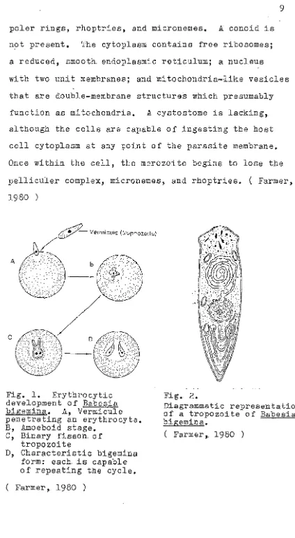
poler rings, rhoptries, and micronemes. 11 conoid is
not present. The cytoplasm contains free ribosomes;
a reduced, smooth. endoplasmic reticulum; a nucleus
with two unit membranes; and mitochondria-like vesicles
that are double-membrane structures which presumably
function as mitochondria. 11 cystostome is lacking,
although the cells are capable of ingesting the host
cell cytoplasm at any point of the parasite membrane.
Once within the cell, the merozoite begins to lose the
pelliculer complex, micronemes, and rhoptries. ( Farmer,
1980 )
__
Z⦅セ@
Vermicule (trophozoite)/@7-• A
Fig. 1. Erythrocytic development of Babesia bigemina. 11, Vermicule penetrating an erythrocyte. B, Amoeboid stage.
C, Binary fisson. of tropozoite
D, Characteristic bigemina form: each. is capable of repeating the cycle.
( Farmer, 1980 )
Fig. 2.
Diagrammatic representation of a tropozoite of Babesia bigemina.
[image:16.546.59.483.13.786.2]D. DIAGNOSIS
Diagnosis of Babesia bigemina infection is based
on. clinical signs of fever, hemoglobinuria, anaemia
and icterus, and confirmed by the detection of
para-sites in the peripheral blood. Both thick and thin.
blood smears may be employed, being stained by one of
the Romanowsky stains; however, the organisms may not
always be present,. and it may be necessary to examine
a number of smears to establish their presence. In
the cerebral form, examination of cerebral capillaries
is necessary (Soulsby, 1978). In the endemic areas,
high fever associated with hemoglobinuria and anaemia
is suggestive of Babesia in£ection, and frequently
ani-mals are treated with recourse to blood examination.
For wider surveys the Complement Fixation Test ( CFT )
of Mahoney (1962) may of value especially in those
animals where the level of parasitemia is low. Babesia
bigemina according to Kreier (1977), can be recognized
in Giemsa-stained kidney smears up to 8 hours after
death and in brain for 16 to 28 hours. Babesiosis can
also be diagnosed from autopsy or necropsy. When the
infection is due to セN@ bigemina, the kidneys are commonly
enlarged and dark, and the bladder may contains
red-brown urine. The blood is almost always thin and watery
(Belschner, 1974). Patches of congestion in varying
carcass shows the general appearence of anaemia, such
as gelatinous exudation in various parts, together
with paleness of the muscular tissue.
To prove diagnosis in. some cases, it may be
ne-cessary to reproduce the disease in. a susceptible
healthy cattle by parentral inoculation of 5 to 10 ml
of fresh or citrated blood from the suspect (Gibbons,
1963) •
E. TRANSMISSION
Under natural conditions transmission is effected
and initiated with the introduction of sporozoites
in-to the blood through the bite of tick (Cheng,
1964).
Numerous species of the genera Boophilus, Haemophysalis,
Ixodes, Rhipicephalus and Hyaloma have been implicated.
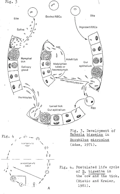
Transovarian transmission (Fig.
3)
is regarded as the commonest method in. most species, the parasite passingthrough the egg to the next generation of the tick.
Ticks feeding on non-susceptible hosts do not lose
their infection.
Transference of parasitaemia blood from an
infect-ed to susceptible animal through the agency of biting
flies or unsterilised surgical instruments is
theori-tically possible but appears to be unimportant.
The ;Babesia survive in. areas of endemic infection
Fig.
3
Bite
9
'" p
Saliva q o P
L>
Fig. 4
"
i
I
e
{;
Salivary gland
INVERTE&R ATE HOS T
(.Ii· .
. )
\
\
Bovine RBCs Bite
A
Malpigl1ian
tubes or
I
Ingested RBCs
Gut
Fig.
3.
Development ofBabesia bigemina. in Boophilus microplus
(Adam, 1971).
Fig.
4.
Postulated life cycleof
B.
bigemina in the-cow and the tick. [image:19.541.54.474.36.712.2]and by carrier cattle providing a source of infection
to the tick population. Moreover ,. ticks are not clean.§.
ed of the infection when feeding on non-susceptible
hosts, and a carrier state probably exists in other
domestic and wild animals (British Veterinary Associai
ion, 1976). Infection. can also be established in a
susceptible host by parenteral inoculation with blood
or with spleenic or hepatic fluid taken from an infeci
ed animal. It can also be transmitted experimentally
from infected to susceptible cattle by intravenous,
subcutaneous, intramuscular,. or intraperitoneal
in-oculation. of blood. A few out-breaks of the disease
have been traced to surgical procedures, such as
de-horning, in which there was no sterilization of
instruments. The blood from animals carrying inactive
infection was thus transferred to susceptible animals
(Gibbons, 1963). According to Farmer (1980), ticks
responsible for transmission of Babesia bigemina are
the ixodids, Boophilus annulatus and セN@ microplus. Schmidt (1981), reported that Babesia bigemina is
transmitted by tick of the genus Boophilus, and the
distribution of babesiosis is limited by the
distri-bution of the tick. The tick after feeding, mature,
and mating on a single host. After engorging and
mating, the female drops to the ground, lays her eggs
that have been inIected with Babesia bigemina through
legged ticks that hatch from the egg climb onto
vege--tation and attach to animal that are brushed by the
plants. In other parts of the world it occurs in a
one-host tick, two and three-host ticks serve as host
and vectors of Babesia bigemina. In these cases,
trans-ovarian transmission is not required and may not occur.
All instars of such ticks can transmit the disease.
Soulsby (1978), stated that one host-tick: Boophilus
annulatus, N. America; セN@ calcaratus, N. Africa; セN@
decoloratus, S. Africa; セN@ microplus, Australia, Panama,
S. America Two-host tick : Rhipicephalus evertsi,
S. Africa;
R.
bursa, S. Africa : Three-host tick :Haemaphysalis puntata, Europe and Eurasia; Rhipicephalus
appendiculus, S. Africa. He further said that there is
'no conclusive evidence that Babesia bigemina can be
transmitted mecanically by blood-sucking arthropods.
On rare occasion it seems that intrautrine transmission
may occur from mother to foetus, but this mode of
in-fection does not seem to be of any significant
impor-tance in the general epidemiology of babesiosis.
F. PATHOGENESIS
Babesiosis is a highly pathogenic disease of
ani-mals and may cause heavy mortality in susceptible stock
(Weinman and Ristic, 1968). On introduction into the
invade the red blood cells and begin. to divide into
paired daughter cells. As the parasitized
erythro-cytes are destroyed, the daughter cells separate and
enter new red blood cells. The destruction of the
red blood cells and loss of hemoglobin into the blood
stream result in anaemia, hemoglobinuria and icterus
(Davis and Anderson,
1971).
In typical acute cases in. adult cattle, the red
cell count may drop to 1 to 2 million per cubic
milli-meter of blood, hemoglobinuria and icterus are marked,
and death may result from anoxemia, particularly under
stress of handling or excercise. In milder infections
of young or partially immune animals and in chronic
infection the destruction of red blood cell is usually
not sufficient to cause hemoglobinuria, although
an-aemia and debilitation may develop in prolonged cases
(Gibbons,.
1963).
According to Burner
(1973),
the inCUbation period following tick transmission varies between 8 to 10 days.The multiplication of Babesia bigemina in the
peri-pheral, reaches a peak with the development of
clinic-ally detectable haemolysis result after an inCUbation
period of
7
to 20 days (Blood et al.,1979).
If the animal survives, it becomes a carrier in which aharm-less, subclinical infection. is maintained by a delicate
immunological balance between protozoa and antibodies.
especially transport and deprivation of food, and in ...
tercurrent disease. iセ@ this carrier state the animal is resistant to infection and it persists for about
a year.
According to Soulsby (1978), the first ev.idence
of the disease is a spectaculer rise in body.tempera'"
ture. The high fever lasts for 2 -
7
days or more andduring this period a profound anaemia frequently 、・カ・セ@
lops. There is hemoglobinuria and cardiac palpitation.
At the hight of fever, up to 75% of red blood cells
may be destroyed, and the mortality may be very high
in acute cases, death occuring 4-8 days after onset of
clinical signs. During the exit of Babesia bigemina
parasites from infected cattle erythrocytes, two or
more parasite-associated proteolytic enzymes are
re-leased (Ristic, 1981) into the plasma. These enzymes
and/or similar parasite metabolic products are beleived
to interact with blood components and are ultimately
responsible for several of the pathologic signs and
symptoms. Ristic (1981), further stated that infection
of セN@ bigemina in spleenectomized calves showed a de-crease in serum potassium levels in some animals
while urine potassium levels were increased in all
animals.
G. CLINICAL SIGNS
The clinical manifestation of babesiosis varies
from a very mild and symptomless infection to acute
and often fatal episodes, depending on the species of
Babesia involved and the susceptibility of the host
animal. In general the most se1rerely pathogenic form
of the disease is seen in infection of highly
suscep-tible adult cattle with セN@ bigemina (British Veterinary
Assosiation, 1976). The first symptom is a sudden rise
in body temperature to 4loC (Schmidt, 1981). This may
persist for a week or more. Infected animal rapidly
become dull and listless and lose their appetite. Up
to 75
%
of the erythrocytes may be destroyed in thefatal cases, but even in milder infections so many
erythrocytes are destroyed that anaemia result.
Me-chanisms for clearence of hemoglobin and its breakdown
products are overloaded, so jaundice results, and much
excess hemoglobin is excreted by the kidneys giving
the urine the red color. Chronically infected animals
remain thin, weak, and out of condition for several
weeks before recovering. According to Belschner (1974),
with milking cattle a drop in. milk-production is
generally the first symptom noted. Rumenation ceases,
respiration is accelerated. There is great weakness,
the animal stands with back arched, head troked out,
the mouth. The animal moves with difficulty and with
staggering gait. It may be constipated and pass hard
dung, often coated with mucous. The mucous membranes
of the eye and_mouth, which are at first very red,
change to white, indicating severe anaemia. Weakness
becomes intense; there is a sUdden drop in temperature
to below normal shortly after death, which commonly
occurs in three to four days Dr even less from the
on-set of the disease. At the stage of jaundice becomes
evident the heartbeat becomes fast and accentuated.
The heartbeat may be so forceful that it may be deteci
ed some distance from the animal (Hall, 1971).
Often the first sign is that the animal isolates
itself from the heard (Seddon, 1966), and becomes
un-easy. Station cattle frequently leave the mob and seek
shade. The febriel stage usually lasts for about
3
weeks (Blood et al., 1979). Pregnant females may abort
(Gibbons, 1963 and Blood et al., 1979). Occasional
animals infected with
f!..
bigemina show cerebralbabe-siosis manifested by incoodination followed by
pos-terior paralysis or by mania, convulsions and coma
(Soulsby, 1978 and Blood セ@ al., 1979).
H. PATHOLOGY ANATOMY
is enlarged, swollen and a soft, pulpy consistency,
the liver is grossly enlarged and dark brown in colour,
and the gall-bladder is distended with thick granular
bile. The kidneys are enlarged (Belschner, 1974 and
Blood セ@ al., 1979). The lungs are swollen: and
oedematous (Seddon and Albiston, 1966). The
pericar-dium contains some blood-stained exudate and there are
petechial haemorrhages under the epicardium and
endo-cardium (Seddon. and Albiston, 1966 and Blood et al.,
1979) •
In animals which have sUffered a more prolonged
illness, acute lesions are absent except that
pete-chial haemorrhages may be present on the heart (Seddon
and Albiston, 1966). The carcase is usually emaciated
and icteric, and the blood is anaemic. A
characteris-tic lesion in cattle which have died from an acute
di-sease is severe intravasculer clotting (Blood et gl.,
1979). In the cerebral form there is perivascular,
perineuranal and interstitial oedema throughout the
brain. and spinal cord (Soulsby, 1978). According to
Gibbons (1963), Burner and Gillespie (1973), Belschner
(1974), RaIl (1977) and Soulsby (1978), oedema often
occurs in the subcutaneus tissue of the ventral part
of the body, and the fatty tissue is yellow and
gela-tinous around the kidneys. Belschner (1974), stated
that patches of congestion in varying amounts are
pre-sent along the intestinal tract, and the blood is thin
I. ECONOMIC LOSS
With a rapidly increasing world population, the
food needs of humans will be critical. Accordingly,
diseases that affect domestic animals must of necessity
be studied, understood, and controlled. Piroplasms
that can be lethal to cattle have a worldwide
distri-bution. For example, Babesia bigemina (Farmer, 1980),
although no longer present in the United States, infects
cattle in Africa, Australia, Europe, and South America.
Piroplasms are important economically because of
their pathogenicity. Death. or loss of productivity in
bovine herd is characteristic of babesiosis. Infected
animals fail to eat and suffer from severe anaemia.
Death usually results from kidney damage and a rapid
accumulation of toxic by-products. Because of economic
impact of Babesia on cattle industry of the world·,
con-siderable money have been allocated towards research·
to produce pharmacological relief (Farmer, 1980).
Serous economic loss in northern Australia,
main-ly through deaths of cattle from the disease, loss of
condition in animals recavering from an acute attack,
loss of milk production ゥセ@ dairy cattle (Seddon and
Albiston, 1966), and the condemnation of carcasses at
abattoirs. In addition the cost of immunization,and
the treatment of clinically affected animals impose
on the country. The losses caused by babesiosis occur
through failure to apply effective methods of
immuni-zation and treatment that are available. Mortality,
production losses, quarantine, and other costs of
con-trolling the spread of babesiosis, opportunity loss,
and loss of markets for live grade or pedegree (Ristic
and Mc Intyre, 1981), are the impact of babesiosis on
development.
The economic analysis in the United States of
America gives some figures that are indicative of the
importance of babesiosis. The study concludes that
once ticks and babesiosis established in the endemic
areas of the United States, annual losses of US$500
million which was all attributed to eradic:"tion of the
tick and babesiosis. While the Australian figures are
derived from a study (Anon, 1975 in Ristic and McIntyre,
1981) in Queensland and New South Wales, the total
estimated annual losses attributed to ticks, tick-borne
disease, and their control amount to A$5.2 ( US$7.8 )
and A$5.1 per head of cattle in, the tick infested
areas of QUeensland and New South Wales respectively.
In Mexico, Beltran (1975 in Ristic and McIntyre,
1981) reported that of 3587 million pesos lost annually
because' of ticks and tick-borne diseases" only 6.3
%
is a result of death, 83.6, 8.5, and 1.6
%
attributedto loss of meat production, milk production, and hides
difficult to ohtain the actual figures because of
li-mited records available to estimate the probable
eco-nomic loss.' However 1. t would' probably be unlch greater
economic loss caused by babesiosis in. many latin
American. countries and in many developing countries
where Babesia bigemina is present (Ristic and McIntyre,
1981).
J. TREATMENT AND CONTROL
Trypan blue is probably the first spesific drug
used successfully t9 treat Babesia bigemina infections
(Weinman and Ristic,
1968
and Ristic and Kreier,1981).
Intravenously injection of trypan blue at the rate
2-3
mg / Kg effectively eliminated セN@ bigemina (Ristic and Kreier,1981).
The drug usually produces 、ゥウ」ッャッセ@ation of the animal flesh (Jones et gl.,
1977),
and in view of availability of new, more effective drugs, itis used less frequently.
Acaprin is the most ーイッュゥョ・ョエセ@ is very effective
against セN@ bigemina at the rate of 1 mg / Kg given su£
cutaneously (Ristic and McIntyre,
1981),
and is the most widely used and is universally accepted as anefficient safe drug (Blood et al.,
1979).
Acridine derivates such as Acriflavin., Flavin,
Euflavin and Gonacrin are also effective, usually a
of
15
to 20 ml per animal or5
ml of a5
%
citratedsolution intramuscularly (Soulsby, 1978).
Berenil is also very effective at the rate of
2-3
mg / Kg by deep intramuscular injection (Soulsby, 1978) •Since the natural transmission of Babesia bigemina
is dependent on certain species of ticks, infection
can be prevented by adequate tick control measures
which keep animals from tick infection. This can be
done by the regular dipping of cattle (Davis and
Anderson, 1971, Jones et al., 1977 and Soulsby, 1978).
Other measures are the immunization of susceptible
sto'ck; treatment of infected animals; and the control
of stock movements (Hall, 1977). Control of infection
may be accomplished by artifical premunization of
young or introduced stock. This procedure may also
be used to prevent or control outbreaks in marginal
zones where ticks infection of stock varies
consider-ably depending on climate condition (Weinman and Ristic,
III. CONCLUSION
Babesia bigemina is an intraerythrocytic parasite
un-der the class Sporozoa and genus Babesia, and is generally
found in the warmer areas infecting a wide variety of
ru-minants, such as deer, water buffalo and zebu, in addition
to cattle; measuring
4- 5
microns long by QセU@- 2.5.
mi.:..·. Grons wide. Diagnosis 0 f
J2.
bigemina infection is basedon clinical signs of fever, hemoglobinuria, anaemia and
icterus, and confirmed by the detection of parasites in
the peripheral blood. It can also be diagnosed from autopsy
or necropsy.
Transmission of
J2.
bigemina is effected and initiatedwith the introduction of sporozoites into the blood through
the bite of infected tick. There are also other possible
modes of infection such as transference of parasitaemia
blood through the agency of biting flies or unsterilised
surgical instruments, by parenteral inoculation with blood.
The pathogenesis of セN@ bigemina is considered as a highly pathogenic disease and may cause heavy mortality in ウオウ」・セ@
tible stock due to the destruction of red blood. Exit of
セN@ bigemina from infected cattle erythrocytes, two or more
parasite-associated proteolytic enzymes are released into
the plasma. These enzymes are ultimately responsible for
the several pathologic and clinical signs of anaemia and
icterus.
III. CONCLUSION.
Babesia bigemina is an intraerythrocytic parasite
un-der the class Sporozoa and genus Babesia, and is generally
found in the warmer areas infecting a wide variety of
ru-minants, such as deer, water buffalo and zebu, in addition
to cattle;, measuring 4 - 5 ·microns J!ong by QセU@ セ@ 2.5 .. ュゥセ@
crons wide. Diagnosis of セN@ bigemina infection is based
on clinical signs of fever, hemoglobinuria, anaemia and
icterus, and confirmed by the detection of parasites in
the peripheral blood. It can also be diagnosed from autopsy
or necropsy.
Transmission of セN@ bigemina is effected and initiated
with the introduction of sporozoites into the blood through
the bite of infected tick. There are also other possible
modes of infection such as transference of parasitaemia
blood through the agency of biting flies or unsterilised
surgical instruments, by parenteral inoculation with blood.
The pathogenesis of セN@ bigemina is considered as a highly pathogenic disease and may cause heavy mortality in susceR
tible stock due to the destruction of red blood. Exit of
セN@ bigemina from infected cattle erythrocytes, two or more parasite-associated proteolytic enzymes are released into
the plasma. These enzymes are ultimately responsible for
the several pathologic and clinical signs of anaemia and
icterus.
omentum, and the spleen, lever, lungs and kidneys are
en-larged, and the bladder is distended with thick granular
bile. The pericardium contains some blood-stained exudat
and petechial haemorrhagic under the epi and endocardium.
Death and the loss of productivity account for the economic
loss due to the pathogenicity of the セN@ bigemina parasites. Acaprin at the rate of 1 mg / Kg administered subcutaneously
and Acridine derivates usually
5
%
solution given intra-venously or intramuscular are the most prominent andeffec-tive against セN@ bigemina. Berenil is also very effective. Control efforts include control of the tick vectors,
immuni-zation of susceptible stock, treatment of infected animals
REFERENCES
1. Adam, K.M •. G., J. Paul and V •. Zaman. 1971. Medical and Veterinary Protozoology. Churchill Livingstone Edinburgh and London. p 116-122.
2.
3.
4.
5.
6.
7.
8.9.
Belschner, H.G. 1974. Cattle Diseases 4th. ed.
Angus and Robertson Publisher, London. p 82_86 Blood, D.C. and J.A. Hendersons am..d O.M. Radostits.
1979. Veterinary Medicine, 5th. ed. Bailliere Tindall, London. p 728-734.
British Veterinary Association. 1976. Handbook on Animal Diseases in the Tropics. Burgess and Son Ltd., Abingdon Oxfordshire. p 166-171.
Burner, D.M. and J.H. Gillespie. 1973. Hogan'sIn-fectious Diseases .oJ Domestic Animals, 6th. ed.
Comstock Publishing Associates Cornell University . Press / Ithaca and London. .p 696-700.
Cheng, T.C. 1964. General Parasitology. Academic Press New York. p 267-268.
Davis, J.W. and R.C. Anderson. 1971. Parasitic Disease of Wild Animals. The Iowa University Press, Ames, Iowa, USA. p 335-341.
Farmer, J.N. 1980. The Protozoa, Introduction to Protozoology. The C.V. Mosby Company ST. Louis Toronto, London. p 487-492.
Gibbons,W.J. 1963. Diseases of Cattle. Revised 2nd. ed. American Veterinary Publication Inc. p 665. 10. Hall, H.T.B. 1977. Diseases and Parasites of Livestock
in the Tropics. Longman Group Ltd., London
p lL,5-149.
11. Hungerford, T.G. 1975. Diseases of Livestock, 8th. ed. McGraw-Hill Book Company, Sydney. p 303-310. 12. Jones, L.M., N. Booth and L.E. McDonald. 1977.
Veterinary Pharmacology and Therapeutics 4th. ed. Oxford and IBH Publishing Co., New Delhi, Bombay, Calcutta. p 1095
13. Jones, T.C. and R.D. Hunt. 1972. Veterinary Pathology 4th. ed. Lea and Febiger, Philadelphia. p 7?3-726. 14. Kreier, J.P. 1977. Parasitic Protozoa Vol. IV. Academic
Press New York, San Francisco. p 1-43. 15. McDiarmid, A. 1969. Diseases in Free-Living Wild
16. Ristic, M. and Ian McIntyre. 19B1. Diseases of Cattle in Tropics. Economics and Zoonotic Relevence. Martinus Nijhoff Publishers, London. 2 pp. 17. Ristic, M. and J.P. Kreier. 19B1. Babesiosis.
Academic Press, New York and London. 443 pp. lB. Schmidt, G.D. and L.S. Roberts. 19B1. Foundations
of Parasitology 2nd. ed. The C.V. Mosby Company, ST. Louis, Toronto, London. p 170-172.
19. Seddon,
H.R.
and H.E. Albiston. 1966. Diseases of Domestics Animals in Australia, IV. Servo Pubs. Dep. Hyg. p 10-30.20. Smyth, J.D. 1976. Introduction to Animal Parasitology 2nd. ed. Hodder and Stoughton London, Sydney, Aukland, Toronto. p 124-125.
21. Soulsby, E.J.L. 1966. Biology of Parasites •. Academic Press, New York and London. p 15-30.
22. Soulsby, E,J.L. 1978. Helminths, Arthropods and Protozoa of Domesticated pセゥュ。ャウ[@ 6th. ed.
Bailliere Tindal and Cassel Ltd., London. p 702. 23. Udall, D.H. 1954. The Practice of Veterinary Medicine,
6th. ed. Ithaca, New York. p 709-713.
24. Weimman, D •. and M. Ristic. 196B. Infectious Blood Diseases of Han and Animals VOL. II (Diseases caused by ProtoZOa). Academic Press, New York .and London. p 219-265.
25. Wells, E.A. 1979. Workshop on Hemoparasites (Anaplas-mosis and Babesiosis). Cali: Centro Internacional de Agricultura Tropical. p 23-92.
, \ I.
,
INFECTION OF BABESIA BIGEMINA IN CATTLE
AND METHODS OF CONTROL
SCRIPT
By
JUANIS YAJUNI B 161100
FACULTY OF VETERINARY MEDICINE
BOGOR AGRICULTURAL UNIVERSITY
Babesia bigemina is the species which occurs mostly
through out the tropics and subtropical areas. It gains
access to the susceptible cattle through the bite of an· ig
fected tick and penetrates an erythrocyte. The
pathogene-sis of セN@ bigemina is considered to be as highly pathogenic and may cause heavy mortality in susceptible stock. Infeci
ed cattle rapidly become dull and listless and lose their
appetite, the erythrocytes are destroyed resulting anaemia,
icterus, emaciation, weakness, and a drop in milk
product-ion. Death and loss of productivity due to セN@ bigemina account for the economic loss.
Trypan blue was the first drug to be used in treating
セN@ bigemina but is now less frequently used because of its
discoloration effect of flesh. Acaprin at the rate of 1 mg
/ Kg given subcutaneously and Acridine derivates given
in-travenously
15
to 20 ml of5
%
solution or5
ml of a5
%
citra ted solution given intramuscular are most widely
used. Berenil is also very effective at the rate of 2 - 3
mg / Kg given by deep intramuscular injection.
Ticks are the only vector. Therefore babesiosis due
to セN@ bigemina is best controlled by eliminating the tick. Immunization of susceptible stock, treatment of infected
animals, and the control of stock movements are other
me-thods of controlling.
The author wishes to place on record his deepest sense
of gratitude to Dr. Gatut Ashadi as the adviser and Drh. Umi
Cahyaningsih as member for their help, encouragement and
guidance in the preparation of this script.
He also thankfully acknowledges his gratefulness to
the dean of the Faculty of Veterinary Medicine and all the
member of his staff for assisting and guiding him during
the course of the study at the university.
The author also wishes to express his sincere thanks
to the Chief Minister's Department in Sabah for awarding
the scholarship and other facilities while studying in
Bogor. To all librarians of the University libraries and
the Research Institute for Animal Disease Library in Bogor,
the author express his gratitude for their assistance in
various ways.
Last but not the least, the author records his
deep-est appreciation to his parents, brothers, sisters and
beloved wife and children for their prayers, encouragment
and help aimed at inspiring the author during the course
of his academic enterprise.
AND 1'lETHODS OF CONTROL
SCRIPT
By
JUANIS YAJUNI, Degree in Veterinary Medicine (
1984 )
B 161100
Drh.
sih
Dr Gatut Asha
A:dvistrr----'
AND METHODS OF CONTROL
SCRIPT
A Script
Presented to the Faculty of Veterinary Medicine
of Bogor Agricultural University
In Partial Fulfillment for the Degree of
Doctor of Veterinary Medicine
Adviser
Member
( Dokter Hewan )
By
JUANIS YAJUNI
Dr. Gatut Ashadi
Drh. Umi Cahyaningsih
FACULTY OF VETERINARY MEDICINE
BOGORAGRICULTURAL UNIVERSITY
I.
II.
I I I .
SUMMARY
• • • • • • • • • • • • " • • , • • • • • • . • ' . s" e_e·e_e·s . . . .e-• e-• e-• e-• e-• e-• e-• e-• e-• e-• e-• e-• e-• e-• e-• e-• e-• e-• e-• e-• e-• • e·' •
• • • • • • • • • • . • . • • • • • • • • •• e· e· • •
,.,
... .
ACKNOWLEDGEMENTS
LIST OF CONTENTS
LIST OF TABLE
LIST OF FIGURES
INTRODUCTION
LITERATURE REVIEW
• • • • • • • • • • • • • • • • • • • • • • • ••
e-• e-• e-• e-• e-• e-• e-• e-• e-• e-• e-• e-• e-• e-• e-• e-• e-• e-• e-• e-• e-• e-• • e-.
.
. .
. . . .
.
. .
. .
.
.
. .
.
.
. . .
.
.
.
A.
B.
C.D.
E.
F. G.H.
1.J .'
HISTORY AND OCCURRENCE
.. • • • • • • • • • e' e" • • • •CLASSIFICATION
.
...
..
'.
MORPHOLOGY
DIAGNOSIS
.
.
. .
.
.
.
. .
.
. . .
. . .
.
. .
.
. . .
".
.
.
• • • • • • •• e" • • • • -s . ' • • • • • • • • • • • • ' .
TRANSMISSION
PATHOGENESIS
CLINICAL SIGNS
• • • • • • •. . . e-.· ••
.
...
... .
.
. . .
.
.
.
. .
. . .
. . . .
.
.
. .
.
.
.
.
.
.
.
.
. . .
. . .
PATHOLOGY ANATOMY
ECONOMIC LOSS
eo • • • • • • • • • • • • • • • '"' • • • • • . ,TREATMENT AND CONTROL
• • • • • • • • • • " • • e·, •CONCLUSION
REFERENCES
e· • • • • • • • • • • • • • • • • • • • • • • • • • • • • • •
. . . • e''''
Table Page
1. Percentage of infection in: cattle
according to age • • • • • • • • • • • • • • • • • • • • • • • • • • • • • •
5
Figures Page
1. Erythrocytic development of Babesia bigemina
...
9 2. Diagrammatic representation of a trophozoiteof Babesia bigemina . . . • • . . . • . . . I - I • •
9
3.
Development of Babesia bigemina in Boophilusrnicroplus
...
...
124.
Postulated life cycle of Babesia bigemina inthe cow and tick • • • • • • • • • • • • • • • • • • • • • • • • • • • • • • • 12
The Babesia are intraerythrocytic, tick--transmitted
protozoan parasites. Babes (1888) in Romania was the
first to describe Babesia parasites ゥセ@ the blood of cattle and sheep. Various species of Babesia are known to
in-fect cattle, however, the most economically important
are those caused by Babesia bovis and Babesia bigemina.
Babesia bigemina was accurately described by Smith and
Kilborne ( Burner, 1973). These workers also made the
discovery of epochal importance because this was the
first protozoan disease shown to be so transmitted. In
1930, Babesia sp. had been described in all of the
domestic animals, the geographical distribution of the
parasite was world wide and ticks were the only vectors.
Babesiosis is presently considered as one of the
most important constraints in production of cattle in
A. HISTORY AND OCCURRENCE
Babesia bigemina was first discovered by Smith.
and Kilborne in. the United States ill. 1893. The cause
of an important disease of cattle called Tick fever,
red water fever, piroplasmosis and formally in North
America, Texas fever. Hosts principally the bovine,
also in zebus, water buffalo (Gibbons, 1963) and deer
(Soulsby, 1978).
Occurs through out the tropics and subtropical
areas including America, The West Indes, Australia,
Africa, tropical mainland Asia, Indonesia, The
Philip-pines (Hall, 1977 and Blood et al., 1979). According
to Gibbons (1963) Babesia bigemina infection in
gene-ral occurs in the warmer areas.
In the cattle body, セN@ bigemina is present in the red blood cells during the fibrile stage in the form
of round or pear-shaped bodies (Udall, 1954).
Accor-ding to Hall (1977) and The British Veterinary ABso.
siations (1976), the incidence of the infection of
セN@ bigemina can. be seasonally related to the occurrence and activity of the vectors. Movement from the free
areas into enzootic areas results in high morbidity
and mortality rates in cattle which have either never