Journal of Toxicology and Environmental Health Sciences Vol. 3(14), pp. 356-366, 5 December, 2011 Available online at http://www.academicjournals.org/JTEHS
DOI: 10.5897/JTEHS11.045
ISSN 2006-9820 ©2011 Academic Journals
Full Length Research Paper
Hot spot biomonitoring of marine pollution effects
using cholinergic and immunity biomarkers of tropical
green mussel (
Perna viridis
) of the Indonesian waters
Khusnul Yaqin
1*, Bibiana Widiati Lay
2, Etty Riani
2, Zainal Alim Masud
2and
Peter-Diedrich Hansen
31
Department of Fisheries, Faculty of Marine science and Fisheries, Hasanuddin University, Jalan Perintis Kemerdekaan Km 10, Makassar 90245, Indonesia.
2
Environmental Science Study Programme, Bogor Agricultural University, Darmaga Campus, Bogor 16680, Indonesia.
3
Department of Ecotoxicology, Technische Universitaet, Faculty VI, Franklin Strasse 29 (OE4), D-10587 Berlin, Germany.
Accepted 13 October, 2011
Selected biomakers, Cholinesterase (ChE) and phagocytic activities have been investigated with the exposed green mussel Perna viridis in Indonesian coastal waters. An operative effect-based monitoring
on two polluted sites and one reference area were investigated for aquaculture enterprises and human health aspects. Between two heavily polluted sites, green mussels from Cilincing indicated a lower level of the ChE activity than those from Kamal Muara. The phagocytic activity of green mussels from the polluted sites demonstrated significant higher activity than that of green mussels from the pristine site, Pangkep. However, there were no significant differences of phagocytic activities between the polluted sites. This might indicate that the existing pollutants in Jakarta Bay were more neurotoxic rather than immonotoxic substances. The results showed clearly that both selected biomarkers were potential valuable tools for effect-based monitoring and pollution impacts in coastal zones of Indonesia.
Key words: Green mussel, biomarkers, coastal zone management, Indonesia.
INTRODUCTION
A biological approach has been used as a counterpart of a classic chemical approach for surveying marine pollution effects in many international programs (Cajaraville et al., 2000; Devier et al., 2005; Lehtonen et al., 2006; Orbea et al., 2006; Minier et al., 2006). A chemical analysis solely is considered as an invaluable analysis for interpretation of the pollutant impact in marine ecosystem since it does not illustrate the harmful effects (Walker, 1998; Damiens et al., 2004) and the fate of chemical compounds on living organism through biotransformation of xenobiotic substances within living organism body (Nicholson and Lam, 2005). In many cases, the biotransformation may increase xenobiotic substances toxicity on organism via producing reactive metabolite compounds that are more toxic than original
*Corresponding author. Email: [email protected].
parent compounds (Belden and Lydy, 2000). Moreover, the chemical approach is costly, usable to only a small proportion of the xenobiotic compounds in the environment, produces a little biologically meaningful data, and consequently simplifies the complexity of the ecosystem under monitoring (Butterworth, 1995). For those reasons, the classic chemical analysis should be accompanied by the biological approach which is so called “biomarker” that elucidates biological responses of environmental pollution.
carbamate pesticides, biomarkers are reliable tools for assessing the impacts of the pollutants on biota even if the existing of the pollutants in water cannot be detected (Sturm et al., 1999). It is because biomarkers can detect persistence responses and/or effects of the pollutants in such duration of biota lifetime (Depledge and Fossi, 1994). Therefore, they have been used enormously in biomonitoring to assess the risk of marine ecosystem pollution (Cajaraville et al., 2000; Martin-Diaz et al., 2004).
Mytilid mussels have received tremendous concerns as a sentinel organism when applying biomarkers in many pollution monitoring programmes (Cajaraville et al., 2000; Dizer et al., 2001a; Livingstone et al., 2000; Castro et al., 2004; Nesto et al., 2004; Leinio and Lethonen 2005; De Luca-Abbott et al., 2005; Halldórsson et al., 2007: Verlecar et al., 2008). As sedentary and filter-feeder animals, marine mussels do not escape from contaminated water where they are living and can accumulate many contaminants to the level higher than existing in water (Widdows and Donkin, 1992). Hence, the behaviors are providing realistic sentinel organisms that indicate the biologically available concentrations. The realistic bioavailability of contaminants in mussels is also demonstrated by the fact that they have inefficient detoxifying enzymes permitting small portion of contaminants that can be transformed within their body (Nicholson and Lam, 2005). Consequently, mussels have been considered as notable eco-sentinel organisms for effect-based monitoring activity and have represented the sensitivity of detection harmful effect of pollutions (Goldberg et al., 1978; Kim et al., 2008).
The extensive use of mussels and biomarkers for that purpose were carried out in temperate region by using blue mussels, Mytilus edulis (Halldórsson et al., 2007; Gagné et al., 2008; Tedesco et al., 2008; Yaqin and Hansen 2010). However, compare to the use of blue mussel in temperate regions there are few studies conducted concerning biomarkers in tropical region by using native species, green mussels, Perna viridis
(Nicholson and Lam, 2005). It has been postulated that genetic and ecosystem differences of two marine mussels generated complicated inherent difficulties, when an extrapolation of M. edulis data to P. viridis was conducted. Therefore, a hot spot investigation of biomarkers in tropical regions by employing P. viridis is required to enhance the understanding of biological response of indigenous species toward contaminants to enforce biomonitoring of marine pollution effects programs in tropical region.
This study applied selected biomarkers which are cholinesterase (ChE) and phagocytic activities to monitor effects of pollution in coastal areas of Indonesia. ChE activity has been widely used as a biomarker (biochemical response) for neurotoxic effects of organophophorous and carbamate pesticides. There are some influences of ChE activity by several metals, PAH
and surfactants exposure (Tabche et al., 1997; Guilhermino et al., 1998; Akcha et al., 2000; Moreira et al., 2004). Moreover, the immune system is a vital part of the organism and associates intimately with the function of many organs and organ system (Fournier et al., 2000). In invertebrate, the phagocytic activity which is part of the immune system can be induced by wide range of xenobiotics. Hence, the phagocytic activity is considered as a less specific early indicator of immunotoxicity or as a biomarker (Oliver and Fisher, 1999; Blaise et al., 2002). The two selected biomarkers were employed in the current study based on microtiterplate techniques in order to provide a rapid, cost-effective, justifiable (Blaise et al., 2002), and well-adapted application in developing countries.
MATERIALS AND METHODS
Chemicals
Acetylthiocholine iodide, 5,5’-Dithio-bis-(-2-Nitrobenzoic acid) (DTNB), γ-globuline, Bovine Serum Albumin,
Fluoresceinisothiocyanate were purchased from Sigma. Bradford reagent was purchased from Bio-Rad Laboratories GmBH, Germany. All others reagents used were analytical grade products.
Study area
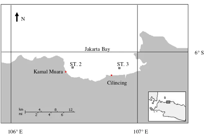
The study was conducted on three different areas of Indonesian coastal zone. A coastal area of Pangkajene Kepulauan (Pangkep) regency in South Sulawesi was chosen as reference site (station 1; Figure 1) because there are relatively minimal anthropogenic activities that were performed in this place such as traditional fisheries, which use static fishing equipment. On the other hand, two sites of Jakarta Bay, Kamal Muara and Cilincing (Figure 2) were chosen and considered as heavily anthropogenic polluted sites (station 2 and 3) since they received almost domestic and industrial wastes from Jakarta and neighboring cities of Jakarta. Moreover, some studies based on chemical analysis indicated that Jakarta Bay was under threatened by anthropogenic pollutants (Williams et al., 2000; Sudaryanto et al., 2002; Munawir, 2005). Whilst, many traditional fisheries activities such as green mussel aquacultures are situated along Jakarta Bay. Hence, Jakarta Bay is considered also as highly valued fisheries resources of coastal area, which plays indispensables role for preserving marine food resources and economic basis of small scales fishermen.
Sample collection and preparation
Thirty two mussels (5 to 6 cm) were handpicked on traditional green mussel cultures along Jakarta Bay at Kamal Muara and Cilincing, and from the Pangkep wild reference population attached naturally on traditional static fishing equipments.
358 J. Toxicol. Environ. Health Sci.
ST.
5° S
119°30’ E
N
Pangkajene Kepulauan
(Pangkep) ST 1
Figure 1. Sampling station (ST) in Pangkajene Kepulauan (Pangkep).
ST. 2 ST. 3
6° S
107° E 106° E
N
Jakarta Bay
Kamal Muara
Cilincing
Figure 2. Sampling stations (ST) in Jakarta Bay.
Germany) using cool box that were filled by dry ice, the tissues were stored at -70°C.
Cholinesterase activity
The enzyme activity was measured following the modified Ellman method (Ellman et al., 1961) for a 96-well plate. A Dounce homogenizer was used to homogenize 0.3 g of each tissue in 2 ml potassium phosphate buffer (0.1 M/pH 8.0). The homogenate was centrifuged for 10 min at 10000 × g and the supernatant was harvested and stored at -80°C prior to the analysis of ChE activity and protein content. The supernatant was diluted in 1:2 of
potassium phosphate buffer (0.1 M/pH 8.0) following the enzyme measurement.
Protein |measurement for cholinesterase assay
Protein content measurement was carried out by diluting the gill extract 1:10 with distilled water. It was measured previously by placing 10 µl of the diluted extract and 10 µl of serial dilutions of γ
-globuline protein standard into a separate well section of the microplate. A blank was made by placing 10 µl of distilled water into the blank section of the microplate. After the addition of 5% Bradford-reagent solutions (200 µl) into the microplate, the samples were left in room temperature for 20 minutes to allow color development. The absorbance was read at 620 nm using photometry (Spectra Thermo TECAN).
The ChE activity is expressed as nmoles of product developed per minute per mg of protein (nmol/min/mg protein). The ChE activity was measured on each tissue to recognize which tissue has the highest ChE activity.
Phagocytic activity
Phagocytic activity of hemocytes was determined by a microplate-based fluorescence measurement method (Hansen, 1992; Anderson and Mora, 1995). This method is based on the number of fluorescence labeled yeast cells that were phagocytosed by mussel hemocytes. The yeast cells were treated and labeled by Fluoresceinisothiocyanate (FITC) (Anderson and Mora, 1995) and kept in aliquots at -80°C. After withdrawing hemolymph from the PAM sinus of the mussels, 100 µl of hemolymph was dropped into 96-microplate wells. Five replicates were used to analyze the phagocytic activity and 3 replicates were used for the protein analysis. The density of hemocytes from each mussel was calculated by using a hemocytometer under a light transmission microscope. After the incubation of the plate for 30 min to allow hemocytes deposition at the bottom of the microplate wells, 25 µl of the FITC-labeled yeast was added into each phagocytic activity section of the microplate wells. A standard was made by adding 100 µl of phosphate buffer saline (PBS) and 25 µl of the FITC-labeled yeast into the microplate wells. One column (8 wells) was used as a blank section by adding 125 µl of PBS. The plate was incubated for 90 min in 21°C at dark condition. At the end of the incubation, 50 µl of 1 % glutaradehyd was added into each phagocytosis section of microplate wells, while 50 µl of methanol was dropped into the protein section of microplate wells. Before transferring to the laboratory in Germany, the plates were covered by a film and wrapped in aluminum and stored at 5°C in darkness. Accordingly, the fixatives were removed carefully and replaced by 125 µl of PBS when samples have arrived in the laboratory. For quenching the fluorescence background of unphagocytosed cells, 25 µl of 0.6 mg/ml trypan blue dissolved in PBS was added to each well of the microplate. The plate was incubated for 20 min prior to removing of all supernatants. The fluorescence of the ingested FITC-labeled yeast cells were read at excitation of 485 nm and emission of 535 nm using a microtiter plate fluorometer (Dynatech, Fluorolite 1000).
Protein measurement for phagocytic assay
A protein content measurement was carried out using hemocytes only. Prior to the measurement, the buffer was removed carefully and hemocytes were lysed with 50 µl of 0.1 N NaOH. After incubating the lysed hemocytes for 10 minutes in a shaking chamber, 10 µl of the lysed hemocytes and the serial dilutions of protein standard (Bovine Serum Albumin) were added to 96-microplate wells. Accordingly, 200 µl of 5% Bradford-reagent solution were added into the plate and incubated for 20 minutes to allow color development. The absorbance of protein was measured at 620 nm using photometer (Spectra Thermo TECAN).
Accordingly, the phagocytic activity was expressed as Relative Fluorescence Units (RFU) and finally calculated as a Phagocytic Index: RFU/mg hemocyte protein.
Statistical analysis
The statistical analyses were performed using non-parametric test, Kruskall-Wallis to distinguish the differences of ChE and phagocytic activity among the sites. If there were differences among the sites (p < 0.05), the test was continued by Dunn’s multiple comparison test to determine the different between two sites. The statistical analyses were conducted using GraphPad Prism trial version 5.0 for Windows, GraphPad Software, San Diego California USA.
RESULTS
Cholinesterase activity
It has been reported that the ChE activity level differed among organs in marine mussels (Bocquené et al., 1990; Brown et al., 2004). The current study was started by recognizing which organ of green mussel, P. viridis that posses the highest ChE activity. It has been performed using P. viridis tissues from expected clean area. The results presented in Figure 3 demonstrated the median of the ChE activity in the gill which had the significant highest ChE activity namely 83.56 ± 12.19 nmol/min/mg protein followed by the foot (46.16 ± 4.18 nmol/min/mg protein), the mantle (27.35± 2.50 nmol/min/mg protein) and the PAM (4.94 ± 4.08 nmol/min/mg protein). Accordingly, the gill was used as a tissue target for measuring the ChE activity since the highest ChE activity of such organ should be the most suitable for measurement of the ChE activity inhibition (Bocquené et al., 1990; Escartin and Porte 1997; Valbonesi et al., 2003; Lau et al., 2004; Brown et al., 2004; Damiens et al., 2007; Taleb et al., 2009; Yaqin 2010).
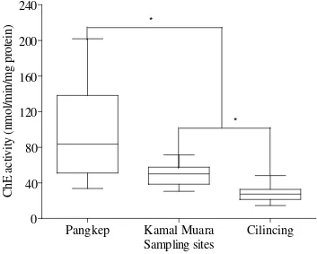
Statistical analysis showed the difference ChE activity in the gills of the samples (p < 0.05) (Figure 4). The animals collected from the reference site had the significant highest ChE activity (83.56 ± 12.19 nmol/min/mg protein) followed by the green mussel collected from heavily polluted areas, Kamal Muara (49.92 ± 3.29 nmol/min/mg protein) and Cilincing (27.20 ±
1.80 nmol/min/mg protein). Between two heavily polluted sites, the animals inhabited in Kamal Muara showed significant less inhibition of the ChE activity than those from Cilincing (p < 0.05).
Phagocytic activity
360 J. Toxicol. Environ. Health Sci.
Gill
Foot
Mantle
PAM
0
20
40
60
80
100
120
140
160
180
200
220
Green mussel's organs
C
h
E
a
ct
iv
it
y
(
n
m
o
l/
m
in
/m
g
p
ro
te
in
)
Figure 3. Cholinesterase activity of different organs of green mussel, Perna viridis from Pangkep Indonesia. Data were expressed as median (25 and 75 % quartile, 5 and 95 % confidence interval).
Pangkep
Kamal Muara
Cilincing
0
40
80
120
160
200
240
*
*
Sampling sites
C
h
E
a
ct
iv
it
y
(
n
m
o
l/
m
in
/m
g
p
ro
te
in
)
Pangkep Kamal Muara Cilincing 1.3××××100 6
1.8××××100 6 2.3××××100 6 2.8××××100 6 3.3××××100 6
Sampling sites
C
el
ls
/m
L
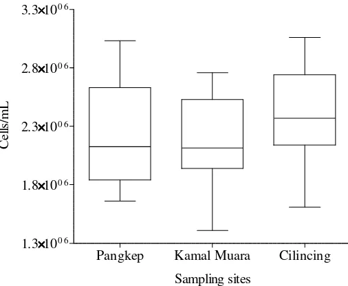
Figure 5. Circular hemocytes of green mussel Perna viridis collected in the selected areas of Indonesia waters. Data were expressed as median (25 and 75% quartile, 5 and 95% confidence interval).
Pangkep Kamal Muara Cilincing 10
100 1000 10000 100000 1000000
*
Sampling sites
P
h
ag
o
cy
to
si
s
in
d
ex
(R
F
U
/m
g
p
ro
te
in
)
Figure 6. Phagocytotic Index of green mussel Perna viridis collected in the selected areas of Indonesia waters. Data were expressed as median (25 and 75% quartile, 5 and 95% confidence interval). *indicate significant difference (p < 0.05) of the hemolymph phagocytic activity. Y-axis is logarithmic scale.
from 2,1250,000 to 2,370,000 cells/ml. In contrast, the median of the phagocytotic index demonstrated significant different phagocytic activities of P. viridis
collected from gradient pollutions of Indonesian coastal waters (p < 0.05). The animals collected from the two heavily polluted sites in Jakarta Bay showed significant higher phagocytic index than those collected from reference sites. Nevertheless, there was no significant
different phagocytic index within the polluted site (p = 0.118). The highest phagocytic index was demonstrated in hemocytes of P. viridis from Cilincing (23410.10 RFU/mg protein) which followed by Kamal Muara (7566.84 RFU/mg protein) and reference site, Pangkep (1714.19 RFU/mg protein).
DISCUSSION
Cholinesterase activity
Cholinesterases (ChEs) are enzymes that hydrolyses and inactivates neural transmitter acetylcholine (ACh) for regulating neural transmission impulse in the synaptic gap of cholinergic synapses and neuromuscular junctions (Soreq and Seidman, 2001). ACh play an important role both as excitatory and inhibitory transmitters of the gill muscle of bivalve (Gainey et al., 2003). In blue mussel,
Mytilus edulis, ciliary movement of the gill is controlled by acetylcholine, dopamine and 5-hydrotryptamine (Aiello, 1990). Organophosphorous and carbamate pesticides inhibit ChE activity which may lead to severe
physiological impairment of marine animals
(Dauberschmidit et al., 1997) such as reduction in feeding efficiency of marine mussels (Donkin et al., 1997).
Since ChEs was purified by Wachtendonk and Neef (1979) in marine mussels hemolymph, a measurement of ChE activity in marine mussels has been used as a biomarker in laboratory test (Galloway et al., 2002; Rickwood and Galloway, 2004; Canty et al., 2007; Yaqin and Hansen 2010) and several international monitoring programs in the field (Narbonne et al., 1999; Cajaraville et al., 2000; Dizer et al., 2001a,b; Roméo et al., 2003; Bocquené et al., 2004; Gagné et al., 2008).
Characterization of ChEs in bivalve has been conducted in some bivalves e.g. in M. galloprovincialis
the ChE specific activity was predominantly localized in the gills compare to others organs (Mora et al., 1999; Porte et al., 2001; Taleb et al., 2009). Moreover, the ChE activity from M. galloprovincialis gill was observed more sensitive to organophosphorous pesticides than that from the digestive gland (Escartin and Porte, 1997). In M. edulis, Bocquené et al. (1990) found that the highest ChE activity occurred in the gill compare to others organs such as the hepatopancreas, the mantle and the adducent muscle. By characterizing and comparing the ChEs in different organ of M. edulis, Brown et al. (2004) found that ‘mitochondrial’ fraction of foot had the highest ChE specific activity with very low recovery of activity.
362 J. Toxicol. Environ. Health Sci.
Compared to the foot, the gill of the bivalve, Scapharca inaequivalvis, demonstrated the higher specific ChE activity level as well (Romani et al., 2005). Eventually, Bonacci et al. (2008) observed that the highest ChE activity also occurred in the gill of scallop, (Pecten jacobaeus) compared to others organs which were the adducent muscle and the digestive gland. Kopecka-Pilarczyk (2010) observed that the ChE activity from gill of Mytilus trossulus was the most sensitive enzyme activity compared to the activity from others organs and whole body tissue when exposed to carbaryl and metals.
The current study compared the ChE activity of green mussel, P. viridis in different organs such as the gill, the foot, the mantle and the PAM. The results demonstrated that the gill of P. viridis had significant higher of the ChE activity compared to the foot, the mantle and the PAM. Porte and Albaiges (2002) demonstrated that the ChE activity from the gill of blue mussels, Mytilus galloprovincialis was more sensitive enzyme activity than that of digestive gland and it revealed a certain correlation with the concentration of fenitrothion in whole mussels. It has been reported that the gill of P. viridis
collected from Hong Kong waters had the higher ChE activity than that of the whole tissue and this ChE activity was not size-dependent (Lau and Wong, 2003). This is conceivable because mussels use their gills not only as a respiratory apparatus but also as filter feeder organ thereby ambient water filtered and managed for gaseous exchanges and sifting food (Bayne et al., 1976). Since the gill are the front line of contact with contaminants and the first line of defense (Lau and Wong, 2003), detoxification compounds such as ChEs are necessary to be produced to protect other organs. Consequently, the production of ChEs not only provides as the control of neurotransmission, but also serves as contaminants detoxification particularly for organophosphorus and carbamate pesticides (Soreq and Seidman, 2001). In addition, it has been reported that the protein level of P. viridis gill was not seasonal dependent which lead to reduce the intrinsic variability of the biochemical responses in different growth phase throughout the year (Lau et al., 2004). Those evidences set up the gill as a
par excellence tissue for biomarkers application to minimize effects caused by the natural reproductive cycles and the dilution effect due to large variation in the total tissue protein (Lau et al., 2004). The selection of the gill as tissue target for conducting biomarkers were also shown by the nature of the gill, which comes into contacts with relatively large volumes of seawater compared to the rest of the animal so that conferring them with the potential for being a suitable target tissue for xenobiotic substance exposure.
The present study used the gill of P. viridis to investigate pollutants effect to ChE activity in some coastal areas of Indonesia. The results suggested that the ChE activity was a sensitive tool to detect neurotoxic effects of pollutants since it could discern different levels
of two heavily polluted areas. It was supported by the evident that the ChE activity of the gill of P. viridis from reference site was significantly higher than that from the gill of P. viridis which inhabit two polluted sites. The inhibition of the ChE activity from the gill of P. viridis
collected from Kamal Muara was about 49.2%. Statistically, the greatest inhibition of the ChE activity was indicated in mussels from Cilincing, which was about 72.41%. By exposing brown mussels, (Perna perna) to furadan (carbamate pesticide), Alves et al. (2002) observed that the ChE activity of the gill was suppressed by 35%.
Ludke et al. (1975) classified the percentage of ChE activity inhibition based on comparison of the individual value with the activity of the normal population for providing the interpretation of the environmental risk. The following are the risk criteria of inhibition percentage of ChE activity that were proposed by Ludke et al. (1975):
0 to 20% = zone of normal variation
20 to 50% = presence of exposure or zone of reversible effects
50 to 100% = life-threatening situation or zone of irreversible effects
data of ChE activity from the research above which ranging from bird to freshwater mussel it is suggested that the green mussels which populated in Kamal Muara indicated reversible effects, while those from Cilincing showed irreversible conditions.
The link between the inhibition of ChE activity of sentinel organism and the discharged neurotoxic compounds from agricultural, urban and industrial activity to aquatic environment has been suggested by many studies (Fulton and Key 2001; Printes and Callaghan 2004; Galloway et al., 2002; Crane et al., 2002; Rickwood and Galloway, 2004; Canty et al., 2007; Warberg et al., 2007). However, the relationship between ChE activity and higher level biomarker such as feeding rate in green mussel has not been studied yet. Therefore, a chronic in vivo study on the response of ChE activity in green mussel and other behavioral biomarkers such as feeding rate to the serial concentrations of pollutants, which picturize suspected pollution area concentrations, is indispensable to translate the inhibition of ChE activity induced by pollutants into ecological perspective. The translatable of ecological consequence of the suppressed ChE activity is a vital consideration in ecological risk assessment in the coastal zone. It is because an appropriate ecological relevance of biomarkers can eliminate the primary source of uncertainty in application of ecological risk assessment (Sibley et al., 2000).
Phagcytosis activity
Green mussel hemolymph contains both hemocyte and humoral defense factors which are responsible for the defense system. Hemocytes circulating in hemolymph are the principal cellular effectors of invertebrate immunity (Mitta et al., 1999) which have a capability to perform phagocytosis of foreign materials (Cheng, 1984; Carballal et al., 1997) and cytotoxycity via the production of radicals (Winston et al., 1996).
Phagocytosis of mussel hemocytes can be affected by various chemical stressors in the aquatic environment (Anderson and Mora, 1995). Biphasic patterns of mussel phagocytic responses induced by xenobiotic have been demonstrated in many laboratory studies (Cole et al., 1994; Pipe et al., 1999; Parry and Pipe, 2004). Theoretically, the phagocytic activity will be stimulated when mussels are exposed to low level of contaminants, while it will be suppressed when mussel are exposed to high level of contaminants. Consequently, measurement of the phagocytic activity, which is as part of immune system of mussel, has been used as a biomarker of xenobiotic substances effect (Anderson and Mora, 1995; Oliver and Fisher, 1999; Blaise et al., 2002; Gagné et al., 2002).
In spite of mussel hemocytes playing an important role in the phagocytic activity, it is difficult to depict the correlation pattern between circulated hemocytes number
and the phagocytic activity of mussel. The current study showed that there was no different numbers of circulating hemocytes of green mussel, which were collected from both polluted and clean sites. However, significant differences of the phagocytic activity between the collected green mussels from polluted sites and those from clean site were evident. The data showed that discharged pollutants in Jakarta Bay have stressed cultivated green mussels, which stimulated significantly their phagocytic activity compared to the phagocytic activity of the green mussels collected from the clean site. The modulation of mussel phagogytic activity was in accordance with Luengen et al. (2004) who observed the elevation of phagocytic activity of mussels that collected from polluted sites. The elevation of phagocytic activity induced by the pollutants may be a part of mussel’s strategy to sequester the toxic materials from vulnerable organs (Oliver et al., 2001). Nevertheless, Dizer et al. (2001b) found that high number of circulating hemocytes of mussels collected from control site followed by relatively low phagocytic activity, while relatively low number of hemocytes from polluted sites had a high phagocytic activity. They could not depict clearly the relationship between hemocytes number and the phagocytic activity of mussels.
The complicated relationship between hemocytes number and the phagocytic activity of mussels may result from dynamic association/dissociation between hemocytes and bivalve tissues that enable to change the total size of the hemocytes population within bivalve body over short time (Ford et al., 1993). The population could not be simply depicted by circulating number of hemocytes, which were drained from the PAM sinus as the mussel has the open circulatory blood system, which circulate the blood to whole organs. In addition, commonly the mussel hemocytes are composed by phagocytotic and unphagocytotic hemocytes which can be altered by xenobiotic substances (Pipe et al., 1999). Unfortunately, most of the techniques to measure the phagocytic activity including the technique used in the present study were based on the mixture of hemocytes sub-population so that an estimation of capability level of each sub-population of hemocytes was not possible.
364 J. Toxicol. Environ. Health Sci.
approach, the ChE activity indicated a more responsive tool compared to the phagocytic activity so that it could distinguish between two heavily polluted sites. However, it is hard to justify that the ChE activity is more sensitive compared to phagocytic activity as was observed by Perez et al. (2004) in ChE activity of invertebrates,
Scrobicularia plana (clam) and Nereis diversicolor
(marine worm). The authors delineated higher sensitivity of ChE activity compared to others biomarkers that were used in biomonitoring of Spain waters. Therefore, the useful results that recorded by the current study are the information on neurotoxicity and immunotoxicity compounds which were present in Jakarta Bay and the magnitude impact of neurotoxicity contaminants to induce an effect is greater than the immunotoxicity contaminants.
Conclusion
Conclusively, the results suggested that the use of the selected biomarkers is a reliable and preferential strategy in the ecological risk assessment of released xenobiotic compounds in coastal waters due to their ability to elucidate bio-effects of neuro-immuno systems disruptors.
ACKNOWLEDGEMENTS
The authors wish to thanks to DAAD (Deutscher Akademischer Austausch Dients/German Academic Exchange Service) for funding the research through the Special Programme for Young Indonesian Marine and Geoscience Researchers. The authors would like also to thank distinguish colleague Arifin from Marine Science and Fisheries Faculty, Hasanuddin University, Makassar for his invaluable assistence for collecting the green mussels in Pangkajene Kepulauan waters.
REFERENCES
Aiello E (1990). Nervous control gill ciliary activity in Mytilus edulis. In: Stefano GB, editor. Neurobiology of Mytilus edulis. Manchester: Manchester University Press, pp. 189-208.
Akcha F, Izuel C, Venier P, Budzinski H, Burgeot T, Narbonne J-F (2000). Enzymatic biomarker measurement and study of DNA adduct formation in benzo[a]pyrene-contaminated mussels, Mytilus
galloprovincialis. Aquat. Toxicol., 49: 269-287.
Alves SRC, Severino PC, Ibbotson DP, da Silva AZ, Lopes FRAS, Saenz LA, Bainy ACD (2002) Effects of furadan in the brown mussel
Perna perna and in the mangrove oyster Crassostrea rhizophorae.
Mar. Environ. Res., 54: 241-245.
Anderson RS, Mora LM (1995). Phagocytosis: A microtiter plate assay. In Stolen JS, Fletcher TC, Smith SA, Zelikoff JT, Kaattari SL, Anderson RS, Söderhäll K, Weeks-Perkins BA. (eds). Techniques in Fish immunology-4. Immunology and Pathology of Aquatic Invertebrates. Fair Haven, NJ, USA: SOS Publication, pp. 109-112. Bayne BL, Thompson RJ, Widdows J (1976). Physiology: I. In B.L.
Bayne (ed). Marine mussels: their ecology and physiology. London: Cambridge University Press, pp. 121-206.
Belden JB, Lydy MJ (2000). Impact of atrazine on organophosphate insecticide toxicity. Environ. Toxicol. Chem., 19: 2266–2274. Blaise C, Trottier S, Gagné F, Lallement C, Hansen PD (2002).
Immunocompetence of bivalve hemocytes as evaluated by a miniaturized phagocytosis assay. Environ. Toxicol., 17: 160-169. Bocquené G, Chantereau S, Clérendeau C, Beausir, E, Ménard D,
Raffin B, Minier C, Burgeot T, Leszkowicz AP, Narbonne JP (2004). Biological effects of the “Erika” oil spill on the common mussel
(Mytilus edulis). Aquat. Living Resour., 17: 309–316.
Bocquené G, Gaglani F, Truquet P (1990). Characterization and assay condition for use of AChE activity from several marine species in pollution monitoring. Mar. Environ. Res., 30: 75-89.
Bonacci S, CorsiI, Focardi S (2008). Cholinesterase activities in the scallop Pecten jacobaeus: Characterization and effects of exposure to aquatic contaminants. Sci. Total. Environ., 392: 99–109.
Brown M, Davies IM, Moffat CF, Redshaw J, Craft JA (2004). Characteristic of choline esterases and their tissue and subcellular distribution in mussel (Mytilus edulis). Mar. Environ. Res., 57: 155-169.
Butterworth FM (1995). Introduction to biomonitors and biomarkers as indicators of environmental change. In Butterworth FM, Corkum LD, Rincon JG (eds). Biomonitor and biomarkers as indicators of environmental change: A handbook. . New York: Plenum Press, pp. 1-8.
Cajaraville MP, Bebianno MJ, Blasco J, Porte C, Sarasquete C, Viarengo A (2000). The use of biomarkers to assess the impact of pollution in coastal environments of the Iberian Peninsula: A practical approach. Sci. Total. Environ., 247: 295-311.
Canty MN, JA Hagger, RTB More, L Cooper, TS Galloway (2007). Sublethal impact of short term exposure to the organophosphate pesticide azamethiphos in the marine Mollusc Mytilus edulis. Mar. Poll. Bull., 54: 396-402.
Carballal MJ, Lopez C, Azevedo C, Villalba A (1997). In vitro study of phagocytotic ability of Mytilus galloprovincialis Lmk. Haemocytes. Fish. Shellfish. Immunol.,7: 403–416.
Castro M, Santos MM, Monteiro NM, Vieira N (2004). Measuring lysosomal stability as an effective tool for marine coastal environmental monitoring. Mar. Environ. Res., 58: 741-745.
Cheng TC (1984). A classification of molluscan hemocytes based on functional envidence. In Cheng TC (ed). Comparative pathobiology vol. 6. Inverterbrate blood cells and serum factors. New York: Plenum Press, pp. 111-146.
Cole JA, Farley SR, Pipe RK (1994). Effects of fluranthene on the immunocompetence of the common marine mussel, Mytilus edulis. Aquat. Toxicol., 30: 367-379.
Coppage DL (1972). Organophospahte pesticides: Specific level of brain AChE inhibition related to death in sheeps head minnows. Transact. Am. Fish. Soc., 101: 534–536.
Coppage DL, Matthews E, Cook GH, Knight J (1975). Brain acetylcholinesterase inhibition in fish as a diagnosis of environmental poisoning by malathion, O,O-dimethyl S-(1,2 dicarbothoxyethyl) phosphorodiyhioate. Pest. Biochem. Physiol., 5: 536-542.
Coppage DL, Matthew E (1975). Brain acetylcholinesterase inhibition in marine teleost during lethal and sublethal exposures to 1,2-dibromo-2,2-dichloroethyl dimetyl phosphate (Naled) in sea water. Toxicol. Appl. Pharmacol., 31: 128 -133.
CorsiI, Bonacci S, Santovito G, Chiantore M, Castagnolo L, Focardi S (2004). Cholinesterase activity in the Antarctic scallop Adamussium
colbecki: Tissue expression and effect of ZnCl2 exposure. Mar. Environ.
Res., 58: 401-406.
Crane M, Sildanchandra W, Kheir R, Callaghan A (2002). Relationship between biomarker activity and developmental endpoints in
Chironomus riparius Meigen exposed to an organophosphate
insecticide. Ecotox. Environ. Safe, 53: 361-369.
Damiens G, Gnassia-Barelli EHM, Quiniou F, Roméo M (2004). Evaluation of biomarkers in oyster larvae in natural and polluted conditions. Comp Biochem. Physiol. C: Toxicol. Pharmacol., 138: 121-128.
Dauberschmidit C, Dietrich DR, Schlatter C (1997). Organophospates in the Zebra Mussel Dreissena polymorpha: subacute exposure, body burdens and organ concentrations. Arc. Environ. Contam. Toxicol., 33: 42-46.
De Luca-Abbott SB, Richardson BJ, McClellan KE, Zheng GJ, Martin M, Lam PKS (2005). Field validation of antioxidant enzyme biomarkers in mussels (Perna viridis) and clams (Ruditapes philippinarum) transplanted in Hong Kong coastal waters. Mar. Pol. Bul., 51: 694-707.
Depledge MH, Fossi MC (1994). The role of biomarkers in environmental assessment (2). Invertebrate. Ecotoxicol., 3: 161-172. Devier MH, Augagneur S, Budzinski H, Le Menach K, Narbonne J-F,
Garrigues P (2005). One-year monitoring survey of organic compounds (PAHs, PCBs, TBT), heavy metals and biomarkers in blue mussels from the Arcachon Bay, France. J. Environ. Monit., 7: 224-240.
Dizer H, de Assis HCS, Hansen PD (2001a). Cholinesterase activity as bioindicator for monitoring marine pollution in the Baltic Sea and the Mediterranean Sea. In Garrigues Ph, Barth H, Walker CH, Narbonne J-F (eds). Biomarkers in marine organisms a practical approach. Amsterdam: Elsevier Science BV, pp. 331-342.
Dizer H, Unruh E, Bissinger V, Hansen PD (2001b). Investigation of genotoxicity and immunotoxicity for monitoring marine pollution in the Baltic Sea and Mediterranian Sea. In Garrigues Ph, Barth H, Walker CH, Narbonne JF (eds). Biomarkers in marine organisms a practical
approach. Amsterdam: Elsevier Science BV, pp. 237-257.
Donkin P, Widdows J, Evans SE, Staff FJ, Yan T (1997). Effect of neurotoxic pesticide on the feeding rate of marine mussels (Mytilus
edulis). Pest. Sci., 49: 196-209.
Ellman GL, Courtney KD, Andres VJr, Featherstone RM (1961). A new and rapid colorimetric determination of acetylcholinesterase activity. Biochem. Pharmocol., 7: 88-95.
Escartin E, Porte C (1997). The use of cholinesterase and carboxylesterase activities from Mytilus Galloprovincialis in pollution monitoring. Environ. Toxicol. Chem., 16: 2090-2095.
Fleming WJ, Augspurger TP, Alderman JA (1995). Freshwater mussel die-off attributed to anticholinesterase poisoning. Environ. Toxicol. Chem., 14: 877–879.
Ford SE, Kanaley SS, Littlewood DTJ (1993). Celluar response of oyster infected with Haplosporidium nelsoni, change in circulating and tissue-infiltrating hemocytes. J. Invert. Phatol., 61: 49-57. Fournier M, Cyr D, Blakley B, Boermans H, Brousseau P (2000).
Phagocytosis as a biomarker of immunotocity in wildlife species exposed to environmental xenobiotics. Am. Zoo., 40: 412-420. Fulton MH, Key PB (2001). Acetylcholinesterase inhibition in estuarine
fish and invertebrate as an indicator of organophosphorus insecticide exposure and effects. Environ. Toxicol. Chem., 20: 37-45.
Gagné F, Blaise C, Aoyama I, Luo R, Gagnon C, Couillard Y, Campbell C, Salazar M (2002). Biomarker study of a Municipal effluent dispersion plume in two species of freshwater mussels. Environ. Toxicol., 17: 149-159.
Gagné F, Burgeot T, Hellou J, St-Jean S, Farcy E, Blaise C (2008). Spatial variations in biomarkers of Mytilus edulis mussels at four polluted regions spanning the Northern Hemisphere. Environ. Res., 107: 201-217.
Gainey LF, Walton JC, Greenberg MJ (2003). Branchial musculature of a venerid clam: pharmacology, distribution, and innervation. Biol. Bull., 204: 81-95.
Galloway TS (2006). Biomarkers in environmental and human health risk assessment. Mar. Pol. Bull., 53: 606–613.
Galloway TS, Millward N, Browne MA, Depledge MH (2002). Rapid assessment of organophosphorous/carbamate exposure in the bivalve mollusc Mytilus edulis using combined esterase activities as biomarkers. Aquat. Toxicol., 61: 169-180.
Goldberg E D, Bowen VT, Farrington JW, Harvey G, Martin JH, Parker PL, Risebrough RW, Robertson W, Schneider E, Gamble E (1978). The mussel watch. Environ. Conserv., 5: 101–125.
Guilhermino L, Barros P, Silva MC, Soares AMVM (1998). Should the use of inhibition of cholinesterases as a specific biomarker for organophosphate and carbamate pesticides be questioned? Biomarker, 3: 157–163.
Halldórsson HP, De Pirro M, Romano C, Svavarsson J, Sarà G (2007).
Immediate biomarker responses to benzo[a]pyrene in polluted and unpolluted populations of the blue mussel (Mytilus edulis L.) at high-latitudes. Environ. Inter., 34: 483-489.
Hansen PD (1992). Phagocytosis in Mytilus edulis, a system for understanding the sublethal effects of anthrophogenic pollutants (xenobiotic) and the use of AOX as an integrating parameter for the study of the equilibrium between chlorinated hydrocarbons in
Dreissena polymorpha following long term exposures. Limnol.
Aktuell, 4: 171-184.
Hansen PD (1995). The pontential and limitation of new technical approaches to ecotoxicology monitoring. In Richardson M (ed). Environmental Toxicology Assessment. Taylor & Francis Inc. London, pp. 13-28.
Kim Y, Powell EN, Wade TL, Presley BJ (2008). Relationship of Parasites and Pathologies to Contaminant Body Burden in Sentinel Bivalves: NOAA Status and Trends ‘Mussel Watch’ Program. Mar. Environ. Res., 65: 101-127.
Kopecka-Pilarczyk J (2010). The effect of pesticides and metals on acetylcholinesterase (AChE) in various tissues of blue mussel
(Mytilus trossulus L.) in short-term in vivo exposures at different
temperatures. J. Environ. Sci. Health Part B, 45: 336 – 346.
Lagadic L (2002). Biomarkers: Useful tools for the monitoring of aquatic environments. Revue Méd. Vét., 153: 581-588.
Lau PS, Wong HL (2003). Effect of size, tissue parts and location on six biochemical markers in the green-lipped mussel, Perna viridis. Mar. Poll. Bull., 46: 1563-1572.
Lau PS, Wong HL, Garrigues P (2004). Seasonal variation in antioxidative responses and acetylcholinesterase activity in Perna
viridis in eastern oceanic and western estuarine of Hong Kong.
Continent. Shelf. Res., 24: 1969-1987.
Lehtonen KK, Schiedek D, Köhler A, Lang T, Vuorinen PJ, Förlin L, Baršien÷, J, Pempkowiak J, Gercken J (2006). The BEEP project in the Baltic Sea: Overview of results and outline for a regional biological effects monitoring strategy. Mar. Pollut. Bull., 53: 523-537. Leinio S, Lehtonen K (2005). Seasonal variability in biomarkers in the
bivalves Mytilus edulis and Macoma balthica from the northern Baltic Sea. Comp. Biochem. Physiol. C, 140: 408-421.
Livingstone DR, Chipman JK, Lowe DM, Minier C, Mitchelmore CL, More MN, Peters LD, Pipe RK (2000). Development of biomarkers to detect the effects of organic pollution on aquatic invertebrate: recent molecular, genotoxic, cellular and immunological studies on the common mussel (Mytilus edulis L.) and other mytilids. Inter. J. Environ. Poll., 13: 56-91.
Ludke JL, Hill EF, Dieter MP (1975). Cholinesterase (ChE) response and related mortality among birds fed ChE inhibitors. Arc. Environ. Contam. Toxicol., 3: 1-21.
Luengen AC, Friedman CS, Raimondi PT, Flegal AR (2004). Evaluation of mussel immune responses as indicators of conta-mination in San Francisco Bay. Mar. Environ. Res., 57: 197–212. Martın-Dıaz ML, Blasco J, Sales D, DelValls TA (2004). Biomarkers as
tools to assess sediment quality: Laboratory and field surveys. Trends. Analyt. Chem., 23: 807-818.
Minier C, Abarnou A, Madoulet AJ, Le Guellec, AM, Tutundjian R, Bocquené G, Leboulenger F (2006). A pollution-monitoring pilot study involving contaminant and biomarker measurements in the seine estuary, france, using zebra mussels (Dreissena polymorpha). Environ. Toxicol. Chem., 25: 112–119.
Mitta G, Vandenbulcke F, Hubert F, Roch P (1999). Mussel defensins are synthesised and processed in granulocytes then released into the
plasma after bacterial challenge. J. Cell. Sci., 112: 4233-4242. Mora P, Fournier D, Narbonne J-F (1999). Cholinesterases from the
marine mussels Mytilus galloprovincialis Lmk. and M. edulis L. and from the freshwater bivalve Corbicula fluminea Müller. Comp. Biochem. Physiol. C., 122: 353–361.
Moreira SM, Santos MM, Ribeiro R, Guilhermino L (2004). The ‘Coral Bulker’ Oil Spil on the North Coast of Portucal: Spatial and temporal biomarker responses in Mytilus galloprovincialis. Ecotoxicol.,13: 619– 630.
Munawir K (2005). Pemantauan Kadar Pestisida Organoklorin Di Beberapa Muara Sungai Di Perairan Teluk Jakarta. Oseanol. Limnol. Indonesia, 37: 13-23.
366 J. Toxicol. Environ. Health Sci.
Blasten P, Pagano G, Porte C, Livingstone D, Hensen P-D, Herbert A (1999). Biological markers of environmental contamination in marine ecosystems: Biomar project. J. Toxicol. Toxin Rev., 18: 205-220. Narbonne JF, Duabeze M, Baumard P, Budzinski H, Clerandeau C,
Akca F, Mora P, Garrigues P (2001). Biochemical markers in mussel, Mytilus sp., and pollution monitoring in European Coasts: Data analysis. In: Garrigues Ph., Barth H, Walker CH, Narbonne J-F (eds). Biomarker in Marine Organisms: A Practical Approach. Amsterdams: Elsevier Sci., pp. 215-236.
Nesto N, Bertoldo M, Nasci C, Da Ros L (2004). Spatial and temporal variation of biomarkers in mussels (Mytilus galloprovincialis) from the Lagoon of Venice, Italy. Mar. Environ. Res., 58: 287-291.
Nicholson S, Lam PKS (2005). Pollution monitoring in Southeast Asia using biomarkers in the mytilid mussel Perna viridis (Mytilidae: Bivalvia). Environ. Inter., 31: 121-132.
Oliver LM, Fisher W (1999). Appraisal of prospective bivalve immunomarkers. Biomarkers, 4: 510-530.
Oliver LM, Fisher WS, Winstead JT, Hemmer BL, Long ER (2001). Relationships between tissue contaminants and defense-related characteristics of oysters (Crassostrea virginica) from five Florida bays. Aquat. Toxicol., 55: 203–222.
Orbea A, Garmendia L, Marigómez I, Cajaraville MP (2006). Effects of the ‘Prestige’ oil spill on cellular biomarkers in intertidal mussels: results of the first year of studies. Mar Ecol Prog Ser., 306: 177-189. Parry HE, Pipe RK (2004). Interactive effects of temperature and copper
on immunocompetence and disease susceptibility in mussels (Mytilus
edulis). Aquat. Toxicol., 69: 311-325.
Perez E, Blasco J, Sole M (2004). Biomarker responses to pollution in two invertebrate species: Scorbicularia plana and Nereis diversicolor from the Cadiz Bay. Mar. Environ. Res., 58: 275-279.
Picado A, Bebianno MJ, Costa MH, Ferreira A, Vale C (2007). Biomarkers: a strategic tool in the assessment of environmental quality of coastal waters. Hydrobiologia, 597: 79-87.
Pipe RK, Coles JA, Carrisan FMM, Ramanathan K (1999). Copper induced immunomodulation in the marine mussel, Mytilus edulis. Aquat. Toxicol.,46: 43-54.
Porte C, Albaiges J (2002). Residues of pesticides in aquatic organisms. Revue. Méd. Vét., 153: 345-350.
Porte C, Escartin E, Borghi V (2001). Biochemical tools for the assessment of pesticide exposure in a deltaic environment: The use of Cholinesterase and Carbaoxylesterases. In Ph. Garrigues, H Barth, CH Walker, JF Narbonne [Editors]. Biomarkers in marine organisms a practical approach. Elsevier Science B.V. Amsterdam, pp. 259-278.
Printes LB, Callaghan A (2004). A comparative study on the relationship between acetylcholinesterase activity and acute toxicity in Daphnia
magna exposed to anticholinesterase insecticides, 23: 1241–1247.
Rickwood CJ, Galloway TS (2004). Acetylcholinesterase inhibition as a biomarker of adverse effect: A study of Mytilus edulis exposed to the priority pollutant chlorfenvinphos. Aquat. Toxicol., 67: 45-56. Romani R, Isani G, De Santis L, Giovannini E, Rosi G (2005). Effects of
chlorpyrifos on the catalytic efficiency and expression level of acetylcholinsterases in the bivalve mollusk Scapharca inaequivalvis. Environ. Toxicol. Chem., 24: 2879-2886.
Roméo M, Mourgaud Y, Geffard Y, Gnassia-Barelli M, Amiard JC, Budzinski H (2003). Multimarker approach in transplanted mussels for evaluating water quality in Charentes, France, coast areas exposed to different anthropogenic conditions. Environ. Toxicol., 18: 295-305. Sandahl JF, Baldwin DH, Jenkins JJ, Scholz NL (2005). Comparative
thresholds for acetylcholinesterase and behaviour impairement in coho salmon exposed to chlorphyrifos. Environ. Toxicol. Chem., 24: 136 – 145.
Sibley PK, Chappel MJ, George TK, Solomon KR, Liber K (2000). Integration effects of stressors across levels of biological organi-zation:examples using organophosphorus insecticides mixtures in field-level exposure. J. Aquat. Ecosyst. Stress. Recov.,7: 117-130. Soreq H, Seidman S (2001). Acetylcholinesterase – new roles for an
old actor. Nature Rev. Neurosci., 2: 8 – 16.
Sturm A, de Assis HCS, Hansen P-D (1999). Cholinesterase of marine teleost fish: Enzymological characterization and potential use in the monitoring of neurotoxic contamination. Mar. Environ. Res., 47: 389 – 398.
Sudaryanto A, Takahashi S, Monirith I, Ismail A, Muchtar M, Zheng J, Richardson BJ, Subramanian A, Prudente M, Hue ND, Tanabe S (2002). Asia-Pacific Mussel Watch: Monitoring of Butyltin Contamination in Coastal Waters of Asian Developing Countries. Exp. Toxicol. Chem., 21: 2119-2130.
Tabche LM, Mora BR, Faz CG, Castelan IG, Ortiz MM, Gonzalez VU, Flores MO (1997). Toxic efect of sodium dodecylbenzenesulfonate, lead, petroleum, and their mixtures on the activity of acetylcholinesterase of Moina macropa in vitro. Environ. Toxicol. Water Quality, 12: 21-215.
Taleb ZM, Benali I, Ykhlef-Allal A, Bachir-Bouiadjra B, Amiard J-D, Boutiba Z (2009). Biomonitoring of environmental pollutionon the Algerian west coast using caged mussels Mytilus galloprovincialis. Oceanologia, 51: 63–84.
Tedesco S, Doyle H, Redmond G, Sheehan D (2008). Gold nanoparticles and oxidative stress in Mytilus edulis. Mar. Environ. Res., 66: 3-131.
Valbonesi P, Sartor G, Fabbri E (2003). Characterization of cholinesterase activity in three bivalves inhabitating the North Ardriatic sea and their possible use as sentinel organisms for biosurveillance programmes. Sci. Total. Environ., 312: 79– 88. Van der Oost R, Goksøyr A, Celander M, Heida H, Vermeulen NPE
(1996). Biomonitoring of aquatic pollution with feral eel (Anguilla
anguilla): II. Biomarkers: Pollution-induced biochemical responses.
Aquat. Toxicol., 36: 189-222.
Verlecar XN, Jena KB, Chainy GBN (2008). Seasonal variation of oxidative biomarkers in gills and digestive gland of green-lipped mussel Perna viridis from Arabian Sea. Estuar. Coast. Shelf Sci., 76: 745-752.
Wachtendonk Von D, Neef J (1979). Isolation, perufication and molecular properties of an acetylcholinesterase (E.C. 3.1.1.7) from the haemolymph of the sea mussel Mytilus edulis. Comp. Biochem. Physiol., 63C: 279-286.
Walker CH (1998). Biomarker strategies to evaluate the environmental effects of chemicals. Environ. Health. Perspect Suppl., 106: 613-620. Warberg MB, Coen LD, John E, Weinstein JE (2007). Acute Toxicity and Acetylcholinesterase Inhibition in Grass Shrimp (Palaemonetes pugio) and Oysters (Crassostrea virginica) Exposed to the Organophosphate Dichlorvos: Laboratory and Field Studies. Arch. Environ. Contam. Toxicol., 52: 207–216.
Widdows J, Donkin P (1992). Mussels and environmental contaminants: Bioaccumulation and physiological aspects. In: Gosling E (ed). The mussel Mytilus: Ecology, physiology, genetics and culture. Amsterdam: Elsevier Science Publishers BV, pp. 383-424.
Williams TM, Rees JG, Setiapermana D (2000). Metals and trace organic compounds in sediments and waters of Jakarta bay and the pulau seribu complex, Indonesia. Mar. Poll. Bull., 40: 277-285. Winston GW, More MN, Kirchin MA, Soverchia C (1996). Production of
reactive oxygen species by hemocytes from the marine mussels,
Mytilus edulis: Lysosomal localization and effect of xenobiotics.
Comp. Biochem. Physiol., 113C: 221-229.
Yaqin K (2010). Potential use of cholinesterase activity from tropical green mussel, Perna viridis as a biomarker in effect-based marine monitoring in Indonesia. Coast. Mar. Sci., 34: 156–164.