Suppl
I
-
1998Direct
counting and detection of
living
Salmonella
in
biological
fluid
Takayuki Ezaki, Kenji Hirose, Li-Cheng Zhao, Khan Abdul
Quayum,
Masaki Miyake, Yoshiaki Kawamura
Abstrak
Untuk mengembangkan metoda cepat untuk menghilung Salmonella hidup dalam cairan biologis, 4 metoda tletel<si yang berbeda telah dievaluasi. Metode Flow Cytometric baru yang dapat menghitung partikel lebih besar dari 0,25 mikromete4 telah berhasil
d1terap-kan untuk menghitung bakteri hidup dengan cepat. Metoda ini dapat digunakan bila jumlah sel lebih
dari
5000 cfit/ml. Metoda ini jugamampu membedakan populasi babert hidup dan mati dalam cairan dalam waktu 30 menit. Untuk dapat secara efektif menemukan bak-teri dengan mikroskop, metoda penghitung dengan laser scanning microscopy memerlukan konsentrasi bakteri yang tinggi, Iebih besar dari 106/ml. Laser scanning microscopy juga digunakan untuk menganalisis populnsi berbecla dalam biakan tunggal. SeI S, typhi ter-pisahkan dalam dua populasi berbeda setelah dibiakkan 8 jam dalam kaldu LB, sedangkan sel S. typhimurium masih tetap dalam bentuk
tunggal meskipun telah 24 jam dibiakkan. Perbedaan populasi sel dari satu galur ini dengan mudah terlihat dengan metoda ini. Kamera photon counting VIM merttpakan alat yang paling sensitif, tetapi adanya reduksi
dai
latar belakang merupakan masalah yang terberat untuk aplikasi alat ini bagi sampel klinis. Metoda PCR kuantitatif juga dipergunakan untuk menghitung jumtah bakteri.piR
clengan metoda siklus cepat dengan menggunakan sekuens V, flagela dan rfb dapat mendetelcsi organisme dengan jumlah 20-100 cfii./ml Selamadilakukan proses amplifikasi, amplikon dimonitor secara sekuensial dengan fluoresensi dan hasilnya dapat diperoleh dalam waktu 20 menit,
Abstract
To establish rapid counting methods of living Salmonella in biological fluids, 4 different detection methods were evaluated. A new
flow
cytometer able to count particles bigger than 0.25 micromite4 was successfuIly applied.for
rapicl viable bacterial counting. Whencell numbers were bigger than 5000 cfu/mL, this method worked. The method was also able to
dffirentiate living
and clead bacterial population influid
within 30 min. l-aser scanning microscopiç counting method needetl high concentration of bacteria more than 106/mIto effectively
find
bacteria under a microscope. The laser scanning microscopy y,tas also used to analyze different popalation within asingle culture. S. typhi cells were separated into two
dffirent
populations after 8 hr culture in LB broth but cells of S. typhimurium remained uniform even after 24 hr culture. This dffirence of cell population in a single strain was easily visualizerJ by this methocl.Photon counting VIM camera was most sensitive but reduction of background was biggest problems
for
actual applicationfor
clinical samples. Quantitative PCR was also appliedfor
bacterial counting. Rapid cycling PCR preparecl from V, ftagella, and rJb sequencescould detect organisms around 20-100 cfii'/ml. During amplification process, amplicon was sequentially monitoretl by fLuorescence and the result was obtained within 20 min.
INTRODUCTION
Many
approaches
to
count pathogenic bacteria in
clinical material
had been developed
through out
thehistory
of
microbiology. Among them, quantitative
culture on a
agar
plate was
used as
a gold
standardfor
this
purpose.
However,
this
method
have
severaldisadvantages.
Culture
method takes at least one
day
to
make countable colonies. Some
fastidious
bacteria
cannot
grow on
laboratory media and thus, colony
count
does
not correlate to the
actual
bacterial
num-bers
in clinical
specimens. To overcome
thesedisad-Dept. Microbiology, Gifi.t Univ., School of Medicine, 40
Tsukasa, Gifu 500 Japan
Diagnostic 49
s3-3
vantages,
we
attempted
to
count pathogenic
bacteria
in
biological
fluid.
Flowcytometric counting,
laser
scanning
counting, counting
by
photon counting
ul-tra-sensitive VIM
camera, and
counting by rapid
cy-cling PCR
method.
MATERIALS
AND METHODS
Bacterinl strains
Salmonella
typhi
GIFU10007
and Salmonella
ty-phimurium GIFU
12142 strains were
cultured in
Lu-ria Broth
or
M9
cysteine
broth
andincubated
for
dif-ferent incubation
time and used
for
counting.
All
preparation
were serially diluted and cultured
onheart infusion
agar plates
to
confirm viable cell
F low cy t ome
tric c o untin g
Culture
broth was serially
diluted
with
saline
and 10scfu/ml cell
suspension wasprepared. The
suspen-sion was stained
with Bact-Live
and Dead
cell
stain-ing
reagent
(Molecular
Probes).
After
30 min
stain-ing, the
suspension was
directly applied to
aflowcy-tometer
(V/in-Bryte, Bio-Rad).
In
case, Texas
red
labeled
rabbit
anti-Vi antibody
was
mixed
with
S.ry-phi
andthen
stained
with Bact-Live cell staining
re-agent.
Laser scanning microscopic counting
Overnight culture
broth were serially
diluted with
saline
and stained
with live
and dead
fluorescence
reagent
(molecular probes)
for
15
min
and
the
mix-ture
was placed
on
a
slide glass and sealed with
cover
glass. Fluorescent pictures
were
collected
through BioRad confocal
laser scanning
microscopy
within 30 min after staining. Pictures were
taken
by
the magnification
of
200x
and were
processed
by
MacScope software
(Mitani,
cop.
Japan)to count
liv-ing
particles.
Photon camera counting
Ultrasensitive single photon counting
camera,
VIM
camera (Hamamatsu
Photo,
Hamamatsu, Japan)
wasused
to count single cells. Bacteria were first mixed
with
alkaline-labeled
anti-Vi
and
filtered on HATF
Millipore filter
membrane.
After
washing
with
saline
several
times,
the
substrate
ASPD was
added
to
themembrane
andthe
filter
was
immediately
placed
un-der
VIM
camera
to count
photons.
Counting by Rapid
Cycles
Table
1.
Detection of major rfb gene of Salmonella serovarCroup
rfb PCR O
antigen
rfbE
rfb S(D) rfb J(B)09 O2a
O4aA
(O2)
Paratyphi At,2,
t2 B(O4)
Schottmuellei 1,4, [5), 12Typhimurium
l,4,
12Cl(O6,7)
ParatyphiC
6, 7,tvil
Choleraesuis 6,7C2(O6,8)
Newport 6,8O.CDP
,$
Schottmuellei
Typhimurium
Primers to amplify,
rfb
(Figure 1), vipR (Figure
2),
and
fliC
(Figure
3) geneswere
used.Sybergreen
fluo-rescentwere
addedin
aPCR
mixture
to
monitor
PCR
product during amplification
process.
Amplification
was designed
to finish within
15min. using capillary
light cycler (Aidaho
Technology,
Tokyo,
Japan).Paratyphi
A
Tyverose
wil
upjiD
Typhi
Enteritidis
Dublin
Figure 1. Primers
for
Salmonella O antigen genesrfbE, rfb S, rfbJ primers were designed for
0
serovar specific am-p lifi cati o n.rf bE-u p: 5-ctt-g gg-agt-aat-ctt-gcc-3, rfbE, rfb down: 5-tat-act-gcc-gta-ctg-cct-3
rf bS-up: 5-cct-act-tcg-aaa-gtc-gac-3,
rf b-S-down: 5-cac-gaa-ttg-ata-tcg-cct-3
rf bJ-u p : 5-aat-tgt-cag-tg g-g gc-ttc-3,
rf bJ-down: 5-tcg-atg-aac-ctg-gca-tca-3
+ +
Dl(o9,12) Typhi Enteritidis Dublin
9, 12,
tvil
1), e,,12l),9,
12,tvil
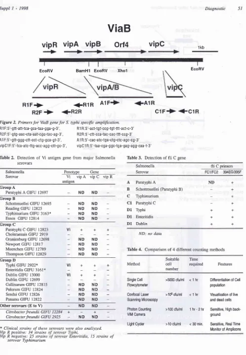
[image:2.595.52.542.92.488.2] [image:2.595.45.287.549.728.2]Suppl
I
-
1998 Diagnostic 51ViaB
vipR
-+
vipA
**
vipB
Orl4
vipC
tt
BamHl
EcoRV1kb I EcoRV I Xhol I EcoRV
RlF+
R2F
->
R 1 F:5'-gtt-att-tca- gca-taa- g ga-g -3', R 2 F:5'-gtg -aac-cta-aat-cg c:ta c-a g - 3', A1 F: 5'- gtt-g g g-ctt-cct-ctg-qca-gt-3',
+R1R
A1F->
<-R2R
clF+
+ClR
+A1R
Rl R :5'-act-tgt-ccg-tgt-ttt-act-c-3' R2 R:5'-ctt-cca-ta c- ca c-ttt-ccg -3'
A1 R : 5'-cac-atc-tga-ctg-ctc-a gc-c g-3'
Salmonella
Serovar
Penotype
Gene@
antigen
Table
3.
Detection offli
C geneSalmonella
Serovar
fli
C primers FCltFC2 394EG/395FParatyphi A
Schottmuellei (Paratyphi B) Typhimurium
Paratyphi C
Typhi Enteritidis Dublin
ND: no data
Tâble
4.
Comparison of 4 different counting methods Group AParatyphi A GIFU 12697 ND ND
A B
c
C1 D1 D1 D1 + + + + + + + ND + + + Group BSchottmuellei GIFU 12695
Reading GIFU 12825
Typhimurium GIFU 3163* Essen GIFU 12814
_NDND
-NDND
-NDND
-NDND
Group CParatyphi C GIFU 12823
Choleraesuis GIFU 2919
Oranienburg GIFU 12698
Newport GIFU 12817
Muenchen GIFU 12789
Thompson GIFU 12829
+
Vi
++
ND
NDND
NDND
NDND
ND Group D'lyphi GIFU 2922* Enteritidis GIFU 3161* Dublin GIFU 13000
Dublin GIFU 12699
Gallinarum GIFU 12815
Pulorum GIFU 12824
Sendai GIFU 12826
Panama GIFU 12822
+ Vi
Vi
Method
Suitable
Timecell
required numberFeatures
++
++
ND
NDND
NDND
NDND
NDSingle Cell Flowcytometer
>5000 cfu/ml
Confocal
Laser
>106 cfu/ml Sænning MicrosæpyPhoton Counting VIM Camera
Light Cyder
< t
hr
Differentiation of Cell population< t
hr
Visualization of live and dead cellsOther serovars (E to V) ND ND
C itrobacte r .fre undii G I F U 1 22 84
C it robac ter .fre undii GI F U 292 5
+
-+
ND
ND>100
cfu/ml
t hr - 2hr
Sensitive, High back-ground>10
cfu/ml
< 30min.
Sensitive, Real ïmeMonitor of Ampliæns
*
Clinical strain.s ofthese
also analiyzed.Vip R positive: 34
strains
hi,Vip R negative: 25
strains
teritidis, 15 strainsof
serovar TyphimuriumFigure 2. Primers
for
VaB genefor
S. typhi specific amplification.vipClF:5'-tca-atc-ttg-acc-agg-ctt-gc-3', vipClR:5'-taa-cga-ggc-tga-gag-agg-caa-t-3'
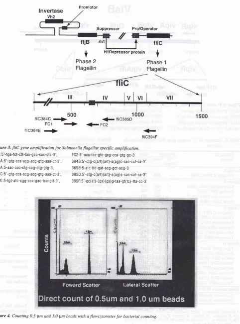
[image:3.595.37.535.46.762.2]Promotor
friB
rh1Suppressor Pro/Operator
ftic
+
Phase
1Flagellin
t
HlRepressor proteinPhase
2
Flagellin
500
fliO384O
r+>
FC1 +>
fllc394E
rf>
1
000
<_
ftic385D!çç2
<r
[image:4.595.59.542.82.733.2]flic394F Figure 3.
fIiC
gene amplificationfor
Salmonella flagellar specific amplffication.FC 1 :5'-tg a-tct-ctt-taa-g ac-ca c-cta
-3',
FC2:5'-aca-tcc-gtc- gcg -cca - gtg -g c- 3'3844:5'-gtg-cca-acg-acg-gtg-aaa-ct-3', 3B4B:5'-ctg-c(a/t)(a/t)-a(ag)c-cac-cat-ca-3'
3 6 9A:5-aac-aac-ctg -ca g -ctg - gtg -3, 3 69 B:5 -atc-ttc - gat-ac S-g ct-acg - 3 3B4C:5'-gtg-cca-acg-acg-gtg-aaa-ct-3', 3B5D:5'-ctg-c(a/t)(a/t)-a(ag)c-cac-cat-ca-S,
394E:5-tgt-atc-cgg-cca-gac-tca-gtt-3', 395F:5'-gc(at)-(ga)(ga)g-taa-gt(rc)-tta-cc-3,
Suppl
I
-
1998RESULTS
AND
DISCUSSION
Direct
andrapid counting of living
pathogenic
bacte-ria
in
biological
fluid is
afinal
goal
of
this
study.
A
new flowcytometric
counting method was able to
count particle bigger than 0.25
pm, while
conven-tional
flowcytometer
only
count particles
bigger
than
2
pm2.
Because sample
flow
on the
surface
of
thin cover
glass surface
in
a
new flowcytometer. As
in
Figure
40.5
pm
and
1pm
particle
were
clearly
dif-ferentiated. The method was very rapid and
simple
and
potential
to
be aroutine
counting method
atclini-cal laboratories. However, number
of
bacteria to
becounted should
behigher
than 5000
cfi/ml.
Theoreti-cally
it
was possible
to
count bacterial lower
than
1000
cfu/ml,
however,
the signal was weak and
nopeak
of
fluorescent was visible when the
organism
was less
than
1000cfu/ml.
We applied
this
method
to
directly count
bacteria
in
urine
(data
not
shown).
Data
from direct plate counting
method
andflow
cy-tometric counting
were
well
correlated
andmixed
in-fection
was predicted
by
their
different
fluorescent
intensities
because
different fluorescent
peaks were
observed
in
caseof
mixed infection.
Laser scanning
counting
method is another technique
directly count
living
pathogenic bacteria
in
clinical
specimens.
Combination
of 2 or 3
emission
lasersand
development
of
computer graphic
software
evo-lutionalized his counting
techniques.
Live
and
deadcell
staining fluorescent were
usedto count metabolic
activities
of
S. typhi and surrrival
of
cell population
during
starvation
andother
stress.Sigma
38deficient
mutant
created
from
S.
typhi GIFU
10007 were
very
sensitive
to
pH
stress,H2O2
stress,starvation
in
M9
cysteine
medium. The
deadcell
andlive cell
popula-tion were quickly counted
and quantitated
within
30minutes
after staining. However, application
of
this
method
to
count cells
in
clinical
fluid
might be
lim-ited
because 106/ml were
usually
required.
Recently,
ChemSan system
was introduced
and
wasable
to detect single
cell
on
afilter.
The
system seemsto
be very simple
and
useful
for
real
time counting
of
bacteria. However, The
method cannot
differenti-ate
pathogenic
bacteria.
Photon counting
VIM
camera8
was used
to
count
cells
emitting
photons
from
thefilter,
because thecell
on
the
filter
wastreated
with alkaline
phosphatasela-beled
antiserum.
The method
was
the most
sensitive
among
the
above mentioned three methods.
How-ever,
reducing
the
background photons emmitted
from
the
filter
was the biggest problem.
Diagnostic
53PCR
amplification of
pathogen
specific
genes are themost sensitive technique. However, the quantitative
detection
of living
cells
were
not
possible
bir
this
method.
RT:PCR
and
quantitative amplification
is
apossible method
to
solve this problem. We
selectedseveral
primers
to
differentiate Salmonella
serovars.rfb
gene7,Vi
regulatory
gene3-sand
flagellin
gene7,9were
selectedfor
this purpose. Primers prepared
from
fliC
gene
of
S. typhi couta amplify.màst^of
Salmo-nella
serovars.
Amplification
was performed
by
alight cycler
and thefluorescent
intensity of
amplicons
were
monitored
in
each
amplification cycle.
The
method was
rapid
becauseamplicons were
quantita-tively
monitored
during
each
amplification
cycle.
When the sample contained more
than
I}2lmI
of
bac-teria, detection
of
S.typhi could
becompleted within
20 min.
REFERENCES
1.
ArmstrongMl
Koziel H, Rose RM, Arena C , Richards FF.Indicators of Pneumocystis carinii viability in short-term cell culture. J Protozoology 1991; 38(6): 88S-90S
2.
Davey HM, KelI DB. Flowcytometry and cell sorting of het-erogeneous Microbial populations: the importânce of single-cell analyses. Microbiol Review 1996;60 641-96.3.
HashimotoXLi
N, Yokoyama H, Ezaki T. Complete nucleo-tide sequence and molecular characterization of via B region encodingVi
antigenin
Salmonella typhi. J Bacteriol 1993;175: 4456-65.
4.
Hashimoto Y, IthoY
et al. Development of nested PCR basedon the
VaB
sequence to detect Salmonella typhi.i
C\)nl/l;i-crobiol 1995; 33: 175-7.
5.
HashimotoI
Khan AQ, Ezaki T. Positive Autoregulation ofvipR Expression in ViaB Region Encoded Vi Antigen of
Sal-monella typhi. J Bacteriol 1996; 1'78(5): 1430-6
6.
Humphreys MJ, Atlman R, Lloyd D. Determinarion of thevi-ability of Trichomonas vaginalis using
flow
cytometry.Cy-lometry 1994; l5(4): 343-8.
7
.
Itoh K, Hirose,EzakiT. Amplification of rfbE and fliC genesby polymerase chain reaction for identification and detection
of
Salmonella serovar Enteritidis, Dublin and Gallinarum-Pullorum. Microbiol Immunol 1997 4l:'791-4.8.
MasukoM,
HosoiS,
HayakawaT.
Rapid detection andcounting
of
single bacteriain
a wide field using a photon-countingTV
camera. FEMS MicrobiolLett
1991; 6'7(2):23t-8.
9.
Selander RK,Li
J, Nelson K. Evolutionary genetics ofSal-monella enterica p2691-2690.
In
FC Neidhardt (eds).Es-cherichia coli and Salmonella. vol. 2 ASM Press Washington